Abstracts from the Department of Biological Sciences
Undergraduate Summer Research Symposium August 8, 2007

Ordered alphabetically by student's last name

Lavender Majid
Millner Neal Ociepa Samant Smith Umoren Wille
Lavoie
McLaughlin Mmagu Nguyen Reisinger Shuman Sun Warren Woody
Madden Michael Mogere Nigro Richards Sloofman Teixeira Warriner Zilora


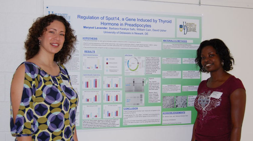
Regulation of Spot14, a Gene Induced by Thyroid Hormone in Preadipocytes
Marysol D. Lavander,
Barbara Kwakye Safo, David Usher
Department of Biological Sciences

Spot14 or Thyroid hormone responsive protein is a transcription factor found in lipogenic tissues such as the liver, brown and white adipose tissues and lactating mammary glands. In previous studies, it has been shown that expression of Spot 14 is regulated by liver X receptor (LXR), peroxisome proliferator-activated receptor gamma (PPARγ) as well as triiodo-L-thyronine (T3). There are two, long term goals of this study. One is to identify Spot14 target genes in adipocytes. Once a gene has been identified, experiments will be done to locate Spot14’s binding sequence. Possible target genes of Spot14 are ATP citrate lyase (Acly), malic enzyme supernatant (Mod1), malate dehydrogenase 1 (Mdh1), acetyl-Coenzyme A carboxylase alpha (Acaca), pyruvate carboxylase (Pcx), and solute carrier family 25 member 1 (Scl25a1). The second goal is to determine regions of the Spot14 promoter, which make Spot14 sensitive to cholesterol concentration. In this study, primers were designed to identify the proximal promoter region of Spot14, a region 2kb upstream of the 5’ end of the gene. Once identified this proximal promoter will be inserted into the expression vector pGL4 Luciferase Reporter from Promega. Primer selection was done by utilizing the Primer3 website to customize the forward and reverse primers for a 2kb product. The reverse primer region was tested along with the HindIII linker that would be attached to the Luciferase gene on the vector by using the NCBI Open Reading Frame (ORF) Finder. Once it was established that the primers worked, we ran RT-PCR on DNA from isolated undifferentiated fibroblast cell line 3T3-L1.  Funded by the Science and Engineering  Program as a Life Science Scholar.


Copy Number Changes in the PLP1 Region in Patients with PMD

Danielle Lavoie
, Karen Sperle, and Grace Hobson1
University
of Delaware
and 1Nemours Biomedical Research, Alfred I. duPont Hospital for Children

Pelizaeus- Merzbacher Disease (PMD) is an X-linked recessive condition that affects the production and function of myelin in the central nervous system.  It is caused by alterations in the proteolipid protein 1 (PLP1) gene on Xq22.  These alterations include copy number changes in the chromosomal region containing the gene, as well as point mutations in the gene itself.  Previous research has shown that the size of the region with copy number changes varies among PMD patients.  We analyzed DNA from nine patients via quantitative multiplex PCR to delineate the region of copy number change in these individuals.  All of these DNAs have duplicated regions, and five have triplications in addition to the duplications.  One patient has DNA regions with a copy number greater than triplicated.  The size of the region of copy number change proximal to PLP1 is variable among these patients, but eight of the nine have their duplications/ triplications ending in the low copy repeat (LCR) region distal to PLP1, suggesting a possible role for this structure in the mechanism by which the rearrangements were formed.  Junction fragments across duplication breakpoints were obtained in two patients, showing tandem head-to-tail orientation.  One patient’s DNA had a simple rearrangement and the other had a complex rearrangement with microhomologies.  One of these patients yielded the same junctions as his affected relative, showing the familial nature of these rearrangements.  These data support previous research suggesting a mechanism of non-homologous end joining in the formation of the copy number changes.  A junction fragment involving a triplicated region is still being pursued. Funded by Nemours Biomedical Research


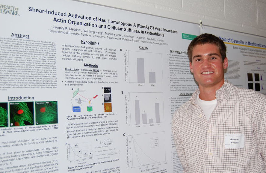
Shear-Induced Activation of Ras Homologous A (RhoA) GTPase
Increases Actin Organization and Cellular Stiffness in Osteoblasts

Gregory Madden,
Weidong Yang, Manisha Malik, Elizabeth L. Adams, Randall L. Duncan
Department
of Biological Sciences 

Osteoblasts respond to mechanical stimuli with increased intracellular signaling and gene expression that ultimately results in bone formation, in vivo, yet skeletal mechanosensitivity is lost upon continued stimulation.  Osteoblasts subjected to fluid shear rapidly polymerize the actin cytoskeleton, forming actin stress fibers that we predict is critical to the loss of mechanosensitivity.  Parathyroid hormone (PTH), a calciotropic hormone that is crucial to the regulation of bone mass, enhances the response of bone to mechanical loading and we have shown that PTH reduces cellular stiffness two-fold by disruption of actin filaments.  We postulated that activation of the Ras homologous A (RhoA) GTPase pathway, a pathway known to promote actin assembly in other tissues, mediates cytoskeletal organization in response to shear.  Using Atomic Force Microscopy (AFM) to quantify osteoblastic stiffness through stress-strain kinetics, we found that exposure of MC3T3-E1 osteoblasts to 12 dynes/cm2 fluid shear for 15 min significantly increased cellular stiffness from 1.1 kPa in static, or non-sheared, controls to 6.0 kPa in sheared osteoblasts.  While RhoA activates several effector proteins, previous studies in our lab have shown that Rho-dependent kinase (ROCK) regulates actin polymerization by phosphorylating the actin severing protein, cofilin, rendering this protein inactive.  Specific inhibition of ROCK with Y27632 significantly attenuated the shear-induced increase in cellular stiffness to 2.6 kPa.  Conversely, the cell stiffness of non-sheared cells increased to values similar to those of sheared cells (5.2 kPa) upon addition of a RhoA GTPase activator, lysophosphatidic acid (LPA).  These studies suggest that fluid shear induces increased actin organization through the activation of the RhoA-ROCK pathway and that the resultant changes in cell stiffness may play a role in the decreased mechanosensitivity of osteoblasts.  (Supported by HHMI and NIH/NIAMS AR043222).


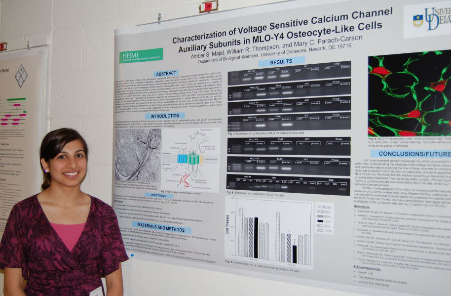
Characterization of Voltage Sensitive Calcium Channel Subunits in MLO-Y4 Osteocyte-Like Cells

Amber S. Majid
, William R. Thompson, Mary C. Farach-Carson
Department of Biological Sciences

There are three major types of bone cells, osteoblasts, osteoclasts, and osteocytes.  Osteocytes are the most abundant cells in bone. While their function is not completely understood, osteocytes sense mechanical signals within bone. Voltage sensitive calcium channels (VSCC) span the cell membrane and open in response to external stimuli resulting in altered Ca2+ permeability.  Once inside the cell, Ca2+ acts as a second messenger eliciting specific cellular responses.  L-type VSCCs are composed of a complex of polypeptide units consisting of a pore-forming α1 subunit, an intracellular β subunit, a dimer of disulfide linked α2 and δ subunits, which form an extracellular auxiliary complex and a γ subunit in some tissues.  Osteocytes have been found to express higher levels of the T-type (Cav3.2) α1H subunit than the L-type (Cav1.2) α1C subunit.  The remaining subunits of the T-type VSCC have not yet been characterized in osteocytes.  This study was designed to examine the expression of L- and T-type VSCC subunits in an osteocytic cell line, MLO-Y4.  We found significant levels of both the α1C subunit transcript and the α1H subunit transcript in the MLO-Y4 cell line.  Additionally, we demonstrated the presence of the α2δ1 subunit transcript but not the α2δ2, α2δ3, or α2δ4 transcripts within MLO-Y4 osteocytes.  Continued studies will determine if the MLO-Y4 α2δ subunits associate with α1 subunits representing L-type and/or T-type VSCCs. Characterization of VSCC structure within osteocytes provides a rationale to conduct functional studies such as the role of these channels in calcium signaling associated with mechanotransduction. Supported by HHMI and COBRE P20 RR016458.


The Role of Caveolin in Mechanotransduction in Osteoblasts
Eamon McLaughlin
, Shyama Majumdar, Randall L. Duncan
Department of Biological Sciences

When subjected to a novel mechanical load, the skeleton will remodel to increase bone mass to sustain this load.  Our lab has shown that when the purinergic receptor P2X7, an ATP-gated ion channel, is deleted in mice, these mice exhibit a skeletal phenotype typical of disuse osteoporosis (paralysis, extended bed rest, or space travel) and demonstrate decreased mechanosensitivity to exogenous mechanical loading compared to normal, or wild-type mice.  We have also shown that activation of the P2X7 receptor promotes the uptake of large solutes such as YO-PRO1, a fluorescent dye weighing 629 daltons.  We hypothesize that caveolins, membrane proteins associated with clathrin-independent endocytosis, are responsible for this uptake and that caveolins act to internalize P2X7 receptors to regulate their mechanical activation.  Western blot analyses of MC3T3-E1 preosteoblasts subjected to 12 dynes/cm2 fluid shear demonstrated that caveolin-1 was phosphorylated within 15 min of the onset of shear. Inhibition of P2X7 with Brilliant Blue G or hydrolysis of extracellular ATP with apyrase significantly attenuated this phosphorylation event.  Stimulation of P2X7 receptors with the specific agonist, BzATP, resulted in increased caveolin-1 phosphorylation.  These data indicate that caveolin-1 is activated in response to shear through P2X7 receptor binding and suggests that this activation may be crucial to the mechanosensitivity of the osteoblast.  Future studies will examine the mechanisms associated with phosphorylation of caveolin and the role of caveolin in mechanotransduction, which could prove to be vital in the treatment of osteoporosis.This project was funded in part by a National Institute of Health INBRE grant and NIH/NIAMS Grant AR043222.


Investigation of long-term effects of Nanoscale TiO2 on Murine Spermatogenesis

Rowan Michael
, Minghai Shao, Genevieve Griffiths, and Patricia A. Martin-DeLeon
Department of Biolgical Sciences

The retention of excess residual cytoplasm (EC), loosely termed cytoplasmic droplets, on the sperm flagellum during spermiogenesis is associated with male infertility in a wide variety of mammalian species, including humans. These abnormal structures are produced by both over-expression of hyaluronidases and environmental exposure to agents creating reactive oxygen species (ROS). Nanoscale titanium dioxide (TiO2) has been shown to form ROS when exposed to ultraviolet light and in aqueous suspension. The objective of this study was to examine the effects of nano-TiO2 in mice, specifically relating to EC formation and other morphological abnormalities. Mice were injected intraperitoneally with either a control, low or high dose of nano-TiO2 for three consecutive days. They were then sacrificed at varying intervals after injection, the cauda and corpus of the epididymis were minced to extract sperm, and the testes were removed and frozen. The sperm from mice exposed to nano-TiO2 were then compared to the control sperm in regards to morphology, motility, ability to acrosome react, and mitochondrial potential. The testes were then sectioned and DNA was extracted and observed for any damage. To date, there have been no significant differences observed between sperm from mice receiving a control, low dose or high dose of nano-TiO2. Funded by Howard Hughes Medical Institute.


Development of an In Vitro Explant Culture System for the Avian Embryonic Lung to Examine
SP-A and SP-B Expression through RT-PCR and Western Blot Analysis
Patricia R. Millner, Department of Biological Sciences, Wesley College, Dover, Delaware,
and Sabrina M. Brougher, Department of Biological Sciences, Delaware State University, Dover, Delaware


Fetal lung cultured as explants, in which intercellular structural design is kept constant, is known to respond to certain hormones and agents that can either promote or inhibit the acquisition of surfactant related properties. Our goal was to develop an in vitro culture system for avian lung explant tissue to examine patterns of differential expression of SP-A and SP-B in response to hormone culture through RT-PCR and Western Blot analysis. We hypothesized that we would see more expression of SP-A and SP-B in the DCI medium compared to the 8-bromo-cAMP + IBMX medium, the Dexamethasone medium, and the Waymouth medium used, because DCI is a combination of all 3 of the other media; having multi-hormones present in its make-up. Fetal lung tissue was obtained on day 15 from 20 fowl embryos. In brief, the tissue was minced into approximately 5 micrometer increments and cultured as explants in a CO2 incubator; temperature was set at 37ºC with an atmosphere of 95% air/ 5% CO2 on a rocker platform (3oscillations/min), gas pressure was set at 15 psi. Explants were maintained for 1-5 days in one of four types of media. The media were changed daily. We have not yet finished Western Blot and RT-PCR analysis on the explant samples. Once we gather results, we will be able to examine the patterns of differential expression of SP-A and SP-B in response to the various hormone cultures. Supported by NIH grant 2 P20 RR016472-07 under the INBRE Program of the National Center for Research Resources (NCRR).


The PPARα Agonist, Fenofibrate, Increases MUC1 Expression in T47D Breast Cancer Cells
Obinna Mmagu, Peng Wang, and Daniel Carson
Department of Biological Sciences

Ninety percent of all breast cancers overexpress the high molecular weight, cell surface mucin, MUC1.  MUC1 is a multifunctional cell surface component that protects normal and tumor cell surfaces from infection, enzymatic attack and immune responses as well as participates in several intracellular signal transduction pathways.  Peroxisome Proliferator-Activated Receptors (PPARs) are ligand-activated transcription factors that enter the nucleus, heterodimerize with retinoid X receptor (RXR), and bind to PPAR response elements.  Recent studies have shown that PPAR agonists display anti-cancer effects, and may modulate progesterone responses.  We hypothesized that fenofibrate, a PPARα agonist, antagonizes progesterone-stimulated MUC1 expression in T47D breast cancer cells.  We confirmed the presence of the subunits of PPAR and RXR by RT-PCR.  After culture and treatment, the T47D cells were lysed, and the proteins were detected by Western Blotting.  However, fenofibrate increased MUC1 expression in T47D breast cancer cells.  It is likely that activation of PPARα causes differential recruitment of transcriptional coactivators and corepressors to produce its effect on MUC1 gene expression, and may vary from cell to cell.  By examining the mechanisms of PPAR action on MUC1 expression, we may be able to exploit these transcription factors as novel targets to reduce MUC1 overexpression in breast cancer.  Funding provided by the Howard Hughes Medical Institute Undergraduate Science Education Program, the Ronald E. McNair Scholars Program, and NIH grant HD29963.


Ion Channel Therapeutics in Prostate Cancer

Brenda Mogere,
Jennifer Ambrose, and Robert A. Sikes
Department of Biological Sciences

Cancer cells have the extraordinary ability to alter their phenotypes and mutate their genotype to attain selective advantage and produce one cell that will survive and colonize at the metastatic site. This is a major cause of treatment failure creating a tremendous hurdle to overcome in designing novel cancer therapeutics. One alternate approach is to examine the interactions between drugs to determine if a previously undiscovered synergy exists. This would enhance tumor cell kill and increase the tolerance of the therapy in the patient by reducing the therapeutic dosage of both drugs. These drugs are chemosensitizers or cooperative chemotherapeutics. Adding drugs at the IC20s helps determine whether synergy with traditional chemotherapy occurs. My research involved obtaining inhibitory concentration curves for different drugs to determine the IC50, IC20 and IC10. Five drugs commonly used to treat prostate cancer Docetaxel, Vinblastine, Cisplatine, Verapamil and Etoposide, were used in this research and two different assays, MTT and crystal violet, were applied to determine cell viability.  LNCaP cells were plated at 360,000 cells per 48 well plate followed by treatment with respective drugs for 5 or 7 days and the results were analyzed. To date we have obtained good dose-response curves for Docetaxel that will allow us to calculate the IC20 and begin synergy experiments with sodium channel blockers. Source of Funding: Department of Defense.


Mutational Analysis of selected residues in the J-Domain and Hinge region of SV40 Large Tumor Antigen
Krista Neal
and Daniel T. Simmons
Department of Biological Sciences

Simian Virus 40 (SV40) provides an exemplary model system to study the highly regulated process of eukaryotic DNA replication. Its genome encodes the multifunctional protein, large T-Antigen (T-Ag), which orchestrates the initiation of replication. T-Ag is known to interact with several cellular proteins including Replication Protein A (RPA), Topoisomerase I, and DNA polymerase alpha/primase. Topo I binds to double-stranded viral DNA ahead of the replication fork and T-Antigen’s helicase activity. It nicks one DNA strand, swivels it around, and reanneals the strands to release torsional strain generated in replication. Topo I has been found to interact specifically with T-Antigen double hexamers at two sites, one N-terminal and one C-terminal. Although these binding domains are approximated, they still remain to be defined. Prior data localized the N-terminal Topo I binding domain to T-Antigen’s J-Domain and adjacent Flexible Linker region. Single point mutations were generated in this region and several mutant proteins have been purified. In vitro replication assays are being performed with mutant T-Antigens to assess their ability to function in DNA replication. In addition, future assays will be performed to characterize the ability of the mutant T-Antigens to bind Topo I as well as to perform various activities involved in DNA replication. We hope to correlate replication deficiency with a Topo I binding deficiency in several mutant T-Antigens. Funded by the HHMI Program and PHS Grant from the National Cancer Institute.


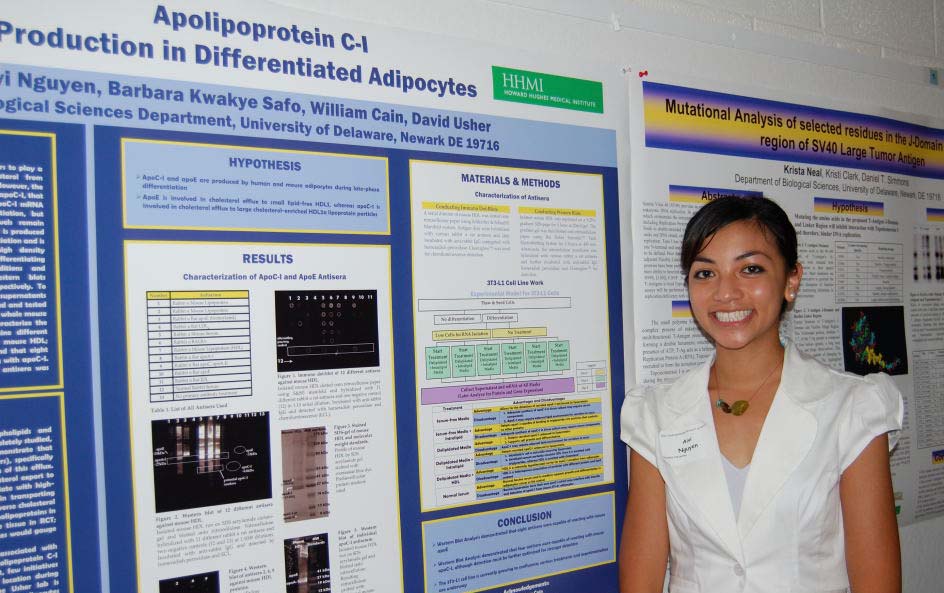
Apolipoprotein C-I Production During Adipocyte Differentiation
Aivi Nguyen, Barbara Kwakye Safo, William Cain, David Usher
Department of Biological Sciences

One apolipoprotein, apoE, appears to play a significant role in the secretion of unesterified cholesterol from adipoctyes. However, the Usher lab has identified another apolipoprotein, apoC-I, which may also be involved. Previous studies have shown that apoC-I mRNA is high in late-phase 3T3-L1 adipocyte differentiation. Thus, in this study it is hypothesized that apoC-I is both synthesized and secreted by human and mouse adipocytes during late-phase differentiation. The hypothesis is to be tested by differentiating human and 3T3-L1 adipocytes under various conditions, and correspondingly, utilizing RT-PCR and western blots to quantify apoC-I mRNA and subsequent expression, respectively. However, to detect apoC-I in the supernatants or within the cells, specific antisera first had to be developed and tested for specificity. A series of immuno-dot and western blots on whole mouse plasma and isolated high density lipoproteins (HDLs) were conducted to characterize the primary antibodies to be used in the study. The immuno-dot blots identified nine different antisera capable of recognizing mouse apoplipoproteins in mouse HDL. Furthermore, Western Blot analysis of these antisera found that eight were capable of reacting with apoE and three for reacting with apoC-I. For detecting human apoC-I, an apoC-1 specific antisera was commercially purchased and tested.  Funded by the Howard Hughes Medical Institute.


Viability and In Situ Intercellular Ca2+ Imaging of Bone Cells
Samantha Nigro1,
Victor Fomin1, Randy Duncan1 and Liyun Wang2
1Department of Biological Sciences, 2Department of Mechanical Engineering,

The goals of this research were i) to study bone cell viability in isolated bone and ii) to image the intracellular Ca2+ responses of the bone cells under chemical and mechanical stimuli. Living bone is capable of adapting its form and mass to meet its requirements of load carrying and calcium homeostasis. The cellular mechanisms of this bone adaptation have been extensively studied using cultured cells and the cells have been shown to respond to external mechanical and chemical stimuli by transient increase of intracellular Ca2+. Due to technical difficulties, experimental study of cellular responses in intact bone has not been undertaken yet. In this study, calvaria, tibia, femur and metatarsals were isolated from B6 mice in order to establish an organ model that allowed for imaging the levels of Ca2+ in situ. Cell viability was measured using the Live/Dead fluorescent assay and the intracellular Ca2+ was imaged using the ratiometric indicator, Fura-2. This research was sponsored by NIH. (P20 RR016458-06 A1)


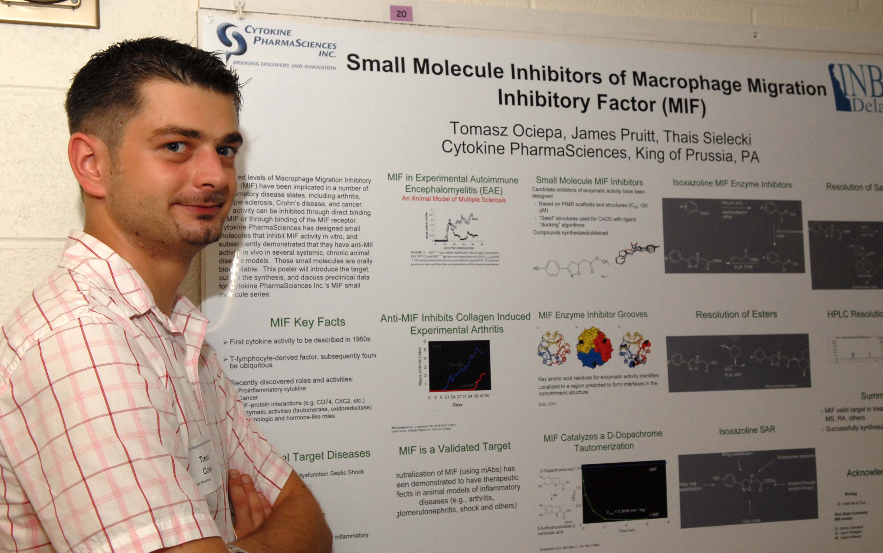
Small Molecule Inhibitors of Macrophage Migration Inhibitory Factor (MIF)
Tomasz Ociepa
, James Pruitt, Thais Sielecki,
Cytokine PharmaSciences, King of Prussia, PA 

Elevated levels of Macrophage Migration Inhibitory Factor (MIF) have been implicated in a number of inflammatory disease states, including arthritis, multiple sclerosis, Crohn’s disease, and cancer.  MIF activity can be inhibited through direct binding of MIF or through binding of the MIF receptor.  Cytokine PharmaSciences has designed small molecules that inhibit MIF activity in vitro, and subsequently demonstrated that they have anti-MIF activity in vivo in several systemic, chronic animal disease models.  These small molecules are orally bioavailable.  This poster will introduce the target, outline the synthesis, and discuss preclinical data for Cytokine PharmaSciences Inc.’s MIF small molecule series.


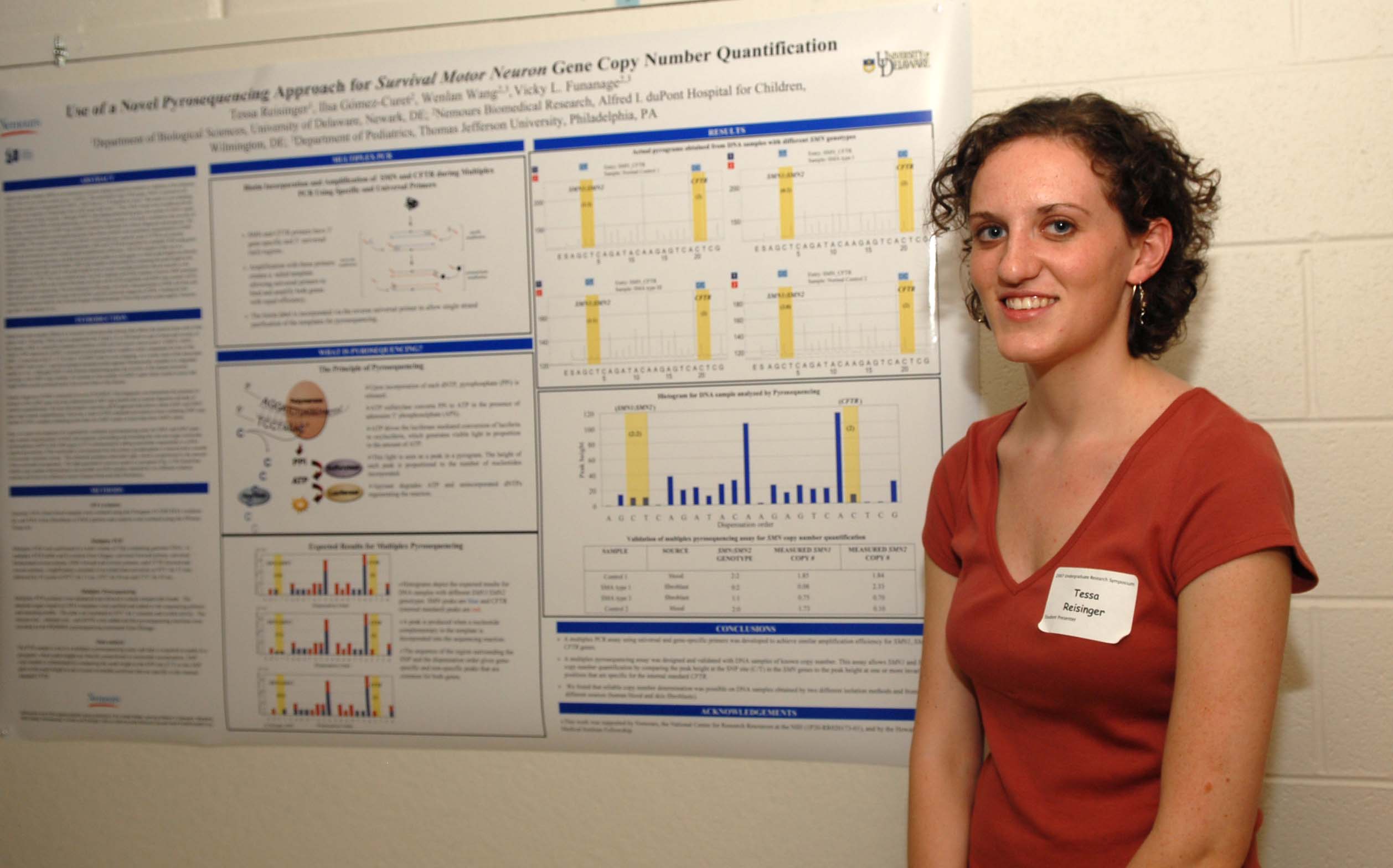
Use of a novel Pyrosequencing approach for Survival Motor Neuron Gene Copy Number quantification

Tessa Reisinger1, Ilsa Gómez-Curet2, Sarah Swain1, Vicky Funanage2,3, Thomas O. Crawford4, Mena Scavina2, Wenlan Wang2,3
1
Department of Biological Science, University of Delaware, Newark, DE; 2Nemours Biomedical Research, Alfred I. duPont Hospital for Children, Wilmington, DE; 3Department of Pediatrics, Thomas Jefferson University, Philadelphia, PA; 4Departments of Neurology and Pediatrics, Johns Hopkins University, Baltimore, MD

Spinal Muscular Atrophy (SMA) is an autosomal recessive disease caused by mutation or deletion of the telomeric copy of the survival motor neuron gene 1 (SMN1). The centromeric copy of the gene, SMN2, is present in all patients but unable to fully compensate for SMN1. SMN2 has a C→T transition in exon 7 that leads to exon skipping. As a result, the majority of transcripts from SMN2 gene lack exon 7. Thus, SMA results from insufficient levels of SMN protein in motor neurons. Although SMN1 is the disease-determining gene, the number of SMN2 copies appears to modulate SMA clinical phenotype. Determination of SMN copy number is therefore important for clinical diagnosis and prognosis. We have developed a Multiplex pyrosequencing assay that allows sensitive and quantitative determination of SMN copy number. This assay includes the cystic fibrosis transmembrane regulator gene (CFTR) as an internal standard of known copy number (2 copies/genome). SMN and CFTR are amplified by multiplex PCR using gene-specific and universal primers to achieve equal amplification efficiency. The PCR sample is then run in a multiplex pyrosequencing assay and data is acquired as peaks in a pyrogram in which peak heights are directly proportional to nucleotide incorporation. SMN copy number is determined by comparing the peak height at the SNP site (C/T) in the SMN gene to the peak height at one or more positions that are specific for the internal standard CFTR. This assay was validated with DNA samples of known SMN copy number and will be used to determine SMN copy number of DNA samples isolated from SMA and control fibroblasts. The development of this assay will allow characterization of SMA cell lines and may also generate a quantitative assay that could be used for molecular diagnosis of SMA. Funding for this research has been provided by the Howard Hughes Medical Institute Fellowship and by grant support: Nemours and NIH 1 P20 RR020173-01.


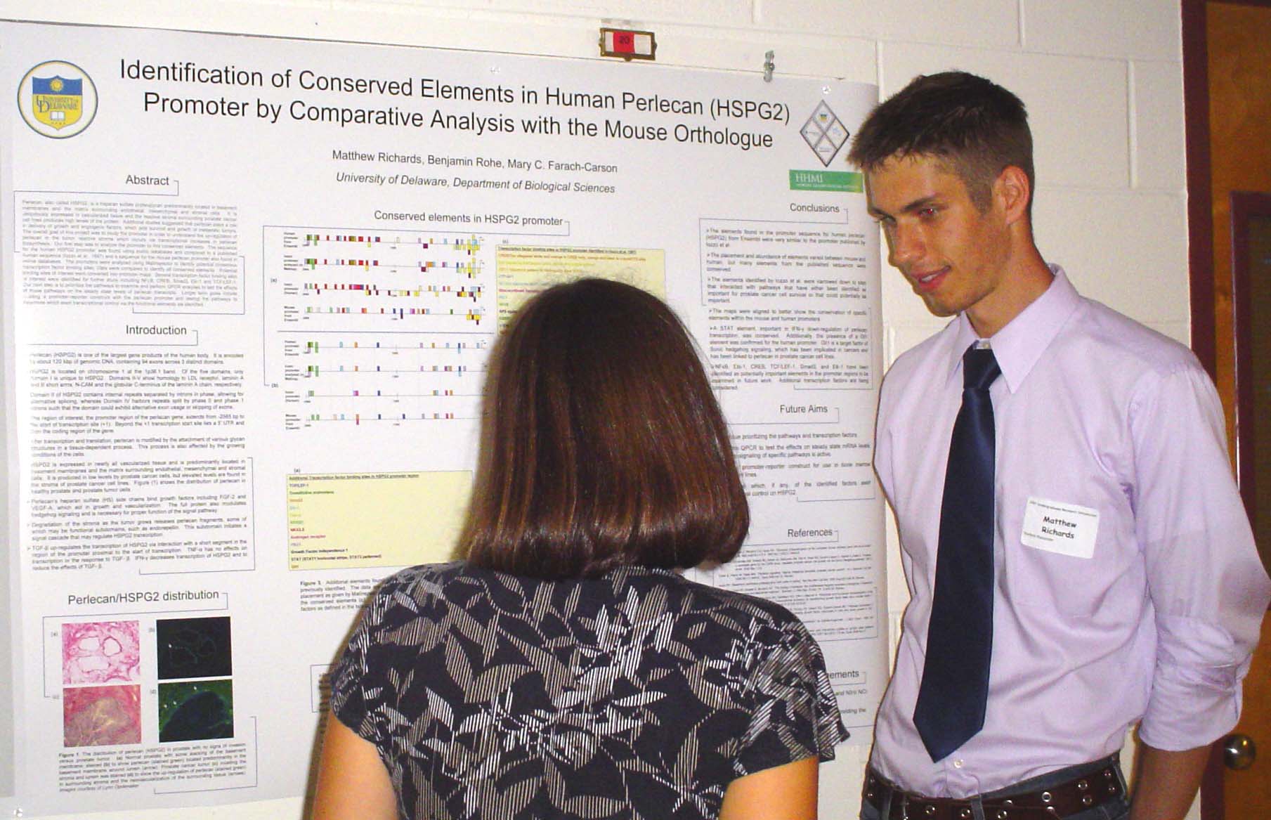
Identification of Conserved Elements in Human Perlecan Promoter by Comparative Analysis with the Mouse Orthologue.

Matthew Richards
, Mary C. Farach-Carson, Benjamin Rohe
Department of Biological Sciences

Perlecan is a heparan sulfate proteoglycan predominantly located in basement membranes and the matrix surrounding endothelial, mesenchymal and stromal cells.  It is ubiquitously expressed in vascularized tissue and the reactive stroma surrounding prostate cancer cell lines produces high levels of the protein.  Additional studies suggested that perlecan plays a role in delivery of growth and angiogenic factors, which aids survival and growth of metastatic tumors.  The overall goal of this project was to study the promoter in order to understand the up-regulaton of perlecan in the tumor reactive stroma which occurs via transcriptional increases in perlecan biosynthesis.  Our first step was to analyze the promoter to find conserved elements.  The sequence for the human perlecan promoter was found using public databases and compared to a published human sequence (Iozzo et al., 1997) and a sequence for the mouse perlecan promoter also found in online databases.  The promoters were analyzed using MatInspector to identify potential consensus transcription factor binding sites. Data were compared to identify all conserved elements.  Potential binding sites of interest were converted into promoter maps.  Several transcription factor binding sites of interest were identified for further study including NFkB, CREB, Smad3, Elk-1 and TCF/LEF-1.  Our next step is to prioritize the pathways to examine and perform QPCR analyses to test the effects of those pathways on the steady state levels of perlecan transcripts.  Longer term goals include building a promoter-reporter construct with the perlecan promoter and testing the pathways to determine which exert transcriptional control via the functional elements we identified.  This project was funded by HHMI and NIH/ NCI P01 CA098912


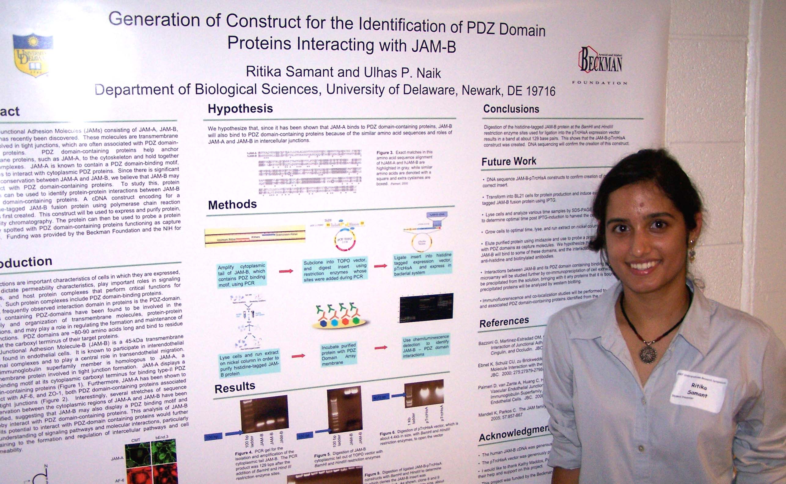
Generation of Construct for the Identification of PDZ Domain Proteins Interacting with JAM-B
Ritika Samant and Ulhas P. Naik
Department of Biological Sciences

A family of Junctional Adhesion Molecules (JAMs) consisting of JAM-A, JAM-B, and JAM-C has recently been discovered.  These molecules are transmembrane proteins involved in tight junctions, which are often associated with PDZ domain-containing proteins.  JAM-A is known to contain a PDZ domain-binding motif, which it uses to interact with cytoplasmic PDZ proteins.  Since there is significant conservation between JAM-A and JAM-B, we believe that JAM-B may also interact with PDZ domain-containing proteins.  To study this, protein microarrays can be used to identify protein-protein interactions between JAM-B and PDZ domain-containing proteins. A cDNA construct encoding for a polyhistidine-tagged JAM-B fusion protein using polymerase chain reaction (PCR) was first created.  This construct will be used to express and purify protein, using affinity chromatography. The protein can then be used to probe a protein microarray spotted with PDZ domain-containing proteins functioning as capture molecules.  Funding was provided by the Beckman Foundation and the NIH.


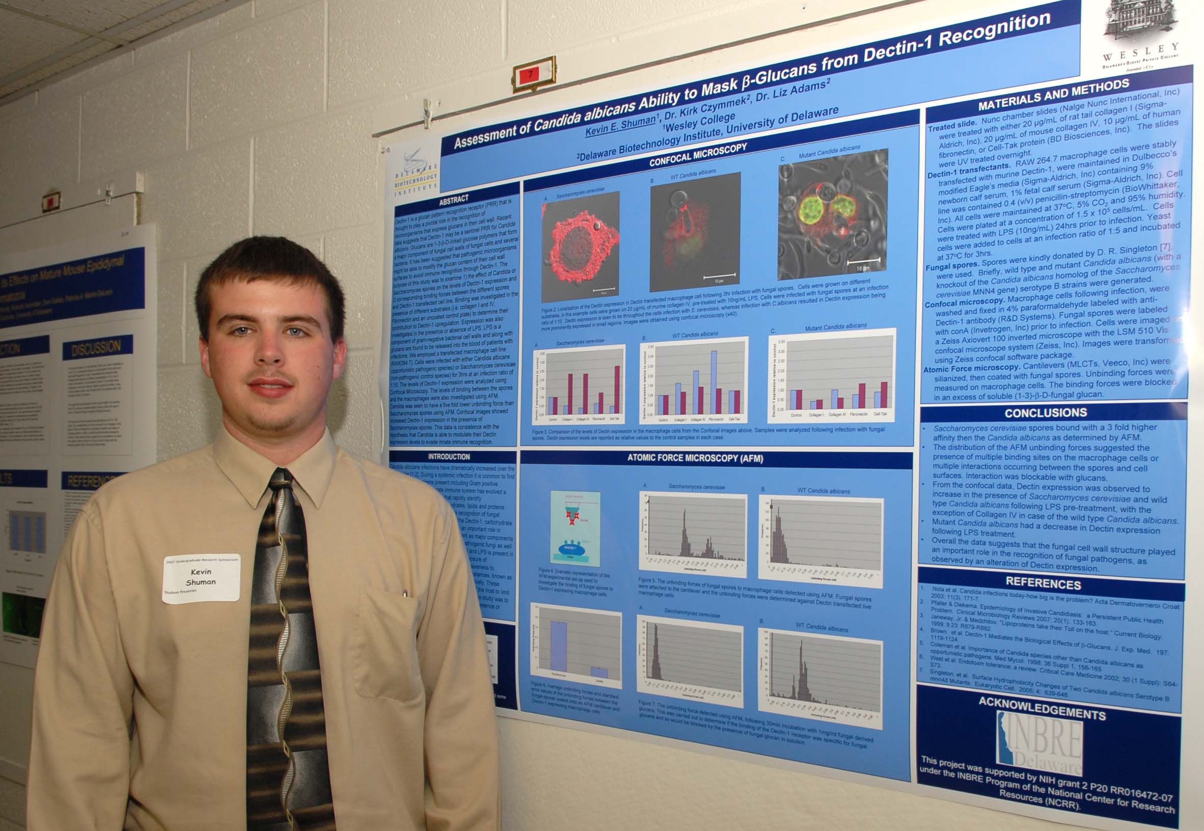
Assessment of Candida albicans Ability to Mask
b-Glucans from Dectin-1 Recognition
Kevin E. Shuman1, Kirk Czymmek2, Liz Adams2
1Wesley College, 2Delaware Biotechnology Institute, University of Delaware

Dectin-1 is a glucan pattern recognition receptor (PRR) that is thought to play a pivotal role in the recognition of microorganisms that express glucans in their cell wall. Recent data suggests that Dectin-1 may be a sentinel PRR for Candida albicans. Glucans are 1-3-b-D-linked glucose polymers that form a major component of fungal cell walls of fungal cells and several bacteria. It has been suggested that pathogenic microorganisms might be able to modify the glucan content of their cell wall surfaces to avoid immune recognition through Dectin-1. The purpose of this study was to examine 1) the effect of Candida or Saccharomyces spores on the levels of Dectin-1 expression and 2) corresponding binding forces between the different spores and Dectin-1 transfected cell line. Binding was investigated in the presence of different substrates (i.e. collagen I and IV, Fibronectin and an uncoated control plate) to determine their contribution to Dectin-1 upregulation. Expression was also investigated in the presence or absence of LPS. LPS is a component of gram-negative bacterial cell walls and along with glucans are found to be released into the blood of patients with infections. We employed a transfected macrophage cell line (RAW264.7). Cells were infected with either Candida albicans (opportunistic pathogenic species) or Saccharomyces cerevisiae (non-pathogenic control species) for 3hrs at an infection ratio of 1:10. Candida was seen to have a five fold lower unbinding force than Saccharomyces spores using AFM. Confocal images showed increased Dectin-1 expression in the presence of Saccharomyces spores compared to Candida spores. This data is consistence with the hypothesis that Candida is able to modulate their Dectin expression levels to evade innate immune recognition. Supported by NIH grant 2 P20 RR016472-07 under the INBRE Program of the National Center for Research Resources (NCRR).


Effects of RPL29 deficiency on bone structure and material properties
Laura Sloofman1, Daniel S. Oristian1, Xiaozhou Zhou2, Liyun Wang2, and Catherine B. Kirn-Safran1
1
Department of Biological Sciences and 2Department of Mechanical Engineering

Ribosomal proteins (RPs) play an important function in the maintenance of a normal protein synthetic rate.  Our group generated the first viable mouse mutant model lacking an individual ribosomal protein.  In these mutants, the absence of RPL29 resulted in global skeletal growth deficiencies that persist during adulthood.  In the current study, we used RPL29 knockout mice to evaluate this protein function in bone structure and rigidity. We hypothesized that a decrease in protein biosynthesis affecting matrix production in RPL29 knockout mice reduces bone mass and increases bone fragility. The bone microstructure of both null and wild type six-month-old femurs at mid-shaft was assessed using micro-computed tomography (microCT). Significant differences in the bone composition were found between the null and control mice.  We found that RPL29-deficient femoral diaphyses exhibit a significant decrease in cortical area (11 % and 19% for males and females, respectively) and polar moment of inertia (39% and 32% for males and females, respectively) compared to control animals.  Three-point bending tests were performed on the femurs to determine the elastic modulus times the moment of inertia (EI), ultimate force, and stiffness.  Interestingly, only RPL29 null female femurs showed significant reduction in their structural parameters when compared to controls.  These data indicate that RPL29 deficiency is associated with decreased bone mechanical properties specifically in females.  In conclusion, bone structure and material properties in females appear to be more sensitive to a perturbation in global protein synthesis when compared to males. This project is supported by the University of Delaware NIH-COBRE P20 RR016458-06.


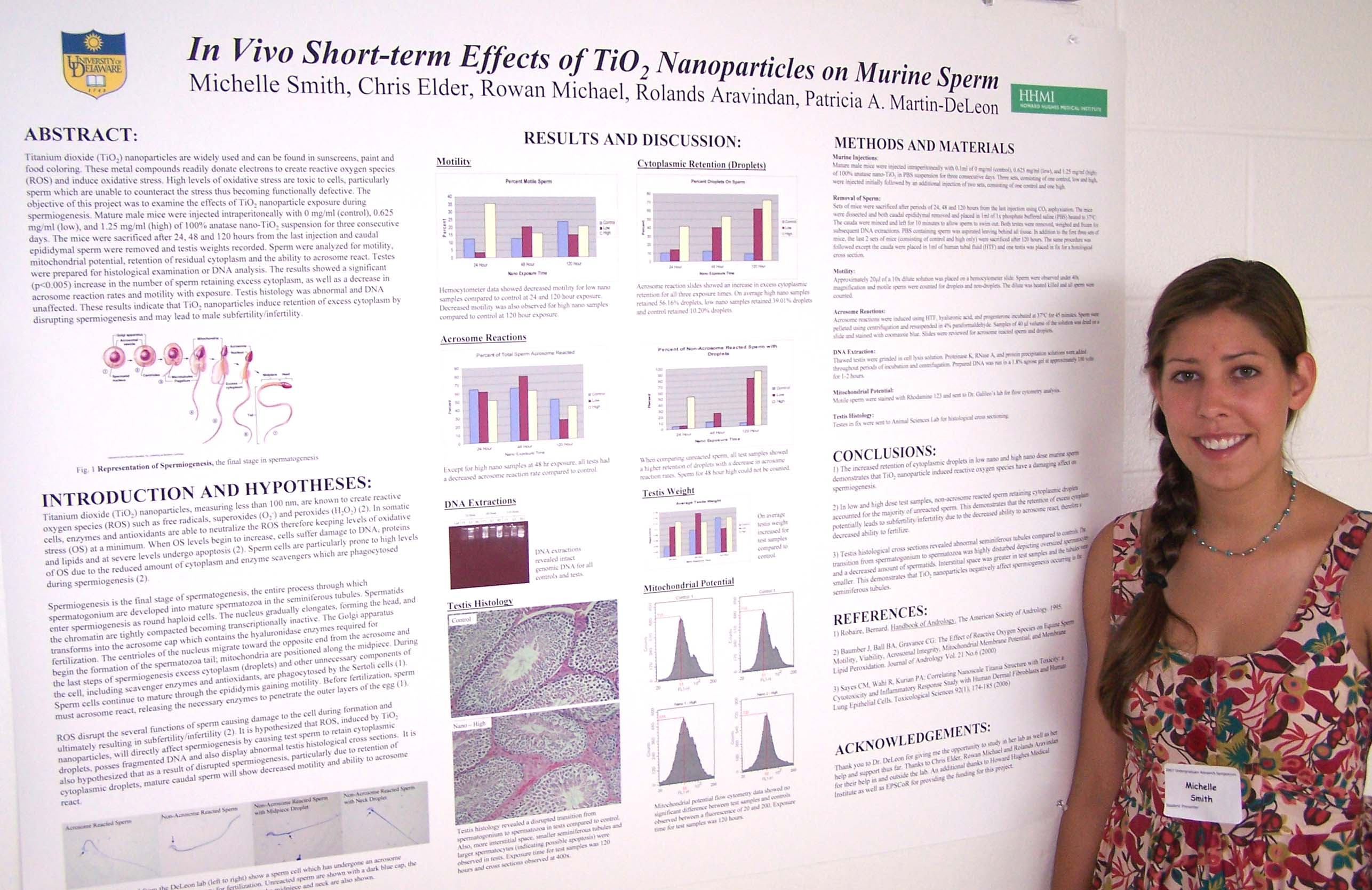
In Vivo Short-term Effects of TiO2 Nanoparticles on Murine Sperm

Michelle Smith, Chris Elder, Rowan Michael, Rolands Aravindan, and Patricia A. Martin-DeLeon
Department of Biological Sciences

Titanium dioxide (TiO2) nanoparticles are widely used and can be found in sunscreens, paint and food coloring. These metal compounds readily donate electrons to create reactive oxygen species (ROS) and induce oxidative stress. High levels of oxidative stress are toxic to cells, particularly sperm which are unable to counteract the stress thus becoming functionally defective. The objective of this project was to examine the effects of TiO2 nanoparticle exposure during spermiogenesis. Mature male mice were injected intraperitoneally with 0 mg/ml, ­­­0.625 mg/ml, and ­1.25 mg/ml of 100% anatase nano-TiO2 suspension for three consecutive days. The mice were sacrificed after 24, 48 and 120 hours from the last injection and caudal epididymal sperm were removed and testis weights recorded. Sperm were analyzed for motility, mitochondrial potential, retention of residual cytoplasm and the ability to acrosome react, and testes were prepared for histological examination or DNA analysis. The results showed a significant (p<0.005) increase in the number of sperm retaining excess cytoplasm, as well as a decrease in acrosome reaction rates and motility with exposure. Testis histology and DNA were unaffected. These results indicate that TiO2 nanoparticles induce retention of excess cytoplasm by disrupting spermiogenesis and may lead to male subfertility/infertility. Funding was obtained by an EPSCOR grant to Patricia A Martin-DeLeon as well as HHMI.


To cleave and to uncleave:
Expression of Heparan Sulfate modifying enzymes, Sulfatases and 6-O-Sulfotransferases during early pregnancy

Pamela Sun1, Sonia S. D’Souza2, Catherine B. Kirn-Safran3, Daniel D. Carson3
1
Imperial College School of Medine, London, SW7 2AZ, UK.
2Department of Chemistry and Biochemistry and 3Department of Biological Sciences, University of Delaware

Many growth factors play essential cell signaling roles during pregnancy and bind strongly to 6-O-sulfated motifs within heparan sulfate (HS) polysaccharides at the cell surface and in the extracellular matrix.  Consequently, modulation of HS 6-O-sulfation is believed to control biological responses to these growth factors.  Sulfatase-1 and Sulfatase-2 (Sulfs) are secreted endosulfatases that cleave the sulfate groups on the 6-O position of GlcNS residue of the HS proteoglycan in order to release the growth factors. Conversely, HS 6-O-Sulfotransferases (6HSOST1-3), encoded by three separate genes, add the sulfate at the same 6-O position of HS during its synthesis. Thus, the balance of Sulf and 6HSOST activities controls the HS-dependent cell signaling paradigm. This research project aims to determine which Sulfs and 6HSOSTs are expressed in the uterus during early pregnancy. Preliminary results obtained using conventional RT-PCR and real-time RT-PCR indicate an increase in mRNA production of both Sulf-1 and Sulf-2 in early pregnancy. Of these, Sulf-2 transcript is found to be more abundant. In addition, immunostaining with Sulf-2 antibodies demonstrated extracellular expression in the antimesometrial region of the decidua. Among the three 6HSOSTs, only 6HSOST-1 mRNA was detected in pregnant (day 8) mouse endometrium.  In conclusion, both Sulfs and 6HSOST-1 are present in pregnant endometrium and are likely to play a significant role in controlling the HS-binding growth factor bioavailabilty during pregnancy. This project is supported by the University of Delaware and Imperial College London Undergraduate Research Opportunity Programme and NIH grant HS25235.


Quantitative Analysis of Breast Cancer Metastasis to Brain

Kathryn Teixeira and Deni S. Galileo
Department of Biological Sciences

Breast cancer is more likely to metastasize to bones, lungs, or the liver, but metastasis to the brain is particularly devastating, and most patients die shortly after diagnosis. Previously, human breast cancer cells were successfully injected into the extra-embryonic vasculature of chick embryos, and metastasis to the brain was documented and quantified. In order to help determine the mechanism of entry into the brain, the sensitivity of the in vivo chick embryo system is being tested. Five day old chick embryos were previously injected with 50,000 MDA-MB-231 cells which were transfected with the lacZ and neor genes. Cancer cells were recovered from dissociated brains several days later, and re-cultured to form colonies in order to determine the percentage of cancer cells that migrated to the brain. I am injecting smaller numbers of cells into the blood vessels to find the minimum number of cells required for tumors to develop in the brain. I am also injecting other, non-tumorigenic cells into blood vessels in order to determine if known metastatic cells preferentially travel to brain. Additionally, I am injecting breast cancer cells directly into the brain to observe metastasis to other organs. Ultimately, these results will be used to investigate the effects of cell adhesion molecule L1 on the rate of metastasis of tumor cells into brain and elsewhere. L1 has been shown to increase cell migration velocity, and it is believed that cell lines transfected with L1 will present higher rates of metastasis. Funding for this research provided by INBRE.


Analysis of Mutated Mitochondrial DNA sequences in Prostate Cancer
and Identification of microRNA through RNA Folding

Mfon Umoren1, John Petros2, Chu Zhang1, M.C. Farach-Carson1
Department of Biological Sciences, University of Delaware1and Emory University School of Medicine, Atlanta, GA2

Protein biosynthesis is influenced by the secondary structure of the coding RNA sequence, which can affect the interactions with RNA binding proteins, microRNAs and ribosomal entry. Secondary structure of RNA is defined by its sequence and the potential for interactions between the nucleotide bases in the RNA sequence which affect folding. Mutation in the RNA sequence can alter intramolecular interactions to create different secondary structures that may predict function.  RNA folding software was used to determine the significance of mutations found in the mitochondrial DNA of prostate cancer patients. One mutation was a non-amino-acid changing sequence in the mtDNA section encoding cytochrome c oxidase II (COII). The second mutation was an amino-acid changing sequence found in the mtDNA encoding cytochrome b, altering leucine to proline. We folded and compared different lengths of the wild type sequence and the mutated sequence. Our hypothesis was that the non amino-acid changing mutation would cause a more significant change in the secondary structure of RNA than would the amino acid changing sequence. Analysis of folded RNA sequences also was used to identify possible microRNAs. MicroRNAs are short RNA sequences, usually 21-23 nucleotides in length that are involved in the regulation of gene expression. When folded, they form a distinct stem-loop structure. With a group of sequences that were either over- or under-expressed in cancerous cells, RNA folding and the presence of the stem-loop structure was used to identify potential miRNAs. Identification of possible miRNAs and further study of their targets will clarify their role in regulation of gene expression, as well as their role in cancer metastasis. Supported by the Department of Defense.


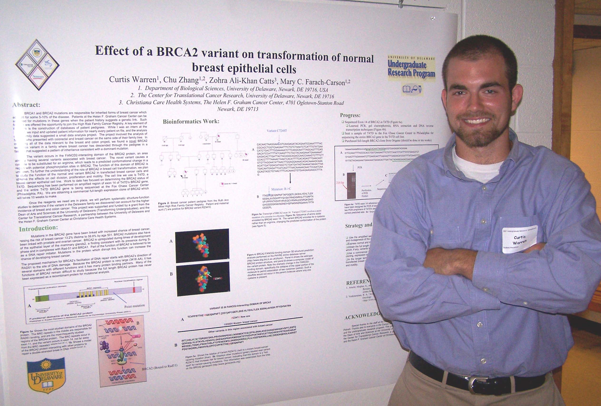
Effect of a novel BRCA2 variant found in a Delaware family on transformation of normal breast epithelial cells
Curtis Warren, Chu Zhang, Zohra Ali-Khan Catts, Mary C. Farach-Carson,
Center for Translational Cancer Research

BRCA1 and BRCA2 mutations are responsible for inherited forms of breast cancer which account for some 5-10% of the disease.  Patients at the Helen F. Graham Cancer Center can be screened for mutations in these genes when the patient history suggests a genetic link.  Such families are offered the opportunity to join the High Risk Family Cancer Registry. A key element of this work is the construction of databases of patient pedigrees.  While I was an intern at the HFGCC, we input and updated information for many patients, and the analysis of the family data suggested a small data analysis project.  In compiling all of the data relevant to the project, we found a novel BRCA2 sequence variant in a family where breast cancer has descended through the pedigree in a fashion that suggested a pattern of inheritance consistent with a dominant mutation.  The novel variant causes a cysteine to be substituted for an arginine, which leads to a predicted conformational change in BRCA2. The function of this domain of BRCA2 is unknown. To further the understanding of the role of BRCA2 in breast cell transformation, we plan to study the function of the normal and variant BRCA2 in transfected breast cancer cells and observe the effects on cell division, proliferation and motility.  Once the reagents we need are in place, we will perform systematic structure-function studies to determine if the variant can account for the higher incidence of breast cancer. Funding provided by: Transforming Undergraduates Grant (UD), and the Center for Translational Cancer Research.


Variations of Gene Repair in sub S-phase Cell Populations

Melissa G. Warriner, Julia U. Engstrom, and Eric B. Kmiec
Department of Biological Sciences and the Delaware Biotechnology Institute 

Targeted nucleotide exchange (TNE) relies on the use of a short synthetic oligonucleotide (ODN) designed to be complementary to a target sequence in the genome except for a centrally located mismatch, which directs the desired base change in the DNA.  The repair reaction is believed to be enhanced by mechanisms that promote a more open conformation of the DNA, thus, enabling the target site to be more accessible for the binding of the oligonucleotide.  One such method is through the modulation of cell cycle progression to increase the population of cells in S phase, a point at which DNA is in a more open structure.  It has been established that targeting cells in S phase leads to more probable integration events than any other phase of the cell cycle; this phenomenon is not only true for the gene repair reaction, where the aim is to direct the exchange of a single base pair, but also in many other systems, where the aim is to insert foreign DNA into a chromosome.  However, it is still unclear if this increase is due to a particular sub-S phase stage or simply S phase as a whole.  Our work aims to discern variations in repair levels in sub-phases of S as cells enter and progress through this phase.  In addition, we evaluate gene expression patterns of several cell cycle genes that may help denote the changes in cell cycle distribution as a function of gene repair events.  Funded by Howard Hughes Medical Institute.


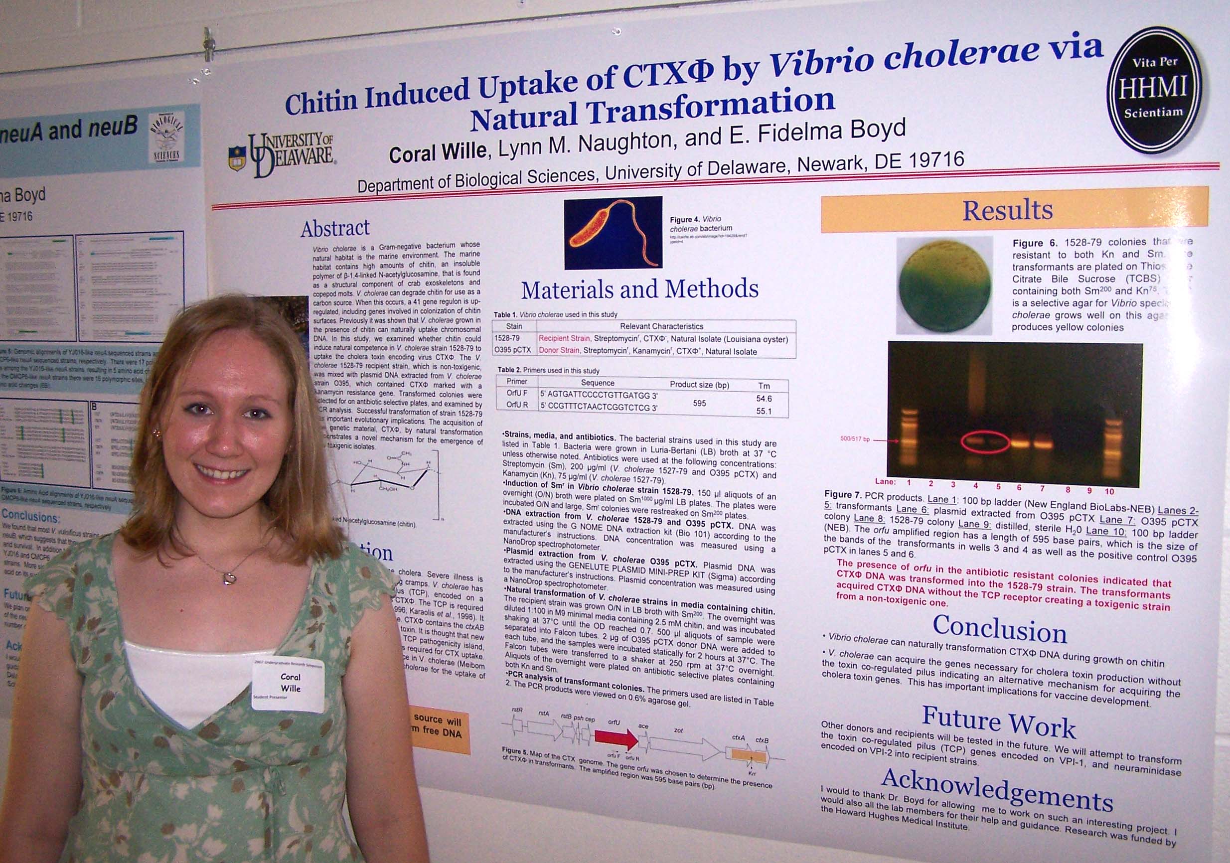
Chitin Induced Uptake of CTXΦ by Vibrio cholerae via Natural Transformation

Coral Wille, Lynn M. Naughton, and E. Fidelma Boyd
Department of Biological Sciences

Vibrio cholerae is a Gram-negative bacterium whose natural habitat is the marine environment. The marine habitat contains high amounts of chitin, an insoluble polymer of β-1,4-linked N-acetylglucosamine, that is found as a structural component of crab exoskeletons and copepod molts. V. cholerae can degrade chitin for use as a carbon source. When this occurs, a 41 gene regulon is up-regulated, including genes involved in colonization of chitin surfaces. Previously it was shown that V. cholerae grown in the presence of chitin can naturally uptake chromosomal DNA. In this study, we examined whether chitin could induce natural competence in V. cholerae strain 1528-79 to uptake the cholera toxin encoding virus CTXΦ. The V. cholerae 1528-79 recipient strain, which is non-toxigenic, was mixed with plasmid DNA extracted from V. cholerae strain O395, which contained CTXΦ marked with a kanamycin resistance gene. Transformed colonies were selected for on antibiotic selective plates, and examined by PCR analysis.  Successful transformation of strain 1528-79 has important evolutionary implications. The acquisition of new genetic material, CTXΦ, by natural transformation demonstrates a novel mechanism for the emergence of new toxigenic isolates. Research was funded by the Howard Hughes Medical Institute.


Variation in Sperm Hyaluronidase Activity in Men Attending an IVF Clinic

Sarah Woody
, Jessica Bruton, Minghai Shao, Patricia A. Martin-Deleon
Department of Biological Sciences

Sperm hyaluronidase activity is necessary for degradation of hyaluronan or hyaluronic acid (HA) in the extracellular matrix surrounding the oocyte in several steps in mammalian fertilization. These steps include the penetration of the cumulus oocyte complex, the zona pellucida, and the perivitelline space. To date, the only known human sperm hyaluronidase is Sperm Adhesion Molecule 1 (SPAM1 or PH-20), a 64 kDa membrane protein which plays multifunctional roles in mammalian fertilization and which has recently been shown to be a marker of sperm maturation in mice (Chen et al. 2006). SPAM1 is unique among the hyaluronidases, with respect to its neutral hyaluronidase activity. Our lab has recently shown that a second sperm hyaluronidase, HYAL3, is present in human sperm. The objective of this study was to compare the levels of hyaluronidase activity in men using assisted reproductive technologies (ART) at Christiana Hospital, a local facility in Newark, DE. Hyaluronic acid substrate gel electrophoresis (HASGE) revealed varying levels of hyaluronidase activity at neutral pH among the subjects, as reflected by variations in band size, width and intensity. Using Western blot analysis, attempts were made to correlate the absence or reduction of hyaluronidase activity with the absence or presence of SPAM1 and HYAL3 isoforms and their relative amounts.  Our data suggests that the presence of HYAL3 is associated with an increase in the intensity of the hyaluronidase activity band from human sperm proteins. Supported by IGERT.


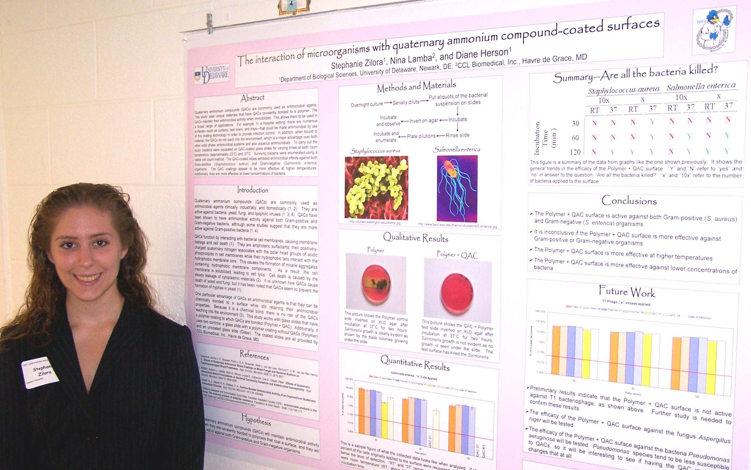
The interaction of microorganisms with quaternary ammonium compound-coated surfaces

Stephanie Zilora1, Nina Lamba2, and Diane Herson1
1
Department of Biological Sciences, University of Delaware, Newark, DE; 2CCL Biomedical, Inc., Havre de Grace, MD

Quaternary ammonium compounds (QACs) are commonly used as antimicrobial agents.  This study uses unique materials that have QACs covalently bonded to a polymer. The QACs maintain their antimicrobial activity when immobilized.  This allows them to be used in a broad range of applications.  For example, in a hospital setting, there are numerous surfaces—such as curtains, bed liners, and trays—that could be made antimicrobial by use of this coating technology in order to provide infection control.  In addition, when bound to material, the QACs do not leach into the environment, which is a major advantage over both other solid phase antimicrobial systems and also aqueous antimicrobials.  To carry out the study, bacteria were incubated on QAC-coated glass slides for varying times at both room temperature and 37°C.  Surviving bacteria were enumerated using a viable cell count method.  The QAC-coated slides exhibited antimicrobial effects against both Gram-positive (Staphylococcus aureus) and Gram-negative (Salmonella enterica) organisms.  The QAC coatings appear to be more effective at higher temperatures.  Additionally, they are more effective at lower concentrations of bacteria.  This project was generously funded by National Science Foundation grant number 0450527 and a Charles Peter White scholarship.


Biology Abstracts for students with names beginning A-K.

Links: Summer 2007 Undergraduate Research Symposium, Symposium Abstracts from other Colleges and Departments,
Undergraduate Research Summer Enrichment ProgramUnversity of Delaware Undergraduate Research Program, Howard Hughes Undergraduate Program.
Created  23 July 2007. Last up dated 15 August 2007 by Hal White
Copyright 2007, University of Delaware