Abstracts from the Department of Biological Sciences
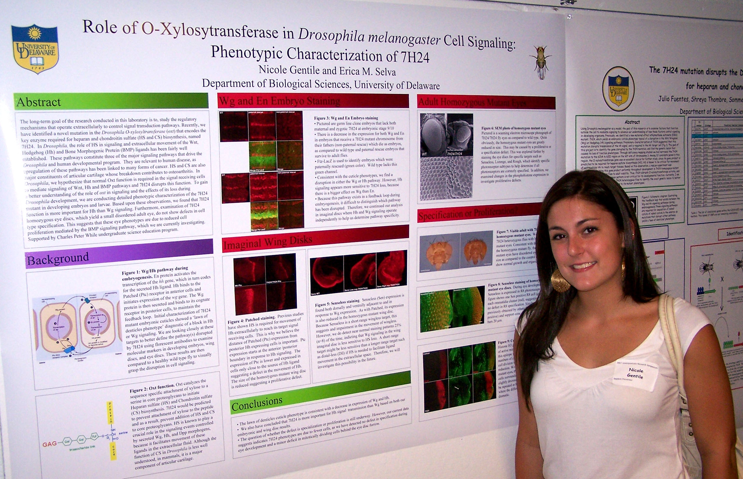
Undergraduate Summer Research Symposium August 8, 2007

Ordered alphabetically by student's last name

Ambrose Brady, A Cooper Desai Edobor Ferrara Fuentes Gibson Grindel Huntley Kasmari
Atchison Brady, M Culver Dominica Elder Fisher Gambogi Greenwood Harmon Isaacs Knox
Berg Cheong Decker Drummer Eppes Frank Gentile Grey Hill Jenkins-
Kabaila
Kuznekoff


Evaluation of Four Cell Viability Assays for the Construction of
Dose-response Curves in Prostate Cancer Cells

Jennifer Ambrose
, Brenda Mogere and Robert A. Sikes
Department of Biological Sciences, Laboratory for Cancer Ontogeny and Therapeutics, Center for Translational Cancer Research Prostate Cancer (PCa) is the second leading cause of cancer related death in US males with over 27,000 deaths this year alone predominantly from aggressive, metastatic disease that is invariably lethal. For this reason, the development and testing of novel therapeutics that target PCa cell growth and metastasis are critical. Several lead compounds known to be voltage sensitive sodium channel (VSSC) blockers were shown by us to be more than 6-fold better than the parent compound phenytoin at inhibiting PCa growth in vitro. Unpublished observations indicated that the compounds may be additive in nature. Other VSSC blockers act as histone deacetylases that often complement traditional chemotherapy. By determining the dose-response of each chemotherapeutic drug individually and then combining treatments at the IC20, we hope to deduce which drugs have super-additive or synergistic effects. Our first objective was to determine the best method for assessing cell viability. We desired a rapid, easily controlled and highly reproducible test. LNCaP cells, an androgen-sensitive PCa cell, were treated with varying concentrations of a drug three times over the duration of a week. Cell viability was assessed using one of four different assays. Through troubleshooting and manipulation of the different procedures, Crystal Violet assay was determined to be the most appropriate method to construct our dose-response curves. Funding provided by the University of Delaware Undergraduate Research Fellows Program and INBRE Delaware P20RR016472-04.


Gα12 Heterotrimeric Subunits and Rho Activation

Keith Atchiso
n and Kenneth van Golen
Department of Biological Sciences

Pancreatic Cancer (PCa) has become the fourth most deadly cancer in the UnitedStates. PCa exists in both a metastatic and a locoregional, non-metastatic variant. The metastatic variant carries a survival rate of less than 1%, and is significantly more painful. Despite these horrifying statistics, the metastatic nature of PCa is understudied. Microarray studies demonstrate that PCa tumor cells overexpress the Gα12 subunit of heterotrimeric GTPases 18-fold. Through guanine exchange factors (GEFs), Gα12 is  known to activate the Rho family of GTPases. The Rho family of small GTPases, through their ability to reorganize the cytoskeleton,are known to play a major role in the invasiveness of metastatic cancers.  While the relationship between RhoA and the heterotrimeric G proteins has been examined in PCa, the relationship and underlying mechanisms of the other 22 members of the Rho family have not been examined. Introduction of constituently active or dominant negative Gα12 subunits in PCa cell lines, can demonstrate differences in Rho activation specifically in RhoC, RhoG, and Rac; all of which are involved in metastatic cancer. Gα12 activation should be directly proportional to Rho GTPase activation.  Analysis of motility assays relate the invasiveness of the cancer cells to the activation. Activation of Rho proteins is expected to increase motility. Finally, the pathways and mechanisms of activation will be studied through the affinity, kinetics, and  inhibition, of the Rho GEFs.  Funding for this project has been provided by Howard Hughes Medical Institution.


Expression of Survival Motor Neuron (SMN) Protein in Baculovirus
Amanda Berg and Wenlan Wang
Department of Biological Sciences and A.I. duPont Hospital for Children

Spinal Muscular Atrophy (SMA) is a neuromuscular disease characterized by the degeneration of spinal motor neurons associated with muscle wasting.  SMA is caused by a deletion or mutation of the Survival Motor Neuron Gene 1 (SMN1).  The encoded SMN protein is ubiquitously expressed, yet lower levels of this protein result in selective loss of motor neurons in patients.  The functions of SMN have been implicated in RNA splicing, cell survival and neurite outgrowth.  The long-term goal of this project is to functionally characterize SMN protein.  As an initial objective towards that end, we proposed to produce a large amount of soluble and correctly post-translationally modified SMN protein in vitro.  The over-expressed SMN protein will be used to search for SMN RNA binding targets and to isolate the SMN-interacting proteins through far western blot or affinity column analyses.  Our previous studies indicated that SMN protein expressed in bacteria is insoluble and highly degraded.  Here, SMN protein fused to His or GST tag and GST control were expressed in insect Sf9 cells through bacoluvirus infection.  We have cloned tagged SMN and GST cDNAs into a pfastbac vector, generated the recombinant bacmid, and transfected the bacmid DNA into Sf9 cells to obtain P1 viral stocks.  Western blotting analyses of lysates from cells infected by these viruses indicated correct expression of tagged SMN and GST proteins.  Currently, large amplifications of these viruses and further analyses of expressed proteins are underway.  Funding for this research was provided by the University of Delaware Undergraduate Research Program’s Science and Engineering Scholarship.


Modeling Tumor Development: the Role of L1

Adam Brady, Deni Galileo, Muhua Yang
Department of Biological Sciences

 L1 is a neural cell adhesion protein involved in the normal development of the nervous system; however it has been implicated in mediating some of the invasive properties of certain cancers including gliomas and breast cancers.  L1 has also been shown to rescue cancerous cells from apoptosis.  It was hypothesized that polyclonal antibodies raised against a 16 amino acid peptide corresponding to a critical integrin binding region of human L1 would slow the migration of rat 9L/LacZ and human T98 G glioma cells in vitro, whereas the peptide alone would stimulate migration.  I have determined that the 16 amino acid peptide actually decreases cellular migration, possibly due to competitive inhibition.  The results from the antibody experiments were inconclusive.  Currently, I am attempting to isolate an L1-Fc fusion protein from transfected QT6 cells.  Once purified, the L1-Fc protein will be applied to 9L/LacZ and T98 G cells and analyzed using automated time-lapse microscopy.  I hypothesize that this will increase the migration rates of these cell lines by mimicking the activity of endogenous L1 ectodomains.  Additionally, a shell-less chicken embryo culture has been developed that may allow in vivo simulation of tumor growth.  Chicks were placed in shell-less culture at day 3 and on day 5 or later were exposed to a dense slurry containing rat C6 glioma cells.  Growth was monitored for signs of tumor vascularization.  This system will be used to examine the relative tumorigenicity of normal QT6 cells and L1-Fc expressing QT6 cells. Supported by HHMI and INBRE.


Development of analog Atlantic horseshoe crab bait for the American eel fishery: results from recent field trials
Marissa G. Brady1
and Dewayne Fox2
Department of 1Biological Sciences and Department of 2Agriculture and Natural Resources, Delaware State University

Atlantic horseshoe crab (Limulus polyphemus) play important economic and ecological roles in the mid-Atlantic region.  Delaware Bay hosts the largest horseshoe crab population which is thought to currently be in decline. Horseshoe crab are truly a multiple-use species providing bait for both commercial American eel and whelk fisheries, vital pharmaceutical products, much needed tourism dollars, as well as being the primary food source for several species of migratory shorebirds.  As a result of increased consumptive needs managers imposed a moratorium on all female harvest and severely reduced the harvest of males which when combined with other competing uses created the need for analog horseshoe crab bait for the commercial American eel fishery.  In this study, we tested the efficacy of a suite of artificial horseshoe crab baits developed by the University of Delaware.  Monthly field trials were carried out from May-July, 2007 in the St. Jones River, DE.  Sampling was conducted with commercial American eel traps at fixed locations and was stratified with respect to the salinity gradient.  A total of 2,411 American eel were collected during all sampling events.  American eel catch rates varied by month and salinity strata with higher catches in the cooler months and higher salinity areas.  In total five analog Atlantic horseshoe crab baits were tested for efficacy against a positive control (1/2 female horseshoe crab).  Results from recent field trials show much progress in achieving a viable alternative horseshoe crab bait for commercial use; through the use of alternative baits we hope to alleviate some of the pressure on this economically and ecologically important species.  Funded by EPSCoR.


Investigating the Neurobiology of Sensory Processing and Learning in an Invertebrate Model System
Ashlee Cooper, Melissa Harrington, and Sohrab Amiri
Biotechnology, Delaware State University

Almost all major advances in the field of neuroscience have come about through work with model systems. While technology advances and our growing understanding of neural function has increased, there is still much that can be learned from working with invertebrate models.  The simple behavior repertoire of the invertebrate Wolf Snail makes its investigation much easier and less problematic than mammals. The goal of this study is to characterize the components of the genetic architecture and target the serotonin gene and its relationship to sensory processing and learning in snails.  Serotonin is a neurotransmitter that is a ligand for a cation channel (5HT3 Receptor). After identification of degenerate serotonin primers through learned molecular methods, amplification of gene parts were shown by electrophoresis.  Electrophoresis of PCR products from a snail DNA template showed bands, indicating amplification of parts of a snail 5HT3 receptor.  Future plans are to sequence the Plasmid DNA and compare it to previous cloned 5HT3 receptors.  This will confirm successful cloning of serotonin receptor from snail allowing further manipulation and evaluation. The project described was supported by Grant Number 2 P20 RR016472-07 under the INBRE Program of the National Center for Research Resources (NCRR), a component of the National Institutes of Health (NIH).


Developmental Expression Pattern of Calcium and Integrin Binding Protein 1 (CIB1)
Colleen M. Cheong and Ulhas P. Naik
Department of Biological Sciences

The calcium and integrin binding protein 1 (CIB1) was named for its initial identified function as a protein that has the ability to bind to calcium ions and to the intracellular domain of the α subunit of the platelet integrin αIIbβ3.  Since its original discovery, it has been found that CIB1 binds to a number of other proteins with a wide array of functions, thus suggesting its importance.  Northern blot analysis has shown that CIB1 mRNA is ubiquitously present in adult tissues.  However, the specific localization of CIB1 in these tissues and the role of CIB1 during development have yet to be studied.  In order to study the expression pattern of CIB1, a series of both immunofluorescence and in situ hybridization experiments were designed.  Based on the immunofluorescence experiments, CIB1 exhibits a unique expression pattern in adult mouse liver, kidney, and heart.  It appears that CIB1 co-localizes with actin in adult liver and is expressed in the blood vessels of the heart and in the periphery of the glomeruli of the kidney.  Even more interestingly, the same patterns do not seem to be exhibited during earlier stages of development.  Furthermore, differences in expression patterns were found between 9.5-16.5 days post coitum (d.p.c.) mouse embryos, which were collected through timed-pregnancies. Further analysis will be required to identify more detailed differences between stages of development.  Additionally, a non-radioactive in situ hybridization protocol is being standardized in order to study the expression pattern of CIB1 mRNA.  From these experiments, we will gain better understanding of the overall expression pattern of CIB1 in various tissues, which may give some indication of the function of CIB1.  This research was supported by the Howard Hughes Medical Institute.


The Effects of Cell Surface NgCAM Expression on Developing Chick Brain Axons in Vitro

Stephanie Culver, Murali Temburni, Deni S. Galileo
Department of Biological Sciences

During embryonic brain development, newly forming axons follow very precise paths guided by the collaborative effects of several guidance cues. The process of axon guidance, which is partly responsible for precise and complex wiring of the nervous system, is not completely understood. NgCAM is a neuronal cell surface membrane protein that has been shown by others to accelerate developmental axonogenesis and increase neurite elongation. My research project aims to study axon outgrowth of developing chick CNS tissue in vitro in order to better understand the role of NgCAM in brain development. Embryonic Day E7 optic tectum (OT) explants were originally used as a preparation to generate growing axons. However, this system failed to be an efficient and reproducible model. As an alternative, multicellular aggregates made from dissociated OT cells were used. These were plated onto various NIH3T3 cell monolayers that either did or did not express cell surface NgCAM. The aggregates were labeled with a fluorescent membrane dye so that the axons could be visualized on top of the cell monolayers. Fluorescent labeling and time-lapse microscopy will be used to follow the axon growth over time to test our hypothesis that NgCAM expression on the cell monolayer will increase rate of outgrowth, length of axons, level of bifurcation, and number of neurite processes. My current research project has demonstrated that neurite outgrowth can be successfully visualized on top of cell monolayers using fluorescent dye labeling techniques and that axons growing on 3T3/NgCAM cells show an increase in both length and number. Supported by a Howard Hughes Medical Institute's Undergraduate Science Education Award. 


Corinne recieved second prize for her oral presentation in the Sigma Xi competition.

The role of bB2-crystallin in epithelial-mesenchymal transition
Corinne Decker
, Kevin Duprey, Yan Wang, Melinda Duncan
Department of
Biological Sciences

The vertebrate lens is composed of epithelial cells and fiber cells, both of which contain crystallins, proteins necessary to maintain the high refractive index of the lens. Mice and humans harboring mutations in bB2-crystallin develop cataracts that have been proposed to be caused by aggregation of mutant βB2-crystallin.  However, we have found that mice harboring the Phy mutation of the bB2-crystallin locus develop cataracts associated with profound fibrosis of the adult lens epithelium, expression of abnormally high levels of asmooth muscle actin (aSMA), and the loss of F-actin from lens fiber cells. Although bB2-crystallin expression in the lens initiates at birth, these observations suggest a role for bB2-crystallin in lens cytoskeletal organization.  Consequently, we undertook a developmental study of this phenotype. While newborn and 2 week-old Crybb2Phil mice exhibited normal lens morphology, the loss of F-actin and upregulation was aSMA was apparent in the 4 week-old mutant.  Upregulation of aSMA in the lens epithelium is associated with epithelial-mesenchymal transitions coupled with the loss of epithelial markers Pax6, Connexin-43, and E-cadherin, as well as aberrant expression of fiber cell markers cMaf and Prox1. Mutant lens epithelial cells which appear fibrotic downregulate Connexin-43 and E-cadherin expression but maintain Pax6. Additionally, neither cMaf nor Prox1 are found to be overexpressed in the mutant epithelium.  Overall, these data suggest that bB2-crystallin plays critical roles in the cytoskeletal organization of the lens and suggest that the pathologies seen in bB2-crystallin mutant lenses are not simply due to protein aggregation. Funding provided by grants from the Howard Hughes Medical Institute, the Fight for Sight Foundation, and the National Eye Institute.


Vivek talking with David Lynn, plenary speaker.

CD44 and Posterior Capsular Opacification (PCO)
Vivek D. Desai, Yan Wang, and Melinda K. Duncan, Department of Biological Sciences 

Posterior capsular opacification (PCO) arises from epithelial-mesenchymal transition (EMT) of lens epithelial cells remaining behind following cataract surgery. In cancer systems, CD44, a receptor for hyaluronan, mediates changes in cellular proliferation and migration that eventually result in EMT, although CD44 function in EMT has not been previously studied in the lens. In the normal adult mouse lens, we found CD44 protein and mRNA only in the lens fiber cells with no detectable expression in the lens epithelium. Following cataract surgery in mice, CD44 expression is highly up-regulated in the remaining lens epithelial cells 12 hours following the surgery and this expression remains elevated during the EMT process. CD44 null mice did not exhibit any qualitative differences in the timing or extent of expression of the EMT marker, -SMA, as compared to wild-type (WT) suggesting that CD44 is not essential for TGFβ mediated EMT. However, the kinetics of CD44 up-regulation in the mouse cataract surgery model suggests that it is one of the earliest molecular markers ever described for lens response to injury and may serve as a good model to understand the early events in the lens epithelial response to injury and the sensitization of these cells to later TGFβ mediated EMT. Notably, CD44 expression is known to be up-regulated in response to hepatocyte growth factor (HGF) mediated signaling and HGF has been reported to be a potent mitogen for lens epithelial cells Future work will investigate the hypothesis that HGF mediated signaling up-regulates in the lens in response to injury leading to up-regulation of CD44 expression and sensitization of lens cells to TGFβ mediated EMT. Supported by Beckman and Barry M. Goldwater Scholarships.


The Function of N-Glycosylation in Drosophila Growth and Development:
Phenotypic Characterization of alg10

Carly Dominica
, Evan Lebois, and Erica Selva
Department of Biological Sciences
 

Proteins that function extracellularly must first undergo the process of secretion, where they are subject to several posttranslational modifications, before cellular signaling can occur. One such modification is N-glycosylation, which is often necessary for proper protein folding, stability of extracellular glycoproteins, and has been linked to functions of the immune system. The goal of this project was to phenotypically characterize mutations in the Drosophila alg10 gene, which produces an enzyme that adds the terminal glucose to a dolichol-linked oligosaccharide in N-glycosylation prior to transfer to nascent polypeptides. Although this residue is not present extracellularly, alg10 mutations that prevent the addition of this glucose yield extreme and pleiotropic effects. In this study, the phenotype of alg10 was characterized on a molecular level in order to better understand the pathway(s) affected by Alg10 loss and the role of N-glycosylation in these pathways. To determine this, the eye discs of both wild type and alg10 mutant Drosophila larvae were removed and molecular markers for cone cells and photoreceptor identity were analyzed. From the data, it was observed that alg10 mutants had a significantly greater number of R7 photoreceptors than the wild type. Specification for R7 photoreceptors is determined by activation of the sevenless receptor, which activates the MAP kinase cascade. MAP kinases also function in other signaling pathways, including the insulin receptor and epidermal growth factor pathways. As these pathways are involved in growth and development, our data suggests that one important target of N-glycosylation acts through the MAP kinase pathway to regulate cell specification in the developing eye. Supported by the Charles Peter White Undergraduate Science Education Program.


Effects of Aging and Exercise on Endothelial Progenitor Cells

Christopher Drummer, David G. Edwards, and Raju Prasad
Health and Nutrition Sciences


With age the endothelial lining of the body’s arteries begin to degenerate and become ridged.   As a result, many individuals become more susceptible to illnesses such as cardiovascular disease.   Endothelial Progenitor Cells (EPCs) are vital in arterial re-endothelizlization and neoangiogenesis; thus, their numbers have been shown to affect the overall health of both healthy individuals and cardiovascular disease patients.  Although arterial health decreases with age, it has been shown that the number of EPCs in peripheral blood and exercise increases endothelial function in not only young but old endurance trained individuals.  The purpose of this study is to quantify the number of EPCs in ones peripheral blood and to find a correlation between this number and endothelial health of healthy young and old endurance trained and sedentary individuals.   To do this we will collect and analysis four groups of subjects.   There will be 15 young (18-30) endurance trained males as well as 15 older (50-75) endurance trained males.  The third and forth groups will consist of 15 young sedentary males and 15 older sedentary males.  Blood is drawn from each patient and the number of EPCs is quantified through flow cytometry.   The following cell surface markers are used to distinguish the  EPCs in the peripheral blood: CD133+/CD34-/KDR+, CD133+/CD34+/KDR+, and CD133-/CD34+/KDR+.  To quantify endothelial function we use Flow mediated dilation (FMD).   We hypothesis endothelial function and the quantity of endothelial progenitor cells will decrease with age however, peripheral EPCs will be greater in endurance trained individuals.   Furthermore, both sedentary and endurance trained older males will have a diminished if not depleted EPC count.   [Funding by NUCLEUS Apprenticeship Program]


Adhesion Mediated Chemoresistance of Pc3 Cells to Docetaxel.
Osemeke Edobor1, Freddie Pruitt, Robert A. Sikes, Kenneth van Golen, Mary C. Farach-Carson, and Carlton Cooper
1Lincoln
University
and UD Department of Biological Sciences

Men who develop metastatic prostate cancer (PCa) and fail androgen ablation therapy rely on docetaxel (taxotere) as the next therapy of choice.  Unfortunately, patients frequently relapse after developing docetaxel chemo-resistance. Understanding the cellular mechanism that underlies chemo-resistance could improve treatment response for bone metastatic PCa. To determine if type I collagen, which comprises 90% of the bone extracellular matrix (ECM), contributes to chemoresistance, we used an androgen independent bone metastatic PCa cell line, PC3. PC3 cells preferentially activate survival pathways during adhesion to type I collagen (kiefer et al, 2001).  We hypothesize that adhesion of PC3 cells to type I collagen, mediates the chemo-resistance to docetaxel.  Our data shows that PC3 cells, when adhered to type I collagen show an increase in p-Akt as compared to PC3 cells on fibronectin and plastic. MTT analysis shows that PC3 cells on type I collagen are more viable in response to increasing concentrations of docetaxel.  Western blot analysis shows that type I collagen inhibits the activation of the apoptosis effector, Caspase 7. We believe that Type I collagen protection is mediated by signaling between P13-Kinase and Akt, and treatment of PC3 with P13-Kinase inhibitor, LY-294002, was able to negate the protective effects of Type I collagen.  In essence, we conclude that adhesion of PCa cells to components of the bone microenvironment may be a critical component of the acquired docetaxel chemo-resistance seen in men with bone metastatic PCa. Funded by: Department of Defense, UDRF


In Vitro TiO2 Nanoparticle Exposure and Its Effects
on Mature Mouse Epididymal Spermatozoa
Chris Elder and Patricia A. DeLeon
Department of Biological Sciences

Environmental nanoparticle contamination is an ever-increasing occurrence in the world today. Nanoparticles are photo-reactive and cause the formation of reactive oxygen species (ROS).  ROS can overwhelm the body’s natural antioxidant supply, resulting in oxidative stress (OS) and damage in cells. This is especially prevalent in spermatozoa due to the relatively low volume of cytoplasm and therefore low amounts of antioxidant scavenger enzyme. OS is known to positively correlate with male infertility and has been found to impair functionality of sperm in vivo. The purpose of this study was to determine what relationship exists, if any, between in vitro nanoparticle exposure and sperm vitality, evaluated with statistical analysis of sperm motility, acrosome reaction rates, retention of excess residual cytoplasmic droplets (ERC), and mitochondrial potential of sperm. Caudal epididymal sperm was extracted from mice and subjected to 45-minute treatments of 0, 50, or 100 μg/mL of 100% anatase TiO2 particles in human tubule fluid capacitation medium. No effect has been determined thus far on motility or mitochondrial potential, with <1% difference in motility and no significant difference in mitochondrial potential among all conditions observed. Acrosome reaction rate and ERC data are currently under analysis. The information elucidated thus far suggests that in vitro TiO2 nanoparticle exposure does not produce damage or adverse effects in mature mouse sperm. This has important implications regarding the determination of the mechanism through which nanoparticles interact with living cells. Supported by the University of Delaware Research Program.


The effects of point mutations in PLP1 intron 3 on RNA splicing

Elisabet Eppes, Jennifer Taube, and Grace Hobson
Alfred I. duPont Hospital for Children

Pelizaeus-Merzbacher Disease (PMD) is generally caused by duplications or mutations altering amino acids in PLP1, however silent mutations including mutations in introns can disturb the ratio of the normal splice forms PLP1 and DM20. The PLP1 form is highly expressed in oligodendrocytes, while DM20 is widely expressed. Intronic mutations can cause disease by disturbing or enhancing the use of a splice site, either directly, or through splicing regulatory proteins. Analysis of a patient’s DNA revealed a point mutation (c.453+55C>T) in intron 3 of PLP1 with unknown significance. When comparing intron 3 of human and other mammals, I found strong conservation in the region, but not at +55 which used C/T, nor at +45 which had C/A. Those species with +55T, like our patient, had +45C; whereas others, including normal human, with +55C contained +45A. I hypothesize that the two simultaneous changes +45C and +55T have no effect on the PLP1:DM20 while a single mutation (as in the patient), changes the ratio. To investigate the effect of mutations on splicing of intron 3, I modified a mini-gene construct to create plasmids containing the mutations: c.453+55C>T, c.453+45A>C, and both c.453+45A>C & +55C>T by site-directed mutagenesis. These and other random mutations were transfected into an oligodendrocyte-like cell line which transcribes both PLP and DM20 from the mini-gene. Analysis by RT-PCR has revealed significantly more PLP due to the +55C>T mutation and significantly less PLP due to a random mutation at +58.


Comparison of Cytokine Levels and Differential WBCs in Synovial Fluid
from Pediatric Patients with Acute and Chronic Lyme Arthritis

Kristen Ferrara, Vicky Maduskuie, Paul Fawcett
Department of Immunology


Lyme Arthritis (LA) inflammation of synovial membranes due to bacterial infection by Borrelia burgdorferi, can be classified as Acute (duration of symptoms < 6 months) or Chronic (duration of > 6 months).  Comparison of cytokine levels and white blood cell differentials in synovial fluid (SF) were done to look for a diagnostic predictor in early stages of the disease. Patients were identified from a database of LA patients seen at a rheumatology clinic.  Synovial fluid samples were collected and stored at -80°C until assayed.  52 samples from 50 patients (34 with Acute LA and 16 with Chronic LA) were tested for levels of cytokine IL-6, determined by enzyme linked immunosorbent assay, ELISA, (R&D Systems).  Differential WBC counts on synovial fluids were performed by cytospin and microscopy. Results obtained showed a statistically significant difference (P=<0.001) of IL-6 in patients with Acute LA (Median= 107,436 pg/ml) versus those with Chronic LA (Median= 28,423 pg/ml).  Comparison of Acute (n=5) and Chronic (n=14) Lyme differentials was inconclusive (See Table).  Differentials of Chronic LA showed clusters of cells resembling small, mature lymphocytes that could be stem cells. Findings indicate testing should continue for an early predictor differentiating between Acute and Chronic Lyme arthritis and the identity and function of cells identified in cytospins should be further investigated. Supported by NIH grant 2 P20 RR016472-07 under the INBRE Program of the National Center for Research Resources (NCRR)


Functional Assessment of Recombinant Human Bikunin
Megan Fisher and John Hoyer
Department of Biological Sciences

Normal human urine is supersaturated in respect to calcium oxalate (CaOx), a major component of kidney stones. However, most individuals do not form stones due to the presence of inhibitors of CaOx crystallization. Urinary proteins contribute the majority of this inhibition. Interestingly, the urinary proteins of stone-forming individuals are less inhibitory than those of non-stone formers. Certain proteins inhibit the nucleation, growth, and aggregation of CaOx crystals, and their adhesion to kidney cells. We are currently studying bikunin, one protein which has been shown to have an inhibitory effect on CaOx crystallization. The functional effects of recombinant human bikunin produced in insect cells were compared to those of human urinary bikunin. Insect cells were infected with a bikunin construct in a viral vector. Three different leader sequences were compared in an effort to evaluate and optimize the productivity of the constructs. Bikunin levels in the supernatants were determined by ELISA. Both recombinant and urinary bikunin were immunopurified using anti-bikunin antibodies coupled to sepharose. Protein functionality was tested before and after purification by comparing the ability of the native and recombinant proteins to inhibit trypsin. It was found that bikunin from both sources inhibit trypsin with similar efficiency, with close to 100% inhibition occurring when the level of bikunin is in the same order of magnitude of that of trypsin. Functionality will be further tested by assaying the ability of the native and recombinant proteins to inhibit CaOx crystallization. Funding provided by HHMI and NIH.


Localizing Compartmental Gene Expression in
Urogenital Sinus (UGS)
Sander Frank, Shengnan Zhang, Qian Chen and Robert A. Sikes
Department of Biological Sciences

The molecular events associated with prostate development are slowly being elucidated. This study seeks to validate the expression profile of earlier microarray (MA) data that showed hundreds of candidate genes in the urogenital sinus (UGS) with significant differences in expression levels in the following sub-compartments: urogenital epithelium (UGE) versus urogenital mesenchyme (UGM); urogenital dorsal (UGD) versus urogenital ventral (UGV) halves.  Of these candidate genes, 10 were chosen with significant differences in both UGM/UGE and UGD/UGV, while 12 were chosen with significant differences in either UGE/UGM or UGD/UGV.2 We collected samples of 16.5 days pc UGS and separated them either into UGE and UGM, or bisecting them into UGD and UGV. RNA was extracted and used to form cDNA.  Oligonucleotide primers were designed for each of the candidate genes for use in Quantitative Real-Time Polymerase Chain Reaction (Q-PCR). Reaction conditions for each primer set were determined by gradient RT-PCR. Vectors and competent cells have been prepared to receive PCR products for generating riboprobes. Validation of microarray results will be done on isolated RNA as described above.  In-situ hybridization (ISH) will be used to physically localize mRNA in the UGS tissue. Immunofluorescence (IF) will be used for protein localization. These techniques should result in validation of the previous microarray results and add critical knowledge about gene segregation and regional expression in the UGS.  Supported byUniversity of Delaware Science and Engineering Scholars program, Undergraduate Research Scholars Program, INBRE P20RR016472-04, 1R01-DK63919-01.


The 7H24 mutation disrupts the
Drosophila O-xylosyltransferase required for Heparan and chondroitin sulfate biosynthesis

Julio Fuentes, Shreya Thombre, Sommer Altvader, Carolyn Lowry and Erica M. Selva.
Department of Biological Sciences

Using Drosophila melanogaster as a model, the goal of this research is to examine factors that function outside the cell to modulate signaling to advance our understanding of how these factors control signaling in developing organisms. Previously, we identified a new maternal effect ethylmethane sulfonate (EMS) mutant, 7H24, which yields an embryonic cuticle phenotype typical of a disruption in the Wnt/Wingless (Wg) or Hedgehog (Hh) signaling pathways. Preliminary characterization of 7H24 suggested that this mutation disrupts transmission of the Hh signal, and is required in the Hh target cell. The goal of this project is to identify the gene disrupted by the 7H24 mutation, and find the genetic lesion that causes its loss of function phenotypes. Through deficiency mapping studies we had narrowed the 7H24 mutation to the 62D4 to 62E1 region on the left arm of chromosome 3. Among 22 candidate genes in this region, the O-xylosyltransferase gene was an excellent choice for further study, since its gene product is predicted to be required for heparan sulfate biosynthesis (HS). HS is known to be critical for movement of extracellular Hh to reach target tissues. Based on this observation, a rescue experiment was performed expressing this gene product uniformly throughout development in 7H24 homozygous mutant flies, which rescued these flies to adult viability. Thus, 7H24 disrupts O-xylosyltransferase activity, and its temporal and spatial expression pattern is not critical for its developmental function. Currently, I am sequencing the genomic DNA from 7H24 heterozygous flies to identify the exact genetic lesion to better understand how the mutation gives rise to its mutant phenotypes. Funding provided by HHMI NUCLEUS Research Apprenticeship Program.


Management of Pulmonary Carcinoids: An Institutional Experience

Alex Gambogi, Thomas L. Bauer, M.D., B. D. Panasuk, M.D., N. Steward, R.N., M.S.N.

The primary objective of this study was to expound upon cases of pulmonary carcinoids as experienced by the Christiana Care Health System over the past 12 years.  This research was accomplished primarily through analysis of the carcinoid subtype as it pertained to the patient’s survival time after treatment and the probability of nodal metastases.  Furthermore, patient demographics along with past medical history were examined to reveal any trends in these cases of tumorous growth in the lungs.  The proportion of patients with atypical carcinoids that exhibited metastases to the lymph nodes was not statistically significant (p=0.08), but a larger study could reveal significance.  Also, a higher proportion of patients identified as having an atypical carcinoid had a history of smoking.  This was not significant (p=0.07), but further data collection and analysis could uncover statistical significance at a 95% confidence interval.  The data obtained points to the importance of carcinoid classification in determining the overall prognosis in patients suffering from pulmonary carcinoids.  The conclusions drawn from this study can be used as guidelines for comparison in future cases of patients with carcinoid tumors in the lungs. Supported by NIH grant 2 P20 RR016472-07 under the INBRE Program of the National Center for Research Resources (NCRR).


Role of O-xylosytransferase in Drosophila melanogaster Cell Signaling: Phenotypic Characterization of 7H24

Nicole Gentile and Erica Selva
Department of Biological Sciences

The long-term goal of the research conducted in this laboratory is to, study the regulatory mechanisms that operate extracellularly to control signal transduction pathways. Recently, we have identified a novel mutation in the Drosophila O-xylosyltransferase (oxt) that encodes the key enzyme required for heparan and chondroitin sulfate (HS and CS) biosynthesis, named 7H24.  In Drosophila, the role of HS in signaling and extracellular movement of the Wnt, Hedgehog (Hh) and Bone Morphogenic Protein (BMP) ligands has been fairly well established.  These pathways constitute three of the major signaling pathways that drive the Drosophila and human developmental program.  They are also relevant to human disease, as upregulation of these pathways has been linked to many forms of cancer. HS and CS are also major constituents of articular cartilage whose breakdown results in osteoarthritis.  In Drosophila, we hypothesize that normal Oxt function is required in the signal receiving cells to mediate signaling of Wnt, Hh and BMP pathways and 7H24 disrupts this function.  To gain a better understanding of the role of oxt in signaling and the effects of its loss during Drosophila development, we are conducting detailed phenotypic characterization of the 7H24 mutant in developing embryos and larva. Based upon these observations, we found that 7H24 function is more important for Hh than Wg signaling. Furthermore, examination of 7H24 homozygous eye discs, which yield a small disordered adult eye, do not show defects in cell type specification. This suggests that these eye phenotypes are due to reduced cell proliferation mediated by the BMP signaling pathway, which we are currently investigating.  Supported by Charles Peter While undergraduate science education program.


H
202-Induced Oxidative Stress in PC3s Increases Adhesion to BMECs
Olivia Gibson, Freddie Pruitt, Carlton R. Cooper
Department of Biological Sciences

Oxidative stress has been implicated to be partly responsible for prostate cancer (PC) metastasis to bone, and has been hypothesized to be caused by obesity and mtDNA mutations, which studies indicate to be prevalent in African Americans.  Reactive oxygen species (ROS) increase in the vascular endothelium when inhibition in the electron transport chain leads to a compensatory upregulation of antioxidants.  Also contributing to free radicals is uncoupled endothelial nitric oxide synthase (eNOS), which is naturally higher in African American men.  Hydrogen peroxide acts as a mediator of eNOS upregulation, allowing ROS to lead to endothelial cell dysfunction.  Here we intend to corroborate existing information suggesting increased PC3 adhesion in the presence of hydrogen peroxide.  Baseline adhesion assays measured metastasis by immunofluorescence to determine the degree of PC3 adherence.  To study the effects of H202 on both PC3s and BMECs, 10mM hydrogen peroxide was administered to each line in separate experimental groups.  Results showed a 3.5% increase in adhesion in the PC3s treated with H2O2, and a 4.4% increase when both the BMECs and PC3s were treated, corroborating the hypthoesis.  Research could be furthered by performing the same experiment with PC3 cybrids, which include mutated mtDNA.  Comparing the effects of H202 on PC3s and the cybrids would illustrate which lines are most greatly affected by H202-induced oxidative stress.  This would also relate to PC metastasis within African American males, since mtDNA mutations are more common in African genes.  Funding and support were provided by the Howard Hughes Medical Institute and NUCLEUS.


Esophageal resections: An institutional experience

Abigail Greenwood
, Christina Perry, and Thomas L. Bauer, M.D.

The primary objective of this research study is to evaluate the charts of 149 esophageal cancer patients to create a data base including the patients’ relevant information to attempt to find a correlation between variables. After finding numerous scientific journals that stated a cause and effect relationship between Barrett’s Esophagus and adenocarcinoma of the esophagus, other journals that were found stated no correlation between the two. This brought about the question of whether or not this is a connection present in Christiana Hospital’s set of esophagogastrectomy patients. This study will also take into account patients’ past medical history, age, sex, race, weight, smoking status, alcohol consumption, occupation, and other pertaining information to compare instances and outcomes based on their charted information. Reasoning for conducting this experiment includes the possible chance of finding a connection between variables and esophageal cancer, which would benefit society in the future. Supported by the NIH NCRR INBRE grant to Delaware, grant number 2 P20 RR016472-0


Pathway to a Career in Genetic Counseling

Becky Grey, David Smith, Zohra Ali-Khan Catts, and Mary C. Farach-Carson
Department of Biological Sciences and Helen F. Graham Cancer Center, Newark, DE

Genetic Counseling is defined as “the process of helping people understand and adapt to the medical, psychological, and familial implications of genetic contributions to disease” [The National Society of Genetic Counselors]. This up-and-coming field is of critical importance to the health field, yet there are only twenty-one fully accredited graduate programs in genetic counseling in the United States. There are none in Delaware. These programs are highly selective, with most admitting approximately five students per year. Hence, there is no guarantee of enrollment into one of these programs, even for qualified students. Undergraduate programs at most institutions have neglected this field in preparing students for later career options. The purpose of my summer research project was to evaluate the existing programs and summarize their general prerequisites for admission. This information could be used to establish an undergraduate curriculum at the University of Delaware which would comprise a pathway for admission and increase the chances for admission. A key component of this proposed pathway is student participation in a clinical internship in genetic counseling along with an area institution having a certified genetics counselor. By following this potential curriculum, students will be able to not only fulfill every course requirement for acceptance into graduate school, but will also gain valuable real world experience in genetic counseling. This project was supported and funded by a grant from the Dean of Arts and Sciences an the University of Delaware (Transforming Undergraduates), and the Center for Translational Cancer Research, a partnership between the University of Delaware and the Helen F. Graham Cancer Center at Christiana Care Health System.


Regulation and expression of ERp57 in hepatocellular carcinoma 
Brian Grindel, Mary C. Farach-Carson, and Joseph Bennett
University
of Delaware, Department ofBiologicalSciences and Helen F. Graham Cancer Center

ERp57, a protein with thiol-oxidoreductase activity, has well known functions in the ER where a majority of the protein resides.  In several cell types, ERp57 has cell 
surface, cytoplasmic and nuclear fractions with functions including binding 1,25(OH)2D3, modulation of calcium and phosphate transport, and interactions with DNA and
transcription factors.  To better understand the expression of ERp57 in hepatocellular carcinoma (HCC),  its steady state distribution was assessed in HepG2 (HCC) cells
using two methods: 1) a GFP tagged ERp57 construct and 2)subcellular fractionation. Additionally, we measured the response to various treatments including the phorbol
ester PMA, 1,25(OH)2D3, or TNF-α, all treatments that induce nuclear localization of ERp57 in other cell types.  Preliminary results with subcellular fractionation showed
that ERp57 does not partition entirely with BiP, a chaperone protein strongly residing in the ER, indicating a cytoplasmic pool of ERp57 may exist.  Confocal images
demonstrate localization of ERp57 in the ER except for structures near the ER membrane. The activity of TNF-α after two separate one hour treatments was inconclusive
because NF-κB was not visualized in the nucleus in these cells.  Co-treatment with PMA and 1,25(OH)2D3 revealed no changes from controls. Given the recent loss of
ERp57 which we recently observed in HCC cells in actual tumor specimens, this may be related to the cell type we chose. Future work will include a time and dose
response for all treatments, methods optimization for cell fractionation, and a comparison of HepG2 results with normal hepatocytes where ERp57 is expressed. Funded
by the Center for Translational Cancer Research and the Howard Hughes Medical Institute.


Andrew was a presenter in the Sigma Xi competition

Investigating Human and Drosophila Sprinter Function
Andrew Harmon
and Erica Selva
Department of Biological Sciences

Within developing tissues, the Wingless (Wg)/Wnt signaling pathway acts as a morphogen, where the extracellular concentration of the Wg ligand governs the level of pathway activation in receiving cells.  Regulated downstream target activation within these receiving cells is essential for proper tissue growth and organization during organismal development.  Our laboratory has identified a novel highly conserved transmembrane protein in Drosophila named Sprinter (Srt), which is required by secretory cells for the release of active Wg ligand.  The specific mechanism of Srt function is unknown, however without it, Wg is retained and accumulates in the cells that produce it.  Functional Wg cannot be effectively disseminated to target cells without complex posttranslational processing and packaging.  Hence, we hypothesize that Srt functions in the secretion of active Wg ligand by supplying a critical activity required for maturation of Wg protein in its route through the secretory pathway.  The focus of this research is to begin to characterize Srt function and determine if the mechanism of function is conserved between humans and Drosophila. Compartment localization studies in Srt-Wg co-transfected cells show that the majority of Drosophila and human Srt reside in an undefined secretory compartment and high level Srt expression causes cells to produce processes that appear to contain vesicles of Wg protein.  Furthermore, we have found that Srt exerts its function by direct physical interaction with the Wg protein and this interaction is conserved across species.  These results suggest that Srt is a multifunctional protein that acts through physical interaction with Wg and perhaps promotes cellular morphological changes that support the dissemination of functional Wg from secretory cells. This data also indicates that these functions are conserved across species. Furthermore, these experiments provide the foundation for evaluating Srt function and conservation in developing Drosophila.  Funding for this project was provided by the University of Delaware Research Foundation.

 
Influence of Androgen on the Stability of IGFBP-2 mRNA
N. Vince Hill, David J. DeGraff and Robert A. Sikes
Department of Biological Sciences, Laboratory for Cancer Ontogeny and Therapeutics, Center for Translational Cancer Research

In 2007, about 27,000 US men will die from metastatic prostate cancer (PCa). IGFBP-2, one of six IGF binding proteins, has shown a direct correlation of PCa progression to hormone insensitivity and facilitating metastasis. The androgen sensitive (AS) cell line LNCaP decreases extracellular IGFBP-2 when treated with androgen (R1881).  To date, no report has identified concretely the role that androgen plays in the control of IGFBP-2 expression.  Our goal is to perform biochemical analyses that will explain the effect of androgen on IGFBP-2 levels. One target is transcriptional control. Preliminary data show IGFBP-2 mRNA levels remain unchanged with R1881 treatment. We designed a new assay based on quantitative PCR to report the half-life of IGFBP-2 mRNA.  Actinomycin D, a general inhibitor of transcription, was added to cells with and without R1881 at different time points over an 8 hour period.  The amount of IGFBP-2 mRNA present at each of those time points was assessed using qPCR.  The half-life was quantified by fitting qPCR data from time point treatments to the standard curve constructed from several doses of IGFBP-2 cDNA template. Contrary to expectations, we found the half-life of IGFBP-2 is decreased in LNCaP cells treated with R1881.  These studies imply that cytoplasmic mRNA levels must be maintained by increasing transcription to account for the previously reported drop in steady state levels of mRNA that were exposed to R1881 for up to 72 hours. Supported by HHMI Undergraduate Award.


Immunofluorescent Staining of Muscle Cell Membrane

Margaret Huntley
, Bobbie Boyce, and Carol Barone HT (ASCP)
Department of Biomedical Research, Histotechnology Core Laboratory
1600 Rockland Road Wilmington, DE 19803

Immunofluorescent staining of muscle membrane proteins is a key element in diagnosing and studying various forms of Muscular Dystrophy. Muscular Dystrophy is a genetic defect that causes improper coding in the production of several structural proteins, found in the cell membrane of muscle cells. Proper titering of the primary antibodies is required to produces tissue slides that fluoresce at a maximum signal-to-noise ratio for evaluation of muscle proteins. This poster describes the importance that proper titer plays in the optimal visualization for evaluation of Muscular Dystrophy and other associated proteins. Funded by NIH and INBRE


Protein-protein Interactions of Arid2 and Prox1

Lauren Isaacs, Xiaoren Chen, Tapan Patel, Melinda K. Duncan
Department of Biological Sciences

Prox1 is a homeodomain transcription factor that is important for the regulation of lens, liver, pancreatic, and lymphatic system development.  Prox1 also regulates the tumor suppressor genes p27KIP1, p57KIP2, and E-cadherin.  Previously, Arid2 (also called BAF200 and zipzap) was isolated from a yeast-two-hybrid assay as a potential Prox1 interacting protein.   Arid2 belongs to the ARID family of proteins that are important for cell development, gene expression, and cell growth regulation.  It is also a vital component of SWI/SNF complexes that function in chromatin remodeling.  This work seeks to test the hypothesis that Prox1 and Arid2 actually interact and to determine the biological relevance of this interaction. A GAL4 yeast two-hybrid assay is used to detect an interaction between proteins by the activation of reporter genes that are transcribed if the two proteins are able to join.  This summer the plasmids Prox1-pGBKT7 and Arid2-pACT2, the yeast two-hybrid shuttle vectors, were transformed into E. coli and minipreps were performed to purify the DNA from the cells.  The plasmids were then transformed into two different yeast strains of two different mating types.  These were mated and plated on selectable media lacking nutrients that the reporter genes are responsible for making to screen for interactions.  Mated yeast cells are still in the process of growing and if colonies are able to grow, this will strongly suggest that Prox1 and Arid2 actually interact.  This study will continue if there is an interaction to determine whether protein-protein interactions between Prox1 and Arid2 occur in vivo and to determine how Arid2 affects Prox1 function.  This research is funded by the Howard Hughes Medical Institute.


The Roles of RhoG, Rac1, and Rac3 GTPases in PC-3 Human Prostarte Cancer Tumor Cell Diapedesis

Makarishi Jenkins-Kabaila, Moumita Chaterjee, and Kenneth L. van Golen

Department of Biological Sciences and Center for Translational Cancer Research
Lincoln University

Based on previous research, the downregulation of the RhoC GTPase in PC-3 human prostate cancer cells derived from bone metastasis leads to increased and sustained levels of Rac GTPase activity. It has been shown that the Rac GTPases are involved in prostate cancer cell migration and invasion particularly through bone marrow epithelial cells. In the currentstudy, we examine the levels of expression, activation, and phenotypic effects of Rac1, Rac3, and RhoG GTPases. The relative and quantitative levels of Rac1, Rac3 and RhoG were compared in PC-3 cells, C3 exotransferance treated PC-3 cells, and siRNA treated cells. A tumor cell diapededis assay across a monolayer of BMECs was done after siRNA treatment of either Rac1, Rac3 or RhoG to determine the individual contributions of each GTPase to a cell's invasive capability. In the future, we will determine the phenotypic and physiological effects of Rac1, Rac3, and RhoG more closely. We plan to calculate changes in morphology and in cell deformation binding strength using Atomic Force Microscopy. Funded by DOD Prostate Cancer Grant.


Identifying the potential gene targets of microRNAs involved in breast and prostate cancer

Allison Kasmari, Chu Zhang, Cheng Lu, Pamela Green, and Mary C. Farach-Carson
Department of Biological Sciences

MicroRNAs recognize and bind to mRNAs of target genes and alter protein expression of those gene products. The first goal of this project was to identify and examine microRNA sequences from prostate cancer (PCa) cell lines representing disease progression from lymph node to bone metastases.  These sequences were generated by large scale sequencing of small RNAs found in LNCaP and its bone metastatic derivative, C4-2B. Various bioinformatics tools and gene databases were used to identify potential targets for both novel and known microRNAs expressed by PCa cells. Identified targets will be prioritized then validated using quantitative PCR. Second, microRNAs that target the gene BRCA2, commonly mutated in inherited forms of breast cancer, also were identified. These microRNAs were matched to the appropriate region in the BRCA2 gene to better understand how they work to regulate gene expression. A third project involved mapping microRNA targeting the gene BMP2, (bone morphogenetic protein 2), a gene known to be involved in bone metastases. Future work on this project is to validate the action of microRNAs on target genes involved in prostate and breast cancer progression. (Supported by Emory and University of Delaware Science and Engineering Scholars Cancer and Genetics Fellowship.)


Regulation of early mechanotransduction responses by osteoblast adhesion to the extracellular matrix
James J. Knox, Patricia A. Jones, Randall L. Duncan
Department of Biological Sciences

Osteoblast adhesion to the extracellular bone matrix (ECM) is critical to the response of these cells to hormonal and mechanical stimuli.  At the site of attachment, the osteoblast forms a focal adhesion complex composed of cytoskeletal proteins and signaling molecules such as focal adhesion kinase (FAK) and Src tyrosine kinases.  We postulate that the ECM controls osteoblastic function and that the intracellular calcium ([Ca2+]i) response, a crucial signaling pathway in MC3T3-E1 preosteoblasts, is unique to the ECM bound by the cell.  Using fura-2 to determine [Ca2+]i, we found that MC3T3-E1 cells respond to binding to both soluble fibronectin (FN) or collagen I (Col 1) with a large increase in [Ca2+]i.  Disruption of microtubules by pretreatment with nocodazole resulted in an increased, two peak [Ca2+]i response to FN binding, while pretreatment with the actin disruption agent, cytochalasin D produced a small increase in peak [Ca2+]i.  Examination of the role of kinases associated with the focal adhesion complex indicated that Src inhibition with PP2 caused MC3T3-E1 cells to detach from their substrate and atomic force microscopy studies suggests that this detachment results from a decrease in the binding force of MC3T3-E1 cells to FN.  Inhibition of the second messenger protein kinase C with the inhibitor GF109203X failed to significantly alter the peak calcium response.  These data suggest that integrin binding to specific ECM proteins directly controls calcium signaling in osteoblasts and is dependent on both an intact cytoskeleton and the function of focal adhesion associated tyrosine kinases. (This project was supported by NIH grant 2 P20 RR016472-07 under the INBRE program of the National Center for Research Resources (NCRR) and NIH/NIAMS R01 AR043222)


Analysis of the sialic acid synthesis genes neuA and neuB from V. vulnificus
Laura Kuznekoff, Ana Luisa V. Cohen, and E. Fidelma Boyd
Department of Biological Sciences

Vibrio vulnificus is a Gram-negative, rod-shaped bacterium that is highly invasive and pathogenic to humans.  Studies suggest that the presence of sialic acid on the bacterial cell surface allows a bacterium to evade the host immune system.  The neuA and neuB genes required for sialic acid synthesis and cell surface sialylation are present in the genome of V. vulnificus strains YJ016 and CMCP6 but are highly variable between the two closely related strains.  In this study, we examined the distribution of neuA and neuB genes among a collection of 67 V. vulnificus isolates using a PCR-based method.  The YJ016-like neuA, a 467-bp PCR product, was present in 23 strains and the CMCP6-like neuA, a 420-bp PCR product, was present in 13 strains.  The results from the neuB gene PCR assays also indicated the presence of two different size bands among the strains, 28 gave a YJ016-like neuB product of 1034-bp and 11 gave CMCP6-like neuB product of 959-bp.  A total of 48 strains contained a neuB allele.  Only 17 of the 67 strains were PCR negative for both neuA and neuB genes.  Of the 23 isolates with a YJ016-like neuA gene, 14 had a YJ016-like neuB gene and of the 13 isolates with a CMCP6-like neuA, 4 had a CMCP6-like neuB gene.  We sequenced the neuA gene from five isolates and significant genetic variability was shown at this locus.  Using a biochemical approach, we tested for the presence of sialic acid on the surface of the cell.  Research funded by a Life Science scholarship.

Biology Abstracts for students with last names starting with L-Z.

Links: Summer 2007 Undergraduate Research Symposium, Symposium Abstracts from other Colleges and Departments,
Undergraduate Research Summer Enrichment ProgramUnversity of Delaware Undergraduate Research Program, Howard Hughes Undergraduate Program.
Created  23 July 2007. Last up dated 21 August 2007 by Hal White
Copyright 2007, University of Delaware