Ordered
alphabetically by student's last name
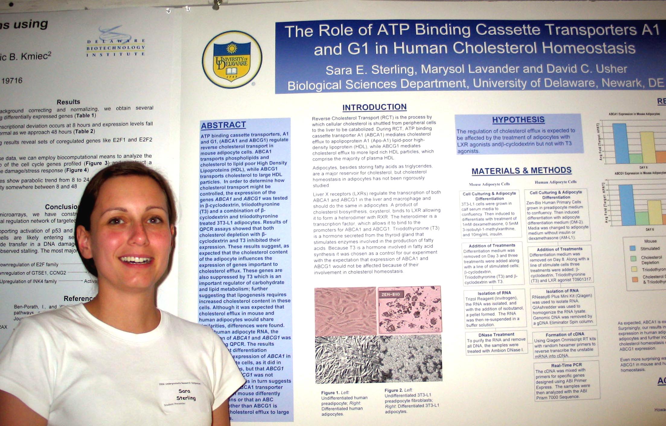
Cholesterol Depletion in Adipocytes and its Affects on Lipoproteins E and C1 Marysol D. Lavander, Sara Sterling, and David Usher Department of Biological Sciences Apolipoprotein (apo) C1,
a 6.6-kD
protein, is a protein constituent of triglyceride (TG) -rich
chylomicrons, very
low density lipoproteins (VLDL) and high density lipoproteins (HDL).
The human APOC1 gene is located 5 kb downstream of
the human APOE gene on chromosome 19
in the same transcriptional orientation. ApoE mediates the uptake of
triglyceride-rich lipoproteins (TRL) by hepatic receptors, where as
apoC-I has
an inhibitory effect. Both these proteins appear to be involved in
cholesterol
efflux in adipocytes. In order to find
how this process is controlled, the expressions of APOE
and APOC1 were
tested in 3T3-L1 mouse adipocytes treated with β-cyclodextrin,
triiodothyronine
(T3) and a combination of β-cyclodextrin and T3. The results showed
that
β-cyclodextrin decreased expression of the two genes 7-10 fold,
T3
decreased expression 2-4-fold and the combination of β-cyclodextrin
and T3 inhibited expression 9-14 fold. The results suggest
that cholesterol depletion or stimulation of carbohydrate metabolism
causes
cells to attempt to increase the cholesterol concentration within the
cell.
This may be due to the formation of lipid droplets which require a
boundary of
cholesterol, phospholipids and protein. Sponsored by INBRE.
|
A Role for N-Glycosylation in Differential Growth Control and Drosophila Development: Phenotypic Analysis of alg10 Evan Lebois and Erica Selva Department of Biological Sciences |
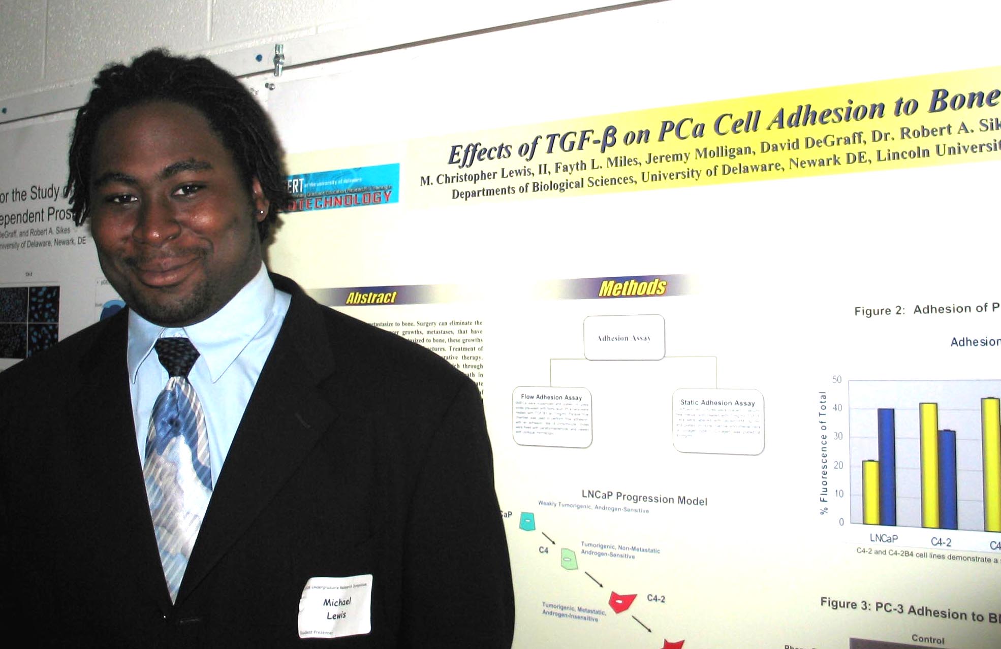
Effects of TGF-β on Prostate Cancer Cell Adhesion to Bone Endothelium under Static and Flow Conditions. M. Christopher Lewis, II1, Fayth L. Miles2, Jeremy Molligan3, David DeGraff4, Robert A. Sikes5, Carlton R. Cooper6 1Biological Sciences, |
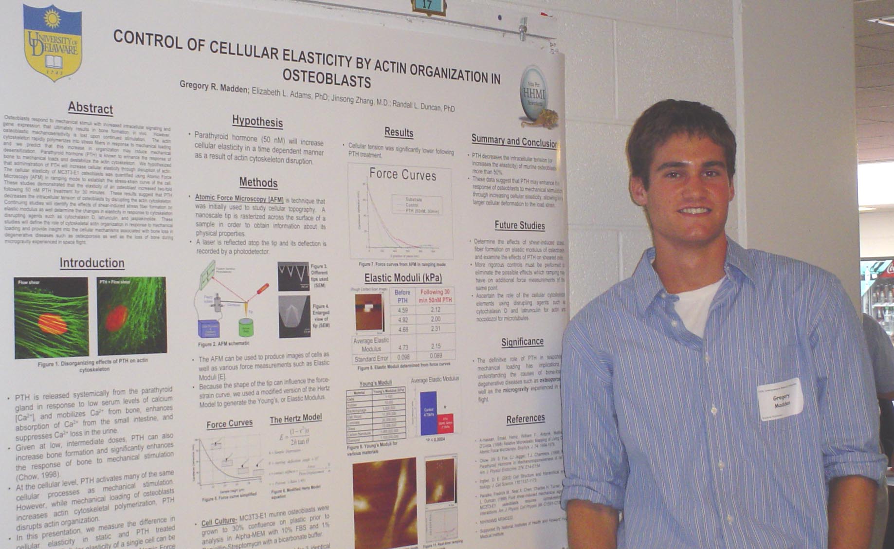
Control of Cellular Elasticity by Actin Organization in Osteoblasts Gregory Madden, Randall L. Duncan, Elizabeth L. Adams, and Jinsong Zhang Department of Biological Sciences Osteoblasts respond to
mechanical stimuli with increased
intracellular signaling and gene expression that ultimately results in
bone
formation, in vivo. However,
osteoblastic mechanosensitivity is lost upon continued stimulation. The actin cytoskeleton rapidly polymerizes
into stress fibers in response to mechanical loading and we predict
that this
increase in organization may induce mechanical desensitization. Parathyroid hormone (PTH) is known to enhance
the response of bone to mechanical loads and destabilize the actin
cytoskeleton. We hypothesized that
administration of PTH will increase cellular elasticity through
disruption of
actin. The cellular elasticity of
MC3T3-E1 osteoblasts was quantified using Atomic Force Microscopy [AFM]
in
ramping mode to establish the stress-strain curve of the cell. These
studies demonstrated that the elasticity of an osteoblast
increased
two-fold following 50 nM PTH treatment for 30 minutes.
These results suggest that PTH decreases the
intracellular tension of osteoblasts by disrupting the actin
cytoskeleton. Continuing studies will
identify the effects
of shear-induced stress fiber formation on elastic modulus as well
determine
the changes in elasticity in response to cytoskeleton disrupting agents
such as
cytochalasin D, latrunculin, and jasplakinolide. These
studies will define the role of
cytoskeletal actin organization in response to mechanical loading and
provide
insight into the cellular mechanisms associated with bone loss in
degenerative
diseases such as osteoporosis as well as the loss of bone during
microgravity
experienced in space flight. Supported
by the Howard Hughes Medical Institute and the National Institutes of
Health.
|
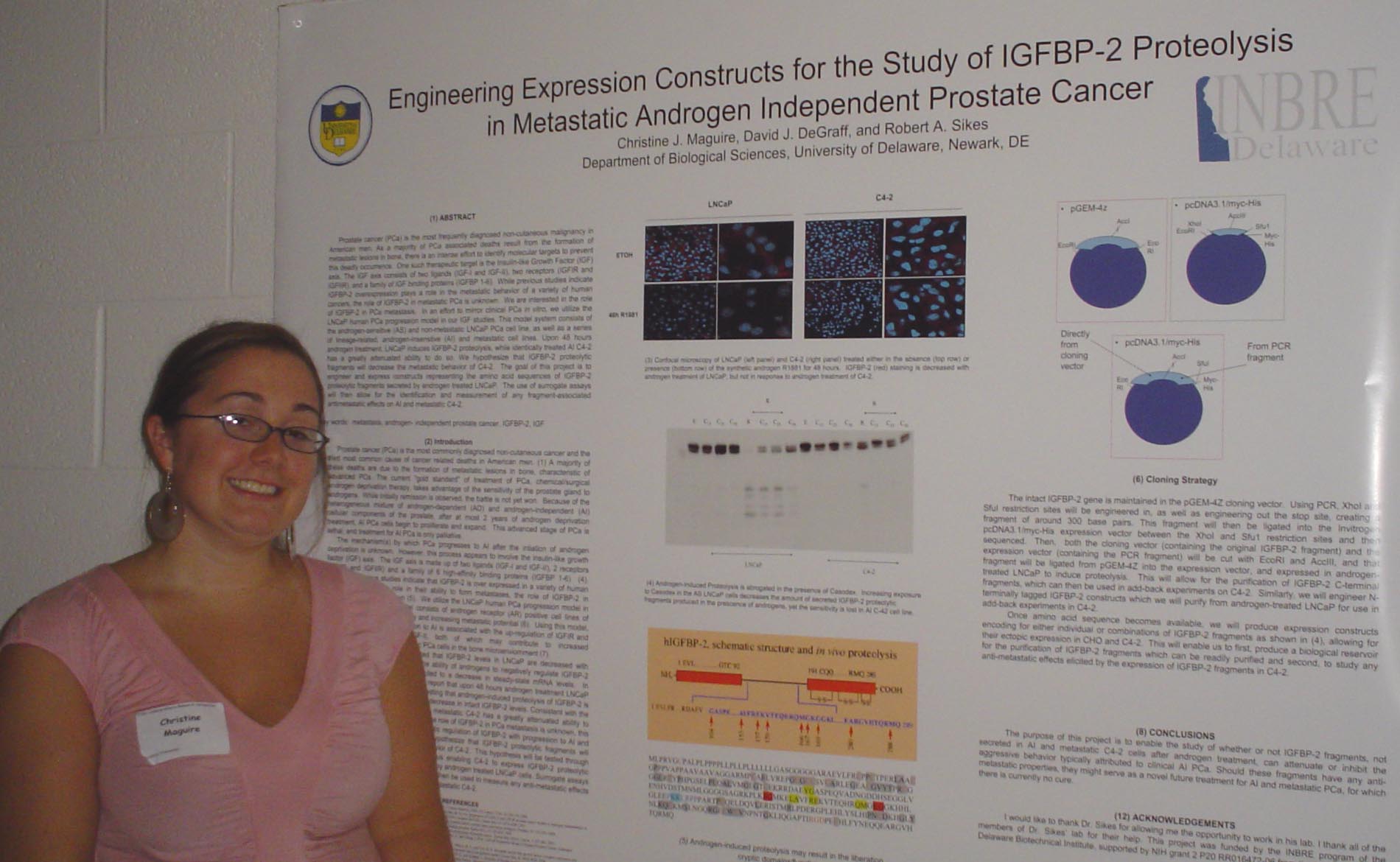
Engineering Expression Constructs for the Study of IGFBP-2 Proteolysis in Metastatic Androgen Independent Prostate Cancer Christine J. Maguire, David J. DeGraff, and Robert A. Sikes Department of Biological Sciences Prostate cancer (PCa) is the most frequently diagnosed non-cutaneous malignancy in American men. As a majority of PCa associated deaths result from the formation of metastatic lesions in bone, there is an intense effort to identify molecular targets to prevent this deadly occurrence. One such therapeutic target is the Insulin-like Growth Factor (IGF) axis. The IGF axis consists of two ligands (IGF-I and IGF-II), two receptors (IGFIR and IGFIIR), and a family of IGF binding proteins (IGFBP 1-6). While previous studies indicate IGFBP-2 overexpression plays a role in the metastatic behavior of a variety of human cancers, the role of IGFBP-2 in metastatic PCa is unknown. We are interested in the role of IGFBP-2 in PCa metastasis. In an effort to mirror clinical PCa in vitro, we utilize the LNCaP human PCa progression model in our IGF studies. This model system consists of the androgen-sensitive (AS) and non-metastatic LNCaP PCa cell line, as well as a series of lineage-related, androgen-insenstive (AI) and metastatic cell lines. Upon 48 hours androgen treatment, LNCaP induces IGFBP-2 proteolysis, while identically treated AI C4-2 has a greatly attenuated ability to do so. We hypothesize that IGFBP-2 proteolytic fragments will decrease the metastatic behavior of C4-2. The goal of this project is to engineer and express constructs representing the amino acid sequences of IGFBP-2 proteolytic fragments secreted by androgen treated LNCaP. The use of surrogate assays will then allow for the identification and measurement of any fragment-associated antimetastatic effects on AI and metastatic C4-2. This project was funded by the INBRE program of the Delaware Biotechnical Institute, supported by NIH grant 2 P20 RR016472-06 from the NCRR. |
Analysis of the Targeted Gene Repair Transcriptome in Mammalian Systems Arjun K. Manrai1, Luciana Ferrara2, Hetal Parekh-Olmedo2, and Eric B. Kmiec2 1Department of Physics, Targeted Gene Repair is
the process in which a modified single-stranded
oligonucleotide directs the exchange of a single mutated DNA base. This
correction is site-specific, utilizing the cell’s endogenous repair
mechanisms.
Upon correction induced by the oligonucleotide, many corrected cells
cease
active replication, and key proteins of senescence pathways have been
implicated through increased activation. We perform the gene repair
reaction on
DLD-1 cells and carry out microarray analysis using cell-cycle specific
oligonucleotide microarrays to profile the transcriptome of
gene-repaired cells
at 8, 24, and 48 h. After mathematically correcting for background and
normalizing the data, we employ biocomputational tools and analyses to
construct a regulatory model and explain the response of cells to gene
repair. We
then use insight gleaned from this experiment to develop and implement
a
framework for further transcriptional analysis of targeted cells.
Specifically,
we create and program a model for global expression analysis of
targeted cells
to be used on data obtained from (Affymetrix) transcriptome-wide
arrays,
capturing more than 47,000 transcripts. The framework integrates the
latest
probe-mapping, normalization algorithms, cluster analysis, ontological
information,
motif finding, and pathway systems-level information into a pipeline
for the
upcoming analysis. Moreover, we tailor our pipeline to be specifically
adapted to
future large-scale gene repair microarray experiments using information
obtained from the DLD-1 study, by developing computational tools to
extract and
compile pathway-specific data. This is the first study to obtain and
analyze
transcriptional profiles of targeted DLD-1 cells and use these analyses
for an
integrated framework and we hope that, in addition to providing
information
about the observed regulatory pathways of targeted cells in
vitro, this work provides valuable insight for future in
vivo gene repair applications. This project
was supported by NIH NCRR INBRE grant to |
Seeing Beyond the Human Eye with Modern Microscopy Eamon McLaughlin1, Kirk Czymmek1,2, Debbie Powell2, Jeanette Miller2, and Liz Adams2 1Department of Biological Sciences and 2Delaware Biotechnology Institute My project
involved the task of creating a way to educate local high school
science
teachers about the microscopy equipment located in the |
Vesicular Transport and Excess Residual Cytoplasm in Sperm Rowan Michael, Minghai Shao, Genevieve Griffiths, and Patricia A. Martion-DeLeon Department of Biological Sciences |
The Gemini virus Coat Protein: Detection of mRNA and Subcellular Localization Stacey L. Mlynarski, Marie Etarock, R.V. Chowda Reddy, Patricia Nugent, Bertrand Hankoua, Christian Felton, Marie Paul-Emile, Marie-Teresa Ananfac, Mastingor Desir, and Vincent N. Fondong Department of Biology, The genome of East
African cassava mosaic
|
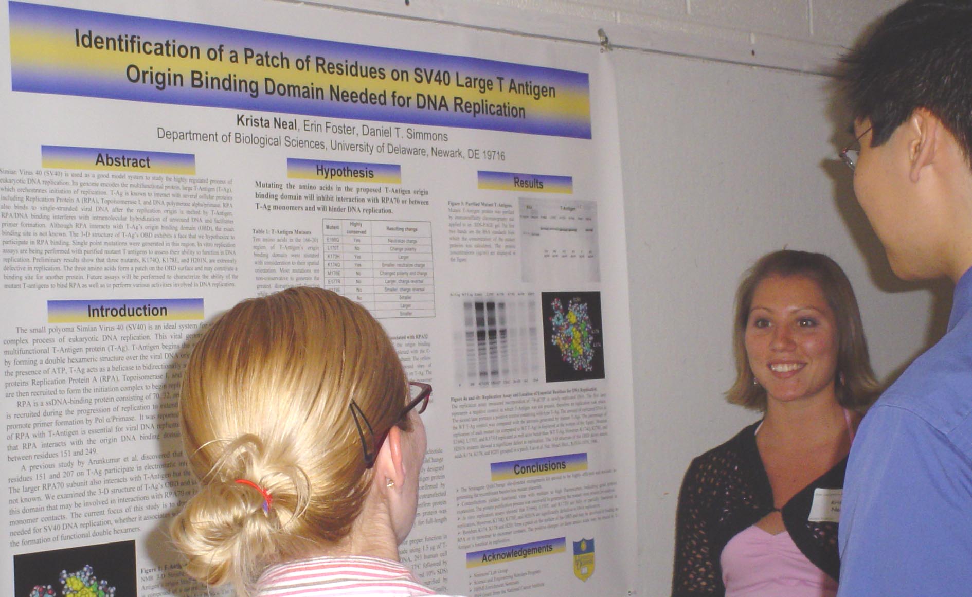
Identification of a Patch of Residues on SV40 T-Antigen Origin Binding Domain Needed for DNA Replication Krista Neal, Erin Foster, Daniel T. Simmons Department of Biological Sciences Simian Virus 40 (SV40) is used as a good model system to study the highly regulated process of eukaryotic DNA replication. Its genome encodes the multifunctional protein, large T-Antigen (T-Ag), which orchestrates initiation of replication. T-Ag is known to interact with several cellular proteins including Replication Protein A (RPA), Topoisomerase I, and DNA polymerase alpha/primase. RPA also binds to single-stranded viral DNA after the replication origin is melted by T-Antigen. RPA/ssDNA binding interferes with intramolecular hybridization of unwound DNA and facilitates primer formation. Although RPA interacts with T-Ag’s origin binding domain (OBD), the exact binding site is not known. The 3-D structure of T-Ag’s OBD exhibits a face of amino acids that we hypothesize to participate in RPA binding. Single point mutations were generated in this region. In vitro replication assays are being performed with purified mutant T antigens to assess their ability to function in DNA replication. Preliminary results show that three mutants, K174Q, K178E, and H201N, are extremely defective in replication. The three original amino acids form a patch on the surface of the OBD and may constitute a binding site for another protein. Future assays will be performed to characterize the ability of the mutant T-antigens to bind RPA as well as to perform various activities involved in DNA replication. This research is funded by a PHS Grant from the National Cancer Institute. |
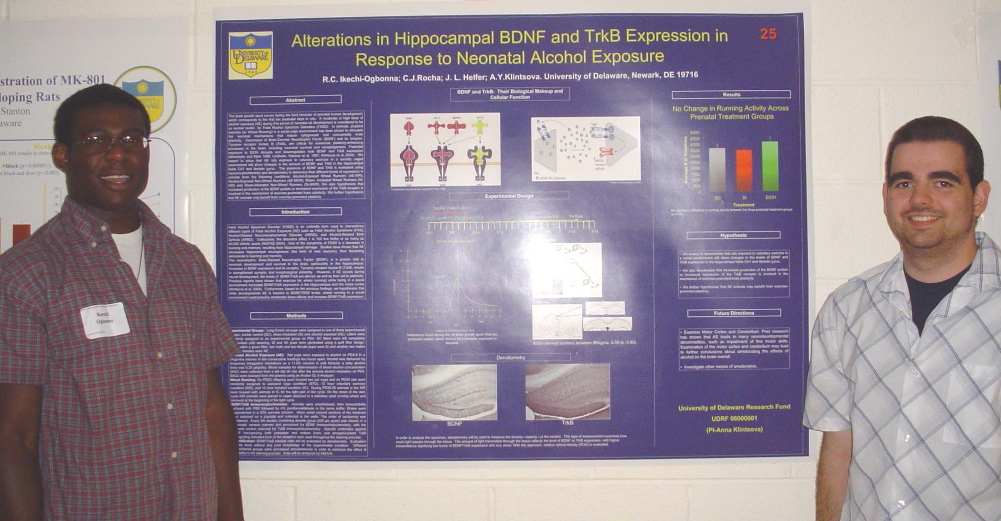
Alterations in BDNF and TrkB Expression in Response to Neonatal Alcohol Exposure Ronald C. Ikechi-Ogbonna, C.J. Rocha, A.Y. Klintsova Behavioral Neuroscience and Psychology |
The Search for Genes in Prostate Morphogenesis Lauretta Ovadje1, Peter S. Nelson2, Cynthia Qian3, Robert Sikes3 1Department of Biology, |
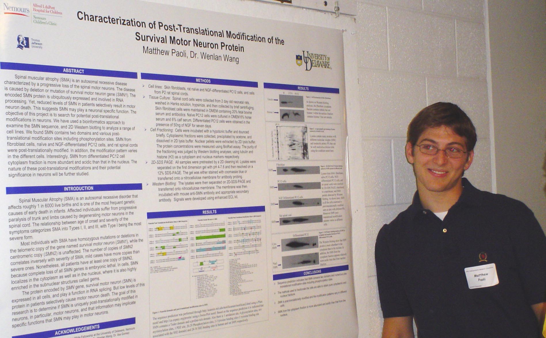
Spinal
muscular atrophy
(SMA) is an autosomal recessive disease characterized by a progressive
loss of
the spinal motor neurons. The disease is caused by deletion or mutation
of
survival motor neuron gene (SMN1). The encoded SMN protein is
ubiquitously expressed and involved in RNA processing. Yet, reduced
levels of
SMN in patients selectively result in motor neuron death. This suggests
SMN may
play a neuronal specific function. The objective of this project is to
search
for potential post-translational modifications in neurons. We have used
a
bioinformatics approach to examine the SMN sequence, and 2D Western
blotting to
analyze a range of cell lines. We found SMN contains two domains and
various
post-translational modification sites including phosphorylation sites.
SMN from
fibroblast cells, naïve and NGF-differentiated PC12 cells, and rat
spinal cords
were post-translationally modified. In addition, the modification
pattern
varies in the different cells. Interestingly, SMN from differentiated
PC12 cell
cytoplasm fraction is more abundant and acidic than that in the
nucleus. The
nature of these post-translational modifications and their potential
significance in neurons will be further studied. Supported by Charles
Peter
White Fellowship. |
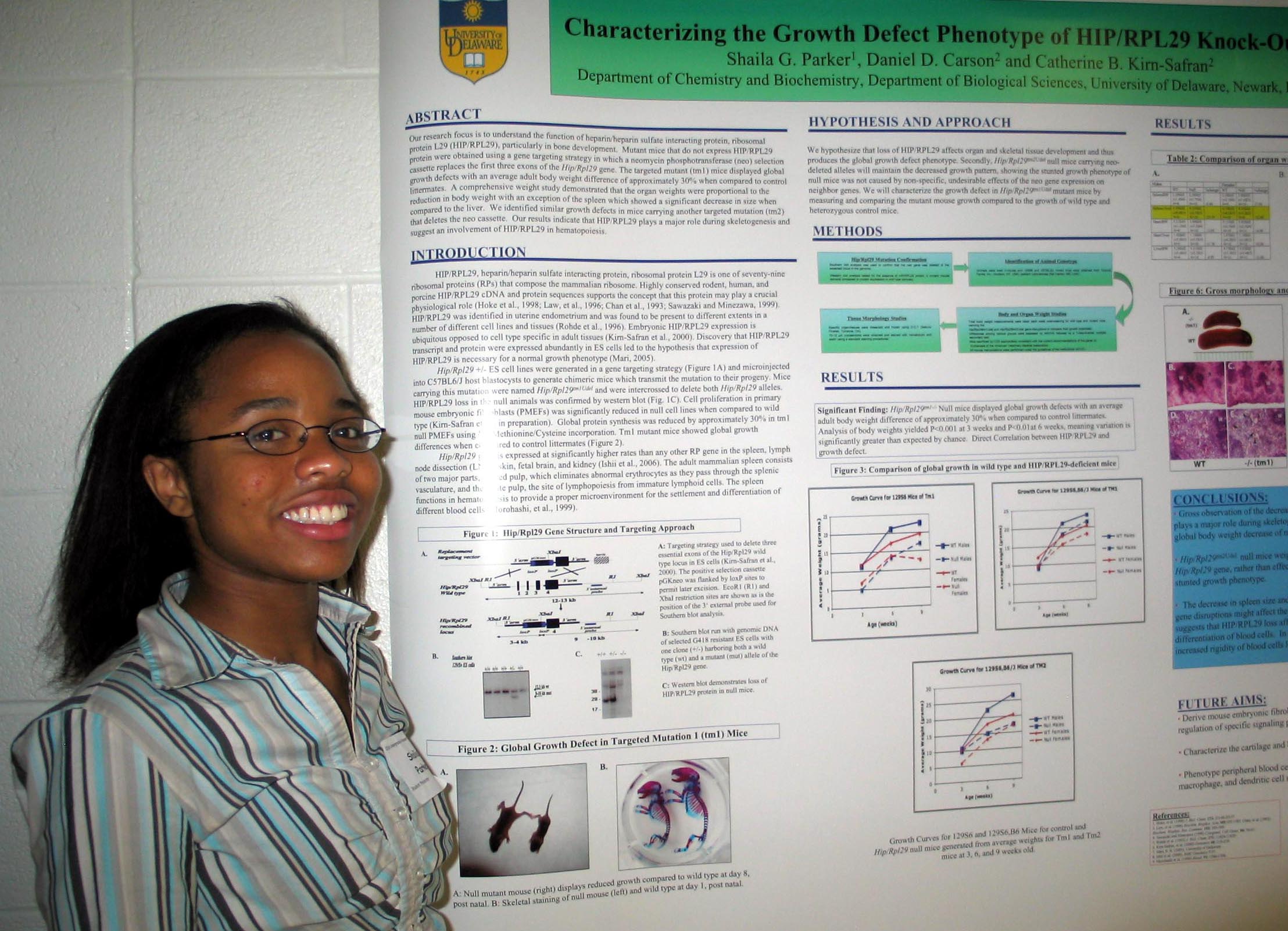
Characterization of the Growth Defect Phenotype of HIP/RPL29 Knock-Out Mice Shaila Parker, Daniel Carson, and Catherine Kirn-Safran Department of Biological Sciences Our
research focus is to understand the function of heparin/heparin sulfate
interacting protein, ribosomal protein L29 (HIP/RPL29), particularly in
bone
development. Mutant mice that do not
express HIP/RPL29 protein were obtained using a gene targeting strategy
in
which a neomycin phosphotransferase (neo) selection cassette replaces
the first
three exons of the Hip/Rpl29 gene. The
targeted mutant (tm1) mice displayed global
growth defects with an average adult body weight difference of
approximately 30%
when compared to control littermates. A comprehensive weight study
demonstrated
that the organ weights were proportional to the reduction in body
weight with
an exception of the spleen which showed a significant decrease in size
when
compared to the liver. We identified
similar growth defects in mice carrying another targeted mutation (tm2)
that
deletes the neo cassette. Our results
indicate that HIP/RPL29 plays a major role during skeletogenesis and
suggest an
involvement of HIP/RPL29 in hematopoiesis. Funding for this project was
provided by the Carson lab and McNair Scholars Program.
|
Biochemistry of Prox1 Tapan Patel1, Xiaoren Chen1, Li Liao2, Melinda K. Duncan1 1Department of Biological Sciences, 2Department of Computer and Information Sciences Prox1 is a vertebrate homeobox transcription factor related to Drosophila Prospero and as such plays many important roles during development such as: lymphangiogenesis, hepatocyte migration during liver development, lens fiber cell differentiation, and differentiation of ganglion cells in retinal development. In certain cell types such as the ocular lens, it acts as a true transcription activator by binding to γF-crystallin and βB1 crystallin promoters and activating their expression. Prox1 can also act as a co-repressor of nuclear hormone receptors SF-1 and LRH1. We hypothesize that these dual functions of Prox1 are achieved through specific protein-protein interactions. A yeast two-hybrid screen of mouse cDNA library led to the discovery of numerous proteins that may potentially interact with Prox1 in vivo. Here we report the interaction of Prox1 with the splicing factor SC35 and the DNA sliding clamp PCNA. Immunofluorescence colocalization data suggest that Prox1-SC35 interaction results in redistribution of intracellular SC35 protein. SC35 is normally clustered in the nucleus giving rise to the famous nuclear speckles; however, in cells expressing Prox1, SC35 is redistributed evenly throughout the nucleoplasm. Furthermore, this interaction leads to the repression of Prox1 mediated transactivation through the PL2 promoter element as determined via CAT assay. Similar repression has been found for Prox1-PCNA interaction as well. To help explain these observations in molecular detail, we propose a docking model of Prox1-PCNA complex whereby the PIP-box of Prox1 binds chemically to the interdomain connecting loop of PCNA. Future in vivo mutagenesis experiments will validate the proposed model. Supported in part by a Beckman and Goldwater Fellowships. |
The Role of MeCP2 in Neuron Development and Bone Homesostasis Donna Pomroy, Carolyn Schanen, Rose Deeter, and Andrea Ham A. |
The Effect of the Scale of an Alien Plant Invasion on Native Insect Communities Erin Reed and Douglas W. Tallamy Department of Entomology and Wildlife Ecology |
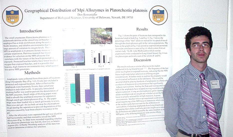
There are myriad examples of polymorphisms that are affected by selection, but few of these are completely understood on a physiological and biochemical ground. The role of MPI in amphipods is to process dietary mannose: After mannose has been converted to mannose-6-phosphate by hexokinase using ATP, mannose-6-phosphate is converted my MPI (the enzyme in question) to fructose-6-phosphate, which is fed into the glycolysis pathway to eventually produce more ATP, especially if there is adequate oxygen. It is possible that hypoxic, high-mannose environments may select against less-active MPI genotypes.
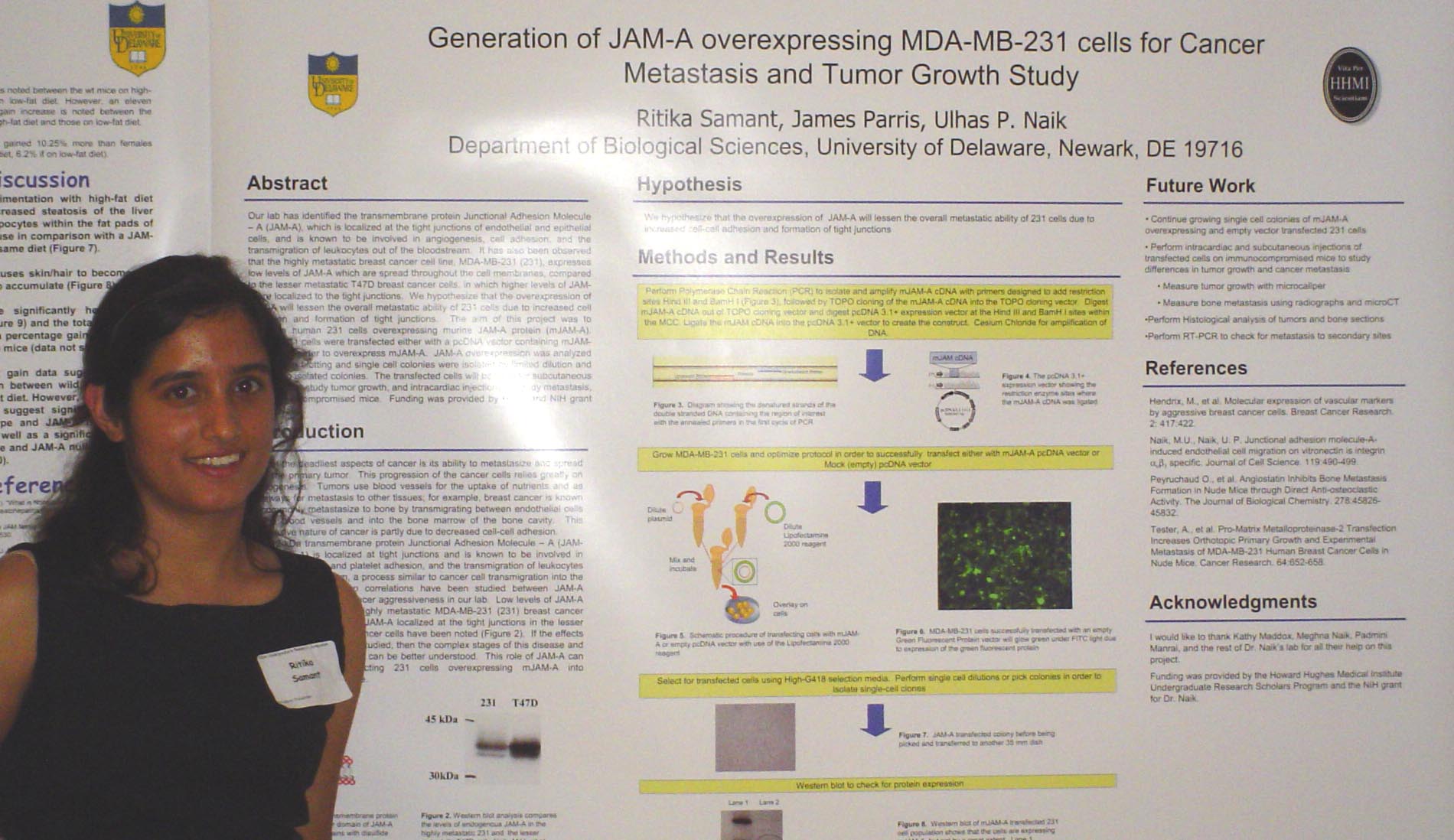
Generation of JAM-A overexpressing MDA-MB-231 Cells for Cancer Metastasis and Tumor Growth Study Ritika Samant, James Parris, Ulhas P. Naik Department of Biological Sciences Our lab has identified the transmembrane protein Junctional Adhesion Molecule – A (JAM-A), which is localized at the tight junctions of endothelial and epithelial cells, and is known to be involved in angiogenesis, cell adhesion, and the transmigration of leukocytes out of the bloodstream. It has also been observed that the highly metastatic breast cancer cell line, MDA-MB-231 (231), expresses low levels of JAM-A which are spread throughout the cell membranes, compared to the lesser metastatic T47D breast cancer cells, in which higher levels of JAM-A are localized to the tight junctions. We hypothesize that the overexpression of JAM-A will lessen the overall metastatic ability of 231 cells due to increased cell adhesion and formation of tight junctions. The aim of this project was to generate human 231 cells overexpressing murine JAM-A protein. Human 231 cells were transfected either with a pcDNA vector containing murine JAM-A (mJAM-A) cDNA in order to overexpress mJAM-A. JAM-A overexpression was analyzed by Western blotting and single cell colonies were isolated by limited dilution and by picking isolated colonies. The transfected cells will be used for subcutaneous injections, to study tumor growth, and intracardiac injections, to study metastasis, on immunocompromised mice. Funding was provided by HHMI. |
Is JAM Making Your Liver Fatty? The Effect of JAM-A on NAFLD Laura S. Shankman1, Vesselina G. Cooke1, William C. Skarnes2 Ulhas P. Naik1 1Department of Biological Sciences, |
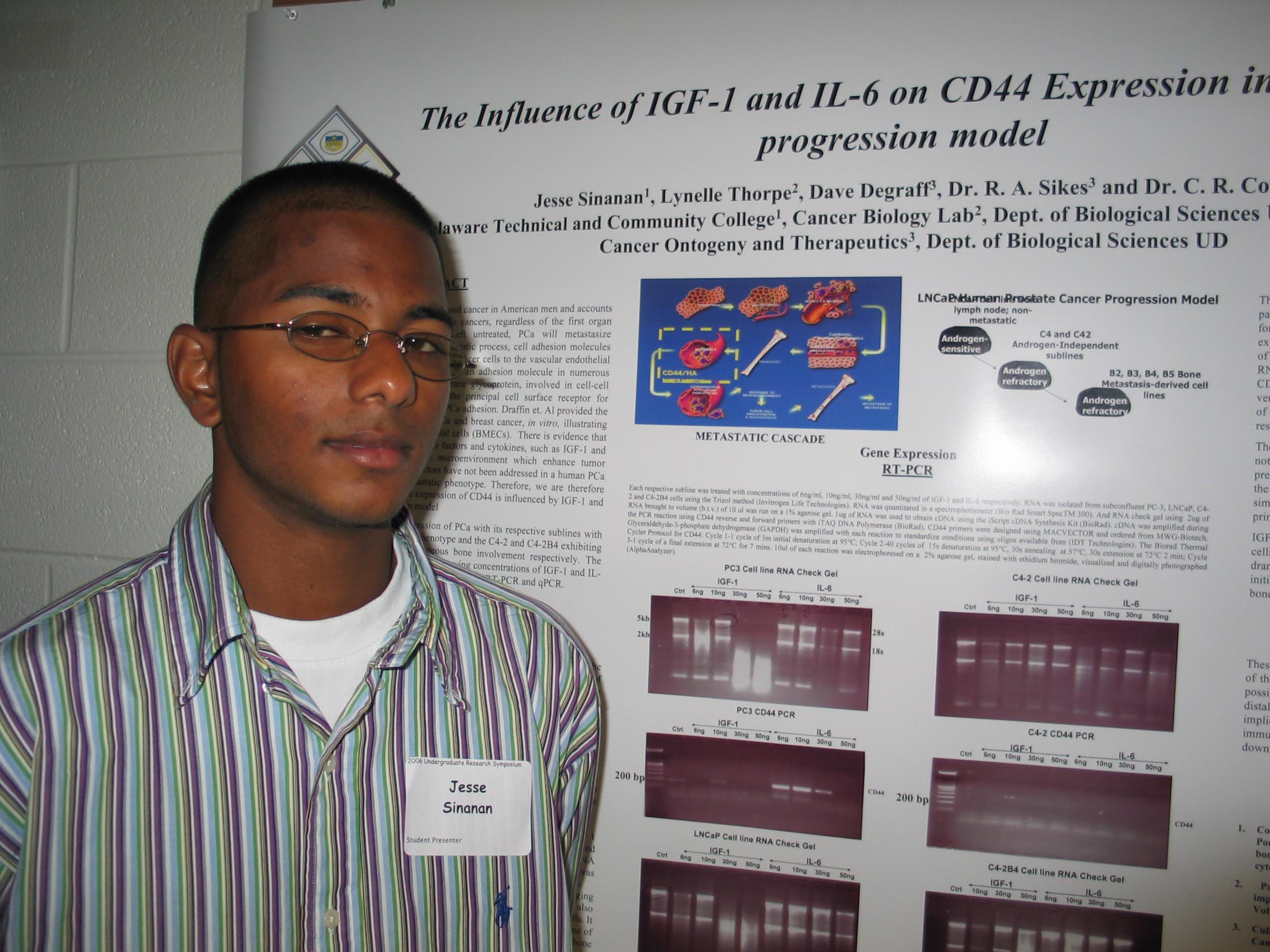
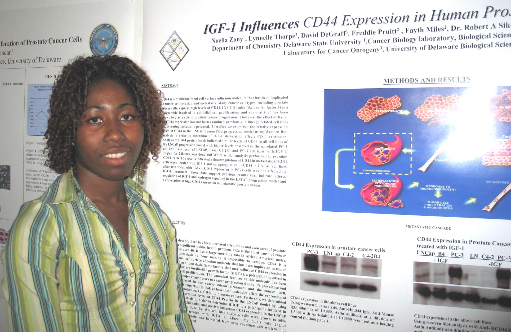
The Influence of IGF-1 and IL-6 on CD44 Expression in the LNCaP Progression Model Jesse Sinanan1, Lynelle Thorpe, Dave Degraff, Robert A. Sikes, and |
Detection of Salmonella in Biosolids Samantha A. Smith, Diane S. Herson, and Yinan Qi Department of Biological Sciences and Department of Civil and Environmental Engineering Biosolids are produced as byproducts of waste water treatment. The presence of Salmonella spp. in this material is of concern because of the illnesses they cause. In the newly proposed EPA Method 1682, selection of Salmonella spp. occurs on modified semi-solid Rappaport Vassiliadis (MSRV), which contains an antibiotic (novobiocin) and a dye (malachite green) to inhibit non-Salmonella species. Method 1682 takes several days due to the multiple cultural steps involved. The standard polymerase chain reaction (PCR) assay is a molecular assay that can be used for the detection of Salmonella spp. This method takes less time, but the results may be inhibited by the presence of substances contained in biosolids. Our studies first compared the results of biosolids samples in the cultural EPA method 1682 and a PCR assay. The results indicated that PCR was inhibited in the most concentrated samples. The use of MSRV as an enrichment step prior to PCR was then tested by removing a sample of cells and media and subjecting the samples to DNA isolation. The results indicated that PCR was inhibited when using this method. The effect of vortexing samples in both MSRV and Trypticase Soy Broth (TSB) after the addition of the Instagene matrix was also tested and was found to have no effect on the results of the PCR assay. We then tested several different ways of isolating the cells or DNA from the MSRV and its potential inhibitors. It was found that heating the sample in a hot water bath for 1-2 minutes to melt the media, followed by centrifugation and removal of the supernatant prior to the addition of Instagene, resulted in the PCR assay agreeing with the results of the cultural method. |
Ontogeny of Surfactant Protein B (SP-B) Expression in Avian Lung Sam Snow and Sabrina M. Brougher |
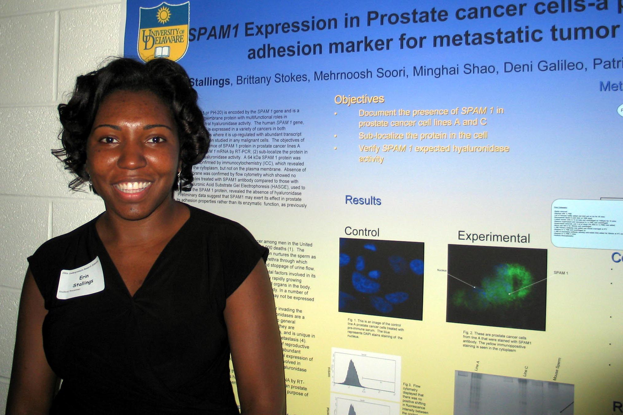
SPAM1 Expression in Prostate Cancer-a Possible Adhesion Marker for Metastatic Tumor Erin Stallings, Brittany Stokes, Mehrnoosh Soori, Minghai Shao, Deni Galileo, Patricia A. Martin-Deleon Department of Biological Sciences Sperm adhesion molecule 1 (SPAM 1 or PH-20) is encoded by the SPAM 1 gene and is a widely conserved mammalian sperm membrane protein with multifunctional roles in fertilization, and it is best known for its neutral hyaluronidase activity. The human SPAM 1 gene, which is located on chromosome 7q31, is expressed in a variety of cancers in both reproductive and non-reproductive organs where it is up-regulated with abundant transcript levels. However, its protein has never been studied in any malignant cells. The objectives of this study are to (1) document the presence of SPAM 1 protein in prostate cancer lines A and C, previously proven to contain SPAM 1 mRNA by RT-PCR; (2) sub-localize the protein in the cells; and (3) verify its expected hyaluronidase activity. A 64 kDa SPAM 1 protein was detected by Western analysis and confirmed by immunocytochemistry (ICC), which revealed the localization of the protein in the cytoplasm, but not on the plasma membrane. Absence of the protein on the plasma membrane was confirmed by Fluorescence Activated Cell Sorting (FACS), which showed no increase in fluorescence in samples treated with SPAM1 antibody compared to pre-immune serum. Lastly, Hyaluronic Acid Substrate Gel Electrophoresis (HASGE), used to detect hyaluronidase activity of the SPAM 1 protein, revealed the absence of hyaluronidase activity at neutral pH. Preliminary data suggest that SPAM1 may exert its effect in prostate cancer metastasis via its adhesion properties rather than its enzymatic function, as previously reported. Funded in part by the Department of Defense Grant (DOD) |
|
SPAM1 Expression in Breast Cancer Cells Is Not Associated with Hyronidase Activity |
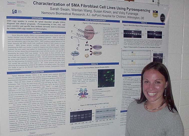
Characterization of SMA Fibroblast Cell Lines Using Pyrosequencing Sarah Swain, Wenlan Wang, Susan Kirwin, and Vicky Funanage Nemours Biomedical Research, Spinal Muscular Atrophy (SMA) is an autosomal motor neuron disease and is the leading inherited cause of infant and early childhood mortality. SMA is caused by mutation or deletion of the telomeric copy of the gene, SMN1, however all patients retain at least one centromeric copy of the gene, SMN2. SMN2 produces reduced amounts of full length transcript and SMA results from insufficient levels of SMN protein in motor neurons. SMA disease severity correlates inversely with SMN2 copy number. Determining the SMN copy number is crucial for diagnosis and clinical prognosis of SMA. Currently, the most widely used method for characterizing cell lines is restriction digest. However, we have shown that pyrosequencing is more selective, sensitive, and specific in determining the relative SMN copy number. In this study we have collected six (6) controls and five (5) SMA type I patient cell lines. It has been shown by restriction digest that all SMA type I patients were deleted for SMN1. We will continue to collect cells from the various types of SMA for characterization. This will enable us to better create a novel assay for drug screening of potential treatments for the disease and to determine the SMN2 copy number that correlates to SMA types I - III. Future projects will also include optimizing the pyrosequencing method for the addition of an internal control. This research was funded in part by the Howard Hughes Medical Institute Undergraduate Science Education program. |
Examination of the Role of JAM-A Homodimerization in Angiogenesis Wen Allen Tseng and Ulhas P. Naik Department of Biological Sciences Junctional adhesion molecule-A (JAM-A) is found at the tight junctions of endothelial and epithelial cells. It has been demonstrated that JAM-A forms cis-homodimers in vitro and in vivo and that this property is needed for the proper maintenance of selective barriers by epithelial monolayers. Our lab has shown that signaling through JAM-A plays an essential role in the induction of angiogenesis by basic fibroblast growth factor (bFGF). This study intends to determine if and how cis-homodimerization of JAM-A is involved in the induction of angiogenesis. To this end, several mutant JAM-A cDNA constructs were generated via site-directed mutagenesis. For some constructs, mutations were designed to impair JAM-A homodimerization. In other constructs, reciprocal mutations were made that were intended to restore cis-homodimerization. An assay for measuring JAM-A dimerization at the cell surface was developed and will be used to quantitate the extent of homodimerization-impairment for the mutant JAM-A constructs. The relationship between JAM-A homodimerization and angiogenesis will be studied by examining the pro-angiogenic behavior of human umbilical vein endothelial cells (HUVECs) over-expressing wild-type or mutant JAM-A. This research was supported by the |
ssDNA Oligonucleotides Arrest Growth of Esophageal Cancer Cells Casey A. Vasta, Timothy R. Schwartz, and Eric B. Kmiec Department of Biological Sciences and the Delaware Biotechnology Institute |
Variation in Sperm Hyaluronidase Activity in Men Attending an IVF Clinic Sarah Woody, Jessica Bruton, Minghai Shao, Patricia A. Martin-Deleon Department of Biological Sciences |
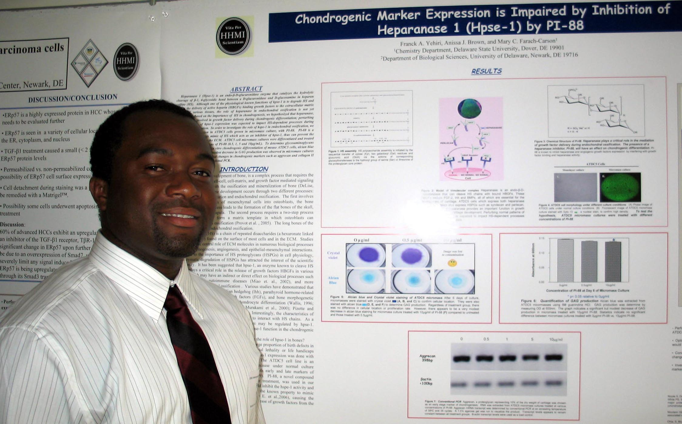
Chondrogenic Marker Expression is Impaired by Franck A. Yehiri, Anissa J. Brown, Mary C. Farach-Carson Department of Biological Sciences |