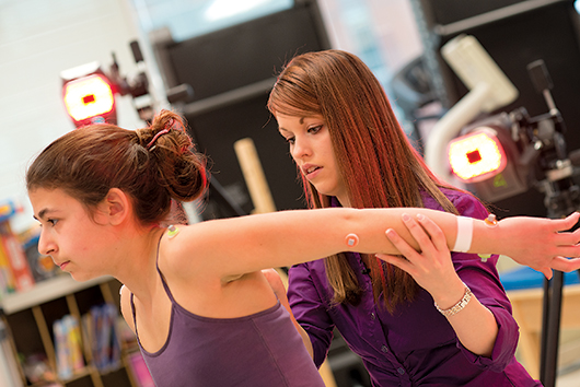
Motion analysis holds promise for children with birth injury

RESEARCH | Researcher Jim Richards has successfully used motion analysis technology to allow elite figure skaters to explore “what-if” scenarios about their jumping technique. Now, the Distinguished Professor of Kinesiology and Applied Physiology hopes that he and his research team can use a similar approach to guide clinicians in treating children with a specific birth injury.
Brachial plexus birth palsy (BPBP) occurs in about four of every 1,000 births and affects nerve roots in the cervical spine, impacting muscle function in the shoulder and the arm.
Most children recover on their own, but about 30 percent are left with lifelong deficits in arm function that require therapy or surgery. The most severe brachial plexus injuries can cause complete paralysis of the arm.
But the answer to a key question has eluded researchers trying to understand exactly what is going on in the musculoskeletal systems of children with BPBP: Where is the scapula, or shoulder blade, at any given moment, and what is it doing? This information would provide valuable insight into a child’s specific defects and enable treatments to be tailored to individual patients, as the location and extent of damage to the nerves and muscles vary from one person to another.
The problem, Richards says, is that the movements of the scapula are incredibly difficult to measure.
“Our motion-capture cameras provide us with reasonable data for the lower extremities,” he says, “but the same approach applied to the upper body fails to tell us much about the movement of the scapula.”
He and his team of doctoral students in the Biomechanics and Movement Science program have taken a systematic approach to filling this gap. If they’re successful, it may someday be possible for surgeons to use the UD simulation to explore what will happen if they move a tendon from one point to another in an individual patient.
The team is working with clinicians at Shriners Hospital for Children in Philadelphia on the first stage of the project. Two of Richards’ students, Kristen Thomas and Stephanie Russo, have collected data on 65 children with BPBP using a motion analysis system.
“We’re fairly confident that we can get accurate scapular measurements under static conditions,” Thomas says. “The question then is: If we put the kids in enough static positions, can we draw conclusions about what happens when they’re moving?”
The researchers’ plan is to build a 3D model to test their early measures and then move on to test a larger pool of subjects. By using the model, they will be able to avoid the need for expensive imaging equipment, Thomas says.
“If it all works,” Richards says, “we’ll be able to go into a clinical setting like Shriners, drop 11 markers onto a patient to find out what’s happening and then do the same after surgery to see what the effects are.”
Ultimately, he envisions clinicians being able to explore what-if scenarios that would enable them to determine how a specific surgical technique will affect a specific patient—and, eventually, to broaden the technique beyond BPBP to other injuries.
Article by Diane Kukich, AS73, 84M


