 |
 EB2011 EXPERIMENTAL BIOLOGY MEETINGS Washington, DC APRIL 9-13, 2011  |
 |
 EB2011 EXPERIMENTAL BIOLOGY MEETINGS Washington, DC APRIL 9-13, 2011  |
University of Delaware students
who presented posters in the Undergraduate Poster Competition sponsored
by ASBMB. Front Row, left to right: Tejal Naik, Ashley Shay, Jean Huynh, Tori Roop, and Erica Boetefuer. Back Row, left to right: Muike Brister, Cory Bovenzi, Bobby Sheehan, Soma Jobbagy, and Matt King. (Dylan Lowe not present) |
|||
| Prof.
Hal White, Chem & Biochem Prof. Dave Usher, Biol. Sci. Prof. Seung Hong, Biol Sci Prof. Gary Laverty, Biol Sci |
Erica
Lee Boetefuer Cory Bovenzi Michael Angelo Brister Jean Huynh |
Soma
Jobbagy Matthew King Dylan Lowe Tejal U Naik |
Victoria
H. Roop Ashley Shay Robert Patrick Sheehan |
|
|
Abstract
No: 2075 The
role of atg18 in signal transduction pathways
during Drosophila development Erica L. Boetefuer, Erica M. Selva, and David Raden Department of Biological Sciences The goal of this research is to clone and characterize two allelic mutations, 8J16 and 9E6. Genetic screens indicated these mutations disrupt Wnt/Wingless (Wg) or Hedgehog (Hh) signaling based upon their embryonic ‘lawn of denticles’ phenotype. Complementation showed 8J16 and 9E6 are autophagy-specific gene 18, atg18, alleles. atg18 plays a role in autophagosome-lysosome fusion for degradation during starvation. In yeast, atg18 negatively regulates endosome-lysosome targeting. Under fed conditions, atg18 mutations should increase endosome-lysosome targeting, as in yeast. In Drosophila, endocytic machinery mutations that block lysosome targeting cause amplified signaling, resulting in mitogenic signaling hyperactivation. This lead to the hypothesis that atg18 mutants would show the reverse; an accumulation of lysosomes at the expense of endosomes and a decrease in Wg signaling. Analysis in Drosophila wing imaginal discs showed no change in endocytic vesicles or Wg signaling in atg188J16 or atg189E6 mutant tissue. To the contrary, Wg signaling was enhanced as measured by the long-range target, Distalless (Dll). Molecular characterization identified atg18 intronic mutations for both alleles. These mutations should not disrupt normal mRNA splicing and RT-PCR revealed no splice variants, supporting this. Future experiments will focus on confirming these mutations are alleles of atg18. EB supported byHHMI undergraduate program. Day of Presentation: Monday April 11, 2011, 1:05 PM - 2:35 PM, Poster Board Number: B311, Program Number: 747.1 |
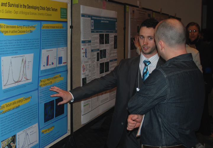 Cory Bovenzi
|
Neuronal
migration and survival in the developing chick optic tectum
Cory Bovenzi and Deni Galileo Department of Biological Science In the developing brain, neurons are born in the ventricular zone and migrate outward via radial glial scaffolds. There also is an early period of widespread programmed cell death (PCD) in the brain, occurring while these neurons are still migrating. In the developing chick optic tectum (OT), we hypothesize there is a relationship between these two processes whereby the cells that die do so because of anoikis: loss of contact with the necessary extracellular matrix produced by radial glia. Specifically, we predict that fibronectin secreted by radial glia interacts with the α8β1 integrin receptors on the neuronal membrane, initiating an intracellular cascade which eventually increases expression of Bcl-2, an anti-apoptotic molecule. Dissociated OT cells were immunostained at different ages and showed a general trend that there is a period (E7.5 through E8) where pro-apoptotic signals such as active Caspase-3 and Bax are present when Bcl-2 is down-regulated. Propidium iodide staining experiments supported immunostaining data, with more fragmented DNA appearing during this same time period. Bcl-2 also was ectopically expressed in the OT using a retroviral vector in hopes to analyze behavior of neurons kept alive artificially. Additionally, OT cells were plated at different densities and cultured to determine whether radial glia contact could suppress neuronal apoptosis and/or increase Bcl-2 levels. CB supported byHHMI undergraduate program.
|
|
Abstract No: 1763 The structural characteristics of
synthetically glycosylated tau protein sequences
Michael Angelo Brister, Agata Bielska, and Neal Zondlo Department of Chemistry and Biochemistry The
microtubule-associated protein tau is the primary constituent of
neurofibrillary tangles, a pathological hallmark of Alzheimer's
Disease. In its diseased state, tau is phosphorylated on over 30
residues, many of which are alternatively modified by O-linked
glycosylation by N-acetylglucosamine (O-GlcNAc) in tau's native state.
We hypothesized that glycosylation of threonine and serine residues on
tau peptides may induce a structural effect different from that of
phosphorylation at these locations. We have employed a scheme for the
synthesis of glycopeptides incorporating OGlcNAc and
isolated the amino acid building block, Fmoc-Thr(β- D-pGlcNAc)-OH.
Furthermore, we have developed a functional group to mimic O-GlcNAc
modification (pseudo-glycosylation). Peptides from the tau proline-rich
domain were synthesized and the conformations were characterized using
circular dichroism and NMR for the unmodified, phosphorylated,
pseudo-glycosylated, and glycosylated peptides. Phosphorylated
sequences exhibitedhigh propensity to form a type II polyproline helix.
In contrast, glycosylated peptides exhibited alternative
structures. These findings suggest a structural role for tau
modification by OGlcNAc. This project was supported by the Howard Hughes
Medical Institute and the Alzheimer's Association. Monday April 11, 2011, 1:05 PM - 2:35 PM, Poster Board Number: B93, Program Number: 708.3 First Prize recipient in the Proteins and Enzymes Division |
|
Abstract
No: 1765 A
Role for JAM-A in Ca2+ Homeostasis and Mammalian Sperm
Motility
Jean
Huynh and Patricia
Martin–DeLeon A
common cause of male infertility is defective sperm motility
(asthenozoospermia) results from the disruption of Ca2+
homeostasis,
including the deletion of the Ca2+ influx channel or the
major efflux
pump, Plasma membrane Ca2+/calmodulin-dependent ATPase
isoform 4b/Cl
(PMCA4b). Pmca4 null
mouse sperm have elevated calcium, decreased hyperactivated
and progressive motility, like Jam-A (Junctional
Adhesion Molecule A) null sperm.
PMCA4b and JAM-A proteins can associate with PDZ (PSD-95/Dlg/ZO1)
domains; Type I and Type II, respectively. PMCA4b can bind
to Ca2+/calmodulin-dependent serine kinase (CASK), a Type
II. Our
lab has hypothesized that each protein interacts with CASK by its PDZ
domain
for Ca2+ efflux. The goal was to show the potential
interactions of
these proteins in murine sperm and the role of JAM-A in human sperm
motility. In silico analysis revealed a high
degree of homology (68- 96%). Immunocytochemistry showed the expression
and
co-localization of human PMCA4b and CASK on the midpiece and principal
piece of
the tail, where all three murine proteins reside and on the murine
acrosome. Phospho-JAM-A
was capacitated and uncapacitated; revealing the presence of the
protein on the
principal piece and the midpiece of the flagellum. Western analysis and
Flow
cytometry were used to analyze human JAM-A, showing its presence on the
plasma
membrane. In a small sample (n=4) of Infertility Clinic patients, Flow
cytometry revealed a correlation between low sperm motility (8-16%) and
low
JAM-A, suggesting that JAM-A may be involved in human subfertility. The
interaction of the proteins will be performed using
co-immunoprecipitation. The
characterization of these proteins and the mechanism of Ca2+
efflux
will increase our understanding of factors leading to male
infertility/subfertility;
improving diagnosis and assisting in reproductive technology. Funded by
NIH-COBRE grant #5P20RR015588-07. All animal experiments were conducted
in
conformance with the FASEB Statement of Principles.
Day of Presentation: Monday April 11, 2011, 1:05 PM - 2:35 PM, Poster Board Number: B304, Program Number: 746.3 |
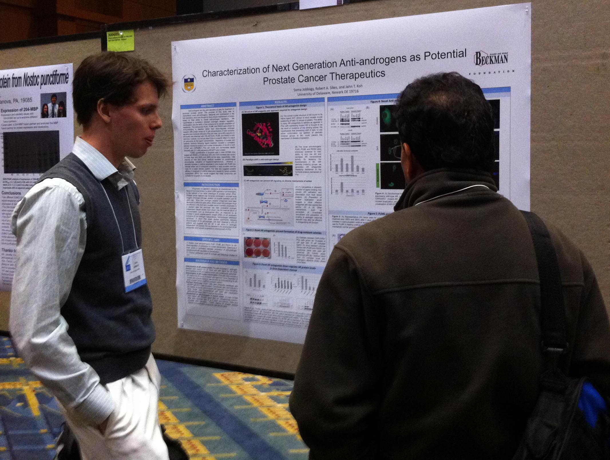 Soma Jobbagy |
Characterization of next-generation anti-androgens as potential
prostate cancer therapeutics Soma Jobbagy1, Robert Allen Sikes2, John Tze-tzun Koh1. 1Chemistry and Biochemistry, 2Biological Sciences Anti-androgens are the gold standard of care for treatment of advanced prostate cancer (PCa). The purpose of this study was to characterize novel anti-androgens PLM1, PLM6, and PAN52 designed and synthesized in the Koh laboratory. PLM6 and PAN52 but not PLM1 are thought to have unique mechanisms of action based on their ability to evade progression to anti-androgen resistance in vitro. Western blot analysis of nuclear fractions showed correlation between prevention of AR nuclear translocation and progression of anti-androgen resistance in prostate cancer cell lines. Some differences in localization effects were also observed between LNCaP and CWR22Rv1 PCa models that may reflect AR mutants present in these cell lines. The compound PLM6 and PAN52 found to down-regulate the androgen receptor (AR) in PCa cell lines. These compounds thus appear to hold promise as therapy for advanced prostate cancer. This work funded by the Beckman Foundation and the National Institutes of Health 5R01DK054257 Day of Presentation: Tuesday April 12, 2011,1:05 PM - 2:35 PM, Poster Board Number: B429,Program Number: 968.2 |
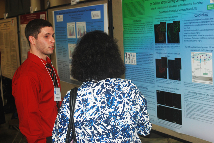 Dylan Lowe |
Effect
of Perlecan/Hspg2 Endoplasmic Reticulum Retention
Presentation: Sunday April 10, 2011,12:30 PM - 2:00
PM, Poster Board Number: C65, Program Number: 492.9on Cellular Stress During Cartilage Development Dylan A. Lowe, Padma P. Srinivasan, and Catherine Kirn-Safran Biological Sciences Perlecan/Hspg2 (PLN) is a scaffolding proteoglycan functioning mainly in the extracellular matrix as a modulator of growth factor bioavailability. PLN gene loss-of-function mutation results in severe skeletal defects and embryonic death. In this study, we use a viable hypomorph mouse model in which PLN expression is reduced. Adult PLN hypomorph mice display a short stature, early osteoarthritis, and altered bone properties. These defects are likely the result of aberrant PLN expression during embryogenesis. Immunostaining of newborn cryosections revealed an absence of columnar organization and severe reduction of PLN secretion in the cartilage matrix of hypomorph versus control bones. Furthermore, the majority of PLN in the hypomorphs co-localized with a strong signal for BiP, an endoplasmic reticulum (ER) chaperone involved in the early steps of the unfolding protein response (UPR). Interestingly, the retention of PLN in the ER was accompanied by increased deposition of another proteoglycan, aggrecan, in the cartilage matrix. Our data suggest that reduced PLN secretion during bone formation induces abnormal patterning that may be partially compensated by increased secretion of other matrix components. Ongoing work further investigates how the UPR and other cellular mechanisms contribute to the abnormal phenotype in PLN hypomorphs. Supported by NIH P20-RR016458. DAL. DAL supported by HHMI undergraduate program. |
|
|
Abstract No:
5569 Development
of a Peptide Nucleic Acid Based siRNA Delivery System
Tejal U. Naik and Millicent O. Sullivan Department of Biological Sciences and Chemical Engineerng Small interfering RNA
(siRNA) is a double stranded RNA
molecule that plays a major role in gene silencing by catalyzing the
degradation of complementary mRNA, and thus inhibiting expression. The ability to harness siRNA for therapeutic
benefit can have a widespread impact on a variety of diseases ranging
from
cancer to HIV/AIDS. However, the
efficacy of such treatments is limited due to the many extracellular
and
intracellular barriers associated with siRNA delivery.
One approach to delivery, involving
siRNA-functionalized surfaces, can improve the cellular response and
specificity by enabling tunable release and creating a
locally-concentrated
microenvironment. In this work, we lay the groundwork for the
development of a
peptide nucleic acid (PNA)-based surface-mediated siRNA delivery system. PNAs are nucleic acid analogs that hybridize
with complementary DNA or RNA sequences, enabling the direct attachment
of
various macromolecules such as peptides. Conjugation
of targeting and protective moieties can
potentially enhance
the delivery of siRNA. To begin
development of this delivery system, we completed two tasks. First, a cell transfection model utilizing
stably transfected B16FO mouse melanoma cells (producing green
fluorescent
protein [GFP]) was established. Anti-GFP
siRNA was designed and its efficacy was evaluated via flow
cytometry and
fluorescence microscopy. Optimization of
cell seeding density, siRNA concentration, use of antibiotics, and time
of
transfections was accomplished, effectively demonstrating gene
silencing capability. Our second task was
to prepare molecular
conjugates for PNA-based siRNA modifications. siRNA-PNA-peptide
conjugates were assembled and purified
through Reverse
Phase High Performance Liquid Chromatography (RP-HPLC).
Future development of this delivery system
will include linkage of the conjugates to surfaces via the
PNA-peptide
tethers, and exploration of the delivery system in B16FO cells.
Presentation: Tuesday April 12, 2011, 1:05 PM - 2:35 PM, Poster Board Number: B75, Program Number: 903.4 Best Thematic Poster Award for the RNA category for the second year in a row. |
|
Mutation
in the βB2-crystallin gene leads to cataracts,
epithelial mesenchymal transition, and upregulation of integrins Victoria
H. Roop, Fahmy Mamuya, Melinda K.
Duncan
The most abundant
protein in the
lens fiber cells is βB2-crystallin (Crybb2). Mutations in the Crybb2
gene have
been shown to cause cataracts in both mice and humans. A twelve
nucleotide
deletion in the lens epithelial cells, known as the Crybb2Phil
mutation, leads to epithelial mesenchymal transition (EMT) during
development.
However, the mechanism behind this phenotype is poorly
understood.
The aim of this project is to understand the
causes and effects of mutations in βB2-crystallin. Tissues obtained
from Crybb2Phil
mutant adult mice were immunostained with αV, α2, α3, α5, α6, β1
integrin
subunits, and phospo-SMAD3. Compared to the wild type lens, β1, αV, and
α5 were
upregulated in the Crybb2Phil mutant. However, no
changes were seen
in α2, α3, and α6 integrin subunits. Phospho-SMAD3 and smooth muscle
actin
(SMA) levels were also upregulated in Crybb2Phil mice. Upregulation
of SMA signifies that the Crybb2phil mice undergo EMT
during
development. Furthermore, over expression of β1 and αV integrin along
with
SMAD3 in these mutants suggest that the TGFβ-SMAD interacting pathway
may be
involved. This project is funded by National Eye Institute grant
EY015279.
VR is a Charles Peter White Scholar.
Presentation: Tuesday April 12, 2011, 12:25 PM - 1:55 PM, Poster Board No: B110 Program Number: 909.10 |
|
Abstract No: 575 Hypoplastic
Left Heart Syndrome: Molecular Consequences of Transcription Factor
Mutations
Ashley Shay1, Susan Kirwin2, Vicky Funanage2 1BiologicalSciences, University of Delaware, Newark, DE, 2Biomedical Research, Nemours, Wilmington, DE. Hypoplastic
Left Heart Syndrome (HLHS; MIM #241550) is a condition of
congenital malformations of the left side of the heart. The left
ventricle is typically underdeveloped and insufficient at supporting
systemic circulation. The mitral valve may be improperly formed or
closed completely and the aorta can be abnormally narrow. These
abnormalities affect the heart’s ability at pumping oxygenated blood to
the body. Normally, the foramen ovale and patent ductus close at birth
to permit blood to be oxygenated by the pulmonary circulation. However,
in HLHS, the blood is pumped through the right ventricle pumping blood
both to the lungs (via the pulmonary artery) and out to the body.
Surgical intervention is required in order to correct these
malformations. HLHS has been found to be more prevalent in males than
females and 10% of patients are diagnosed with other birth defects. To
date, it has been theorized that there is a genetic cause for HLHS;
however, no known monogenic cause has been determined. Current research
is investigating changes in genes known to be involved in cardiac
development: TBX5, NKX2.5, GJA1, GATA4, Hand1 and Hand2. TBX5 has been
linked to Holt-Oram Syndrome, which is a heart-limb disease that has
similar cardiac defects to HLHS. DNA from patients with HLHS will be
analyzed to determine whether a genomic mutation is a cause of the
cardiac defect. These results will be compared within a cohort of HLHS
patients to determine if there is a genetic basis for their abnormal
cardiovascular development.
Presentation: Monday April 11, 2011, 1:05 PM - 2:35 PM,Poster Board Number: B43, Program Number: 698.16 |
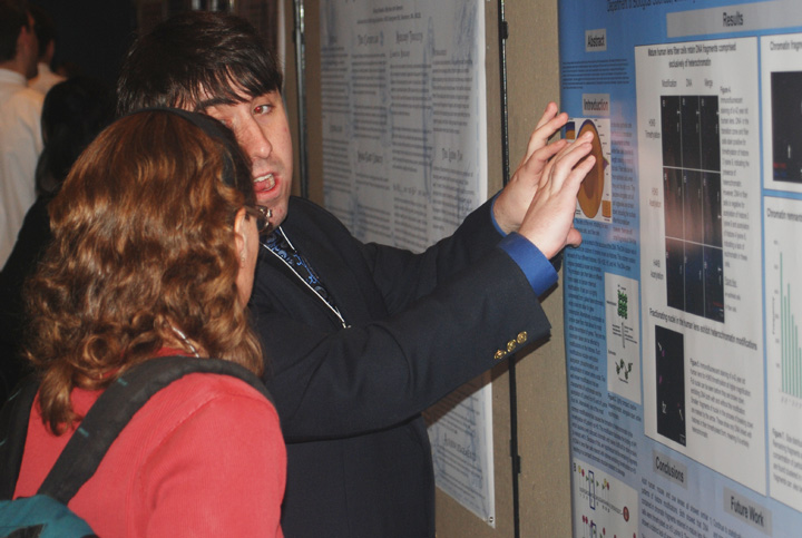 Robert Patrick Sheehan |
Heterochromatin
is retained during lens fiber cell differentiation
Robert Patrick Sheehan and Melinda K Duncan Department of Biological Sciences, The lens of the eye
contains both epithelial and fiber cells, with fibers
cells deriving from the equatorial epithelium. When these
cells differentiate, the nucleus and cellular organelles are broken
down to facilitate transparency. However, small fragments of DNA
can remain in fully mature lens fiber cells, although their structureis
unknown. We found that histone H3, trimethylated on Lysine 9,
as well as 5-Methylcytosine, which are both associated with
compact heterochromatin, co-localize with these DNA fragments.
In contrast, the DNA remnants are not associated with either
histone H3 Lysine 9 acetylation or histone H4 lysine 8 acetylation,
which are predominant in the more accessible euchromatin. This
data suggests that the DNA fragments persisting in mature lens
fiber cells are entirely composed of heterochromatin, likely
because their highly compacted state prevented access of the
nucleases responsible for DNA degradation during lens fiber cell
differentiation. This led to the further hypothesis, which is
currently being tested, that these fragments predominantly contain
transcriptionally silent genes, which are likely to be found in
heterochromatin. Supported by Howard Hughes Medical Institute
and the National Eye Institute.
Presentation: Tuesday, April 12, 12:25-1:55pm. Poster Board Number B53, Program Number 896.7 |
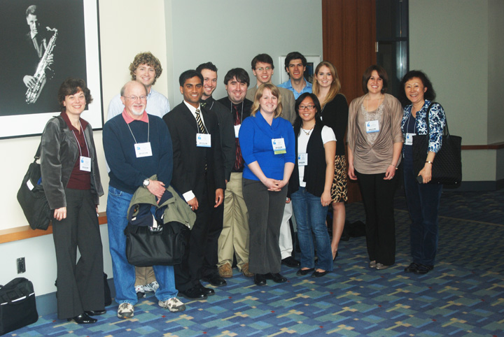 |
After the Awards Ceremony on Sunday, April 10, the UD group gathered outside the hall for a photo with Dr. Kathleen Cornelly who was one of the organizers of the event. Left to right: Dr. Cornelly, Dr. Usher, Mike Brister, Tejal Naik, Cory Bovenzi, Bobby Sheehan, Soma Jobbagy (back), Ashley Shay (front), Matt King (back),l Jean Huynh (front) Tori Roop, Erica Boetefuer, and Dr. Seung Hong. (not shown: Dr. Hal White, Gary Laverty, and Dylan Lowe). Dylan Lowe particpated in the Anatomy Undergraduate Poster session. |
|
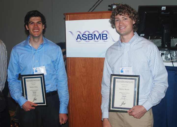 Matt King and Mike
Brister
with their award plaques. |
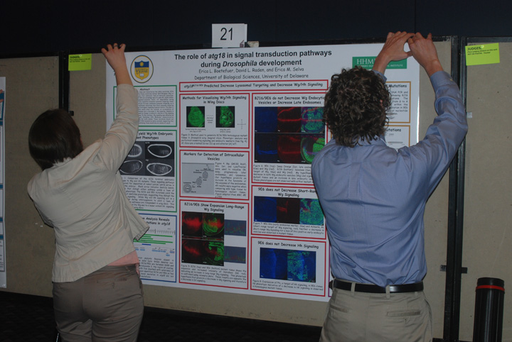 Erica Boetefuer and Mike Brister putting up Erica's poster. |
 Tori Roop posing as a Knockout mouse. |
 The Washington Convention Center where EB2011 was held. |
 Half of the Convention Center's huge ventor and poster display floor. |
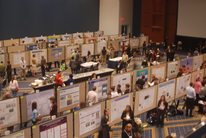 The ASBMB Undergraduate Poster Competition |
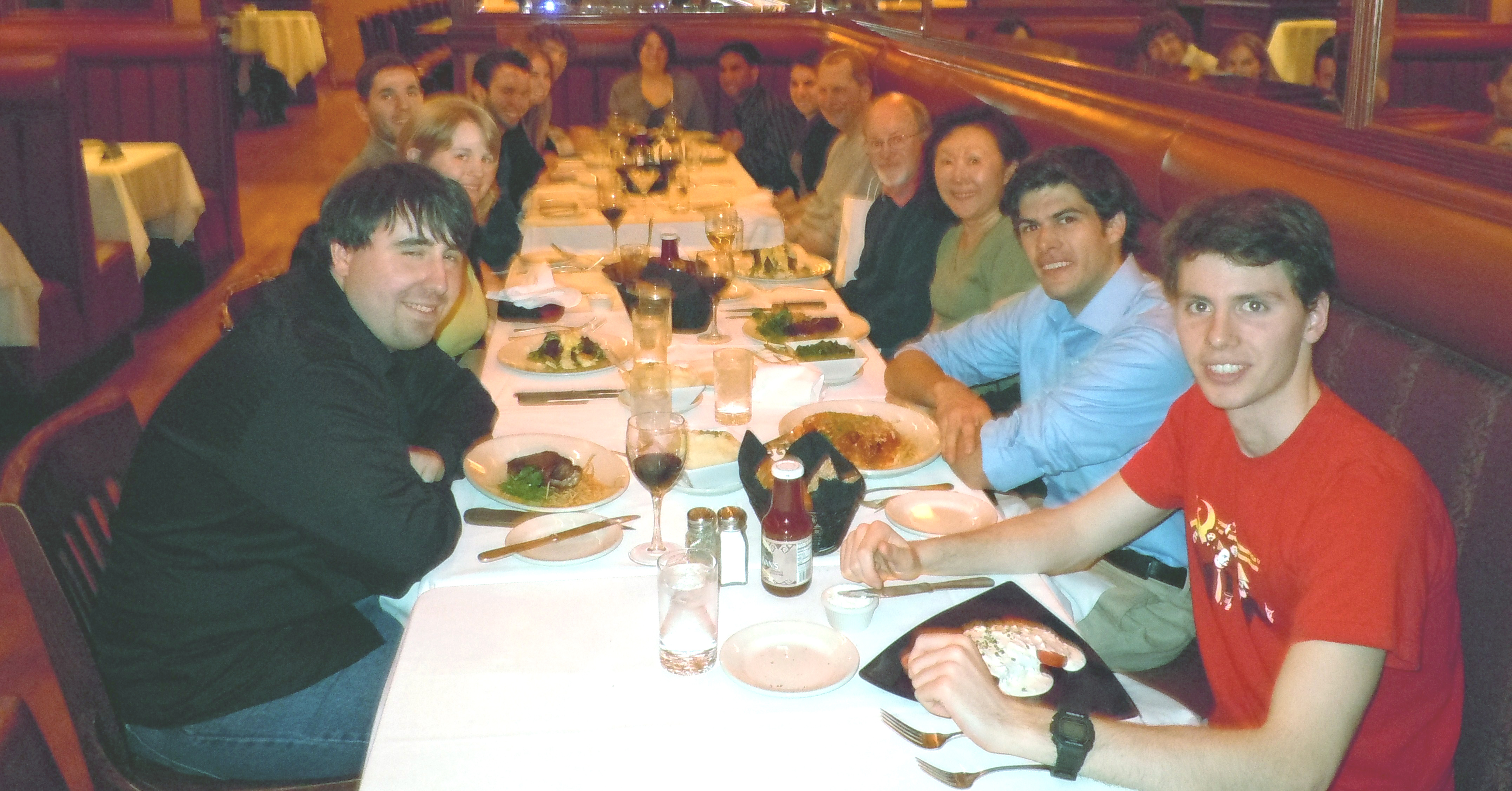 Dinner on the town.
|
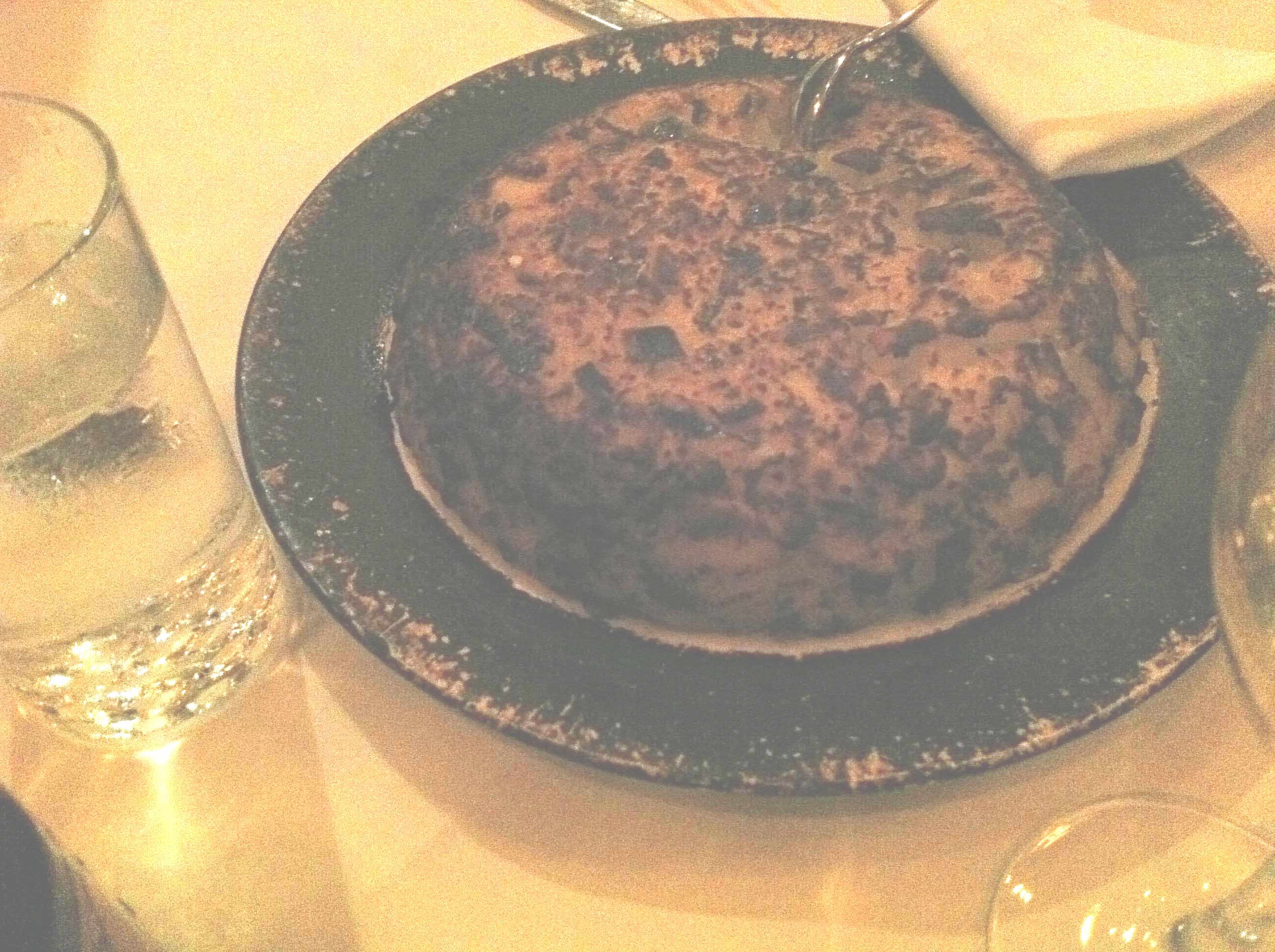 Hashbrowns an Bobby
and Van's
|
|
The trip to the Experimental Biology
2011 Meetings
in Washington, DC was organized by the University of Delaware HHMI
Undergraduate
Science Education Program with additional support from travel grants
from
the American
Society for Biochemistry and Molecular Biology,
Arnold
and Mabel Beckman
Scholars Program, and the Women
Scholars Program. The HHMI
Undergradaute Science Education Program, the Arnold
and Mabel Beckman
Scholars Program, Charles Peter White
Fund, Undergraduate Research
Program, NIH, and NSF, supported
research by the students.
website
for last
year's meeting in Anaheim.