|
Sequence Conservation and Differential Expression of Marek’s Disease Virus MicroRNAs Grace Lagasse, Emily Huang, Amy Anderson, Erin Bernberg, Sachin Kamboj, Grace Isaacs, Mark Parcells, Blake Meyers, Pamela Green, Joan Burnside, Robin Morgan Delaware Biotechnology Institute, Department of Plant and Soil Sciences, and Department of Computer and Information Sciences Abstract withheld |
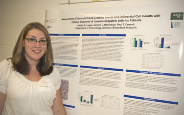 Assessment of Synovial Fluid Cytokine Levels and Differential Cell Counts with Clinical Outcome of Juvenile Idiopathic Arthritis Patients Ashley E. Logan1, Victoria L. Maduskuie2, and Paul T. Fawcett2 1Department of Allied Health, Delaware Technical and Community College; 2Nemours Biomedical Research, A.I. duPont Hospital for Children Juvenile Idiopathic Arthritis
is an uncommon disease characterized by
inflammation of joints or connective tissue in children. The cause is
unknown. This study was performed to determine whether cytokine
profiles and cell counts in samples of synovial fluid (SF) obtained
from pediatric patients correlates with a Good or Bad clinical outcome.
Cell counts were determined for each SF sample and cytospin/pulled
smear slide preparations were made for differential analysis (volume
dependent). Slides were stained (Wright-Giemsa) and analyzed
microscopically. One hundred consecutive nucleated cells were counted
and classified according to their morphology as neutrophils (PMN),
lymphocytes, and monocytes. Levels of selected cytokines (IL-6, IL-10,
IL-8 and TNF-alpha) were determined by enzyme linked immunosorbent
assay (ELISA) from the cell-free aliquots of each SF sample. Patients
were assigned to 1 of 2 groups based on clinical outcome. Forty-five
patients were assigned to the Good outcome group (no flair for > 6
months post treatment) and 23 patients to the Bad outcome group
(patients experiencing a flair of disease activity < 6 months post
treatment). All samples were obtained at the time of baseline treatment
during arthrocentesis. Cell-free aliquots of each SF sample were stored
at -80°C until assayed. This study was approved by the
institutional
review board (IRB). Results of differential cell counts, determinations
of cytokine levels and clinical outcome determinations were analyzed
statistically using Sigma Stat software (Mann Whitney-Rank Sum,
Student’s t test, and Multiple Linear Regression). This project was
supported by Grant Number 2 P20 RR016472-08 under the INBRE Program of
the National Center for Research Resources (NCRR), National Institutes
of Health (NIH).
|
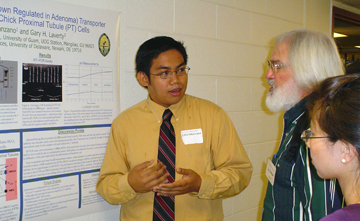 The Potential Role of DRA (Down Regulated in Adenoma) Transporter in HCO3– Secretion of Chick Proximal Tubule (PT) Cells Eiffel John Q. Manzano1 and Gary H. Laverty2 1College of Natural and Applied Sciences, University of Guam, Mangilao, Guam; 2Department of Biological Sciences, University of Delaware, Newark, Delaware The proximal tubule (PT) is a
major transport segment of nephrons in
higher vertebrates. Our laboratory studies the role of parathyroid
hormone (PTH) using chick PT cells in primary cell culture. PTH in
mammals affects PT cells by inhibiting reabsorption (lumen to blood
transport) of phosphate and bicarbonate ions, resulting in increased
urine pH and excretion of these ions. PTH in birds is similar and also
causes increased bicarbonate and phosphate excretion. Moreover, a novel
phosphate secretory mechanism is stimulated. This study focused on a
hypothetical parallel system for bicarbonate secretion in the chick PT.
Previous work had shown that PTH stimulated chloride secretion in PT
cells via a cystic fibrosis transmembrane regulator (CFTR)-like
chloride channel. Based on a bicarbonate secretion model in the
mammalian colon, we hypothesized that the ion exchanger down regulated
in adenoma (DRA) is present in chick PT cells. Colon studies showed
that DRA works with CFTR to reabsorb secreted chlorine from the lumen
and simultaneously secrete bicarbonate into the lumen. PT cells were
cultured on membrane filters and in culture flasks to investigate the
presence of DRA by immunoblotting methods, RT-PCR, and pH measurements.
Immunoblotting studies were unable to detect DRA protein in the PT cell
lysates. However, RT-PCR studies indicate that chicken mRNA for the DRA
gene is present. pH studies suggest that bicarbonate is transported
across the PT cells, resulting in an increase in apical pH.
Optimization of these processes and electrophysiological studies on
monolayer PT cells can further elucidate the presence of the DRA
transporter. Research support from University of Delaware HHMI
Undergraduate Research Program, Office of the Dean of Arts and Sciences
and UOG NIH RISE Grant # GM063682.
|
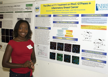 The Effect of FTI Treatment on RhoC GTPase Expression in Inflammatory Breast Cancer Breonna Martin, Kenneth van Golen, and Kirk Czymmek Delaware Biotechnology Institute, Bio-imaging Center Inflammatory Breast Cancer
(IBC) is one of the most fatal forms of breast cancer, claiming about
80% of its victims within 10 years. Studies conducted on IBC have found
a strong relationship between RhoC GTPase expression and the highly
metastatic qualities of IBC cells. We have evidence that RhoB, and AKT
are involved in the activation of RhoC. Post-translational
modification of RhoB, an intracellular transportation protein, by
farnesyl transferase leads to the transportation of Akt to the plasma
membrane. Farnesylated RhoB is found in cancer cells. We
hypothesize that Akt phosphorylates RhoC, leading to activation.
When active, RhoC is capable of mediating the metastatic activities of
IBC. RhoB can also be geranylgeranylated (gg), a similar type of
modification that leads to relocalization within the cell. ggRhoB
should lead to re-distribution of Akt in the cell where it cannot
contact RhoC. Farnesyl transferase inhibitors (FTI) interfere
with the RhoB farnesylation, which leads to accumulation of ggRhoB and
in turn alters the activation of RhoC. Utilizing a scanning
confocal laser microscope we can study the subcellular localization of
RhoB, RhoC, and AKT at different time intervals after FTI
treatment. After 48 hours, we expect to find increased RhoB in
the cell, and a decrease in activated RhoC. If we can stop RhoC
from being activated, we can greatly decrease the metastatic capability
of IBC cells. This project was supported by Grant Number 2 P20
RR016472-08 under the INBRE Program of the National Center for Research
Resources (NCRR), National Institutes of Health (NIH).
|
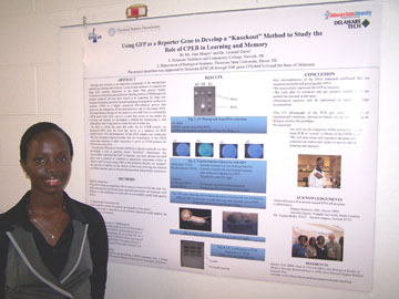 Using GFP as a Reporter Gene to Develop a "Knockout" Method to Study the Role of CPEB in Learning and Memory Joan Mogire, Leonard Davis, Gianna Brisbone, and Jennifer Ukpabi Delaware State University Making new proteins is an
important component of the mechanisms underlying learning and memory.
Local protein synthesis is required for long term memory formation in
the brain. One protein family, Cytoplasmic Polyadenylation Element
Binding protein (CPEB) regulates protein synthesis and has been found
to be important for long term memory formation, possibly through
regulating local protein synthesis in neurons. CPEB is a highly
conserved RNA-binding protein that promotes the elongation of the
polyadenosine tail of messenger RNA. The experimental design for this
project is two-fold. First we need to isolate the CPEB gene from Helix
aspersa, a snail that serves as our model for learning and second, we
developed a method for introducing it, and other genes, into living
snails to study the role in behaviors. In order to isolate the gene
that codes for the CPEB protein, we extracted DNA from the snail that
serves as a template for PCR amplification. The DNA from an organism
rich in mucous materials like the snail was difficult to purify;
however, using repeated extractions and treatment with RNase A, free of
DNase we were able to successfully purify high quality DNA. Gel
electrophoresis of the DNA sample was used to evaluate the quality and
quantity of the extract. We then designed oligonucleotides that are
homologous to the CPEB conserved sequence in other organisms to serve
as CPEB primers for PCR of the snail DNA. Gel electrophoresis was used
to determine if the primers identified could amplify the DNA region of
interest. The UV photograph of the gel, after a number of experimental
variations showed no bands; we are still working on trying to resolve
this problem. Using Green Fluorescent Protein (GFP) as a reporter
molecule, we also developed a tool to perform future "knockout"
experiments. This method involves
incorporating a DNA sequence into a living organism that can be used to
block or over-express a test gene of interest. We successfully
expressed the GFP in bacteria, and then utilized the GFP gene and a
plasmid to construct a eukaryotic expression vector to observe GFP in
snails using CMV as the promoter. Finally, we "painted"
the plasmid of interest on the surface of the snail, allowing the
plasmid to diffuse into the snail to observe incorporation and possible
expression. Histological analysis will be performed to identify
cellular incorporation. Funded by EPSCoR.
|
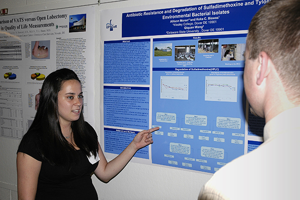 Antibiotic Resistance and Degradation of Sulfamethioxine and Tylosin by Environmental Isolates Allison Moran1, Qiquan Wang2 and Keka C. Biswas1 1Wesley College and 2Delaware State University Veterinary antibiotics are
routinely administered to animals not only
for therapeutical treatments but also for growth promotion. After
administration, these substances are partially metabolized and are
excreted in the urine and feces. It has been evidenced that
sub-therapeutic feeding of food animals for growth promotion along with
casual use of antibiotics in household products, such as soaps and
creams, is contributing to increased antimicrobial resistance in the
environment. If steps are not taken to minimize selective pressure on
bacteria, the effectiveness of antibiotics (hailed as `magic bullets')
may be marginalized. The excretion of feces and urine from medicated
animals and subsequent application of contaminated manure as fertilizer
into agricultural land is one of the major routes through which
veterinary antibiotics enter the environment. This exposes of
microorganisms in the soil to low levels of the antibiotics create
perfect conditions for selectively proliferating resistant bacteria.
This leads humans and animals susceptible to infection by resistant
pathogens either though direct contact or by indirect means such as
through food and water supply putting animal and human health at high
risk. . The research project aims at better understanding the type and
activities of microorganisms present in the poultry litter and bedding
material. The presented representative strains of bacteria were
isolated and physiologically characterized. The degradation of two
widely used veterinary antibiotics, sulfadimethoxine and tylosin, by
the isolates was investigated. This project described was supported by
Delaware EPSCoR, through National Science Foundation Grant EPS-0447610
and the State of Delaware.
|
 Identification and Characterization of the Role of HYAL2 in Mammalian Reproduction Tejal U. Naik, Diniece Barran, Rolands Aravindan and Patricia A. Martin-DeLeon Department of Biological Sciences Fertilization, or the
fusion of gametes, is an essential process in the development of a new
organism. In mammals, the sperm has to be able to penetrate the
barriers surrounding the egg in order to effect fertilization. Sperm
accomplish this task by using enzymes called hyaluronidases (hyase) to
break down hyaluronan, the major component found in the cumulus cell
matrix and the zona pellucida that surround the oocyte. Although Hyal2
been classified as a somatic hyase, three pieces of evidence suggest
that it may be involved in reproduction: HYAL2 is abundantly expressed
in the testes, HYAL3, a closely-related acid-active hyase, is present
in sperm where another acid-active hyase is also present, and RT-PCR
shows that Hyal2 transcripts are present in the uterus where the
protein may be secreted and acquired by sperm during sperm transit.
Based on all of the above, Ii is hypothesized that HYAL2 is found on
sperm and plays a role in fertilization. Thus this project is to
determine the presence and the role of HYAL2 in fertilization and in
the female tract. A Western Blot analysis was conducted to detect the
presence of HYAL2 in sperm proteins.. Results indicate the presence of
a 54 kDa HYAL2 band in sperm in different subcellular locations. Thus
zymography will be performed to determine of the protein is active. /
Supported by INBRE/DBI /
|
 ApoC-I and ApoE Production and Regulation during Cholesterol Efflux from Adipocytes Aivi Nguyen, Marysol Lavander, Barbara Kwakye Safo, William Cain, and David Usher Department of Biological Sciences One apolipoprotein, apoE, has
already been shown to play a significant
role in cholesterol efflux from adipoctyes. The Usher lab has
discovered another protein, apolipoprotein C-I, which may also be
involved in cholesterol efflux. Previous studies have shown that apoC-I
mRNA is high in late-phase 3T3-L1 adipocyte differentiation and is
regulated in a similar manner as apoE. This study examines the
production and detection of apoC-I and apoE during late-phase human and
mouse adipocyte differentiation. Human preadipocytes and mouse 3T3-L1
fibroblasts were grown to confluency and induced to differentiate 48
hours post-confluency. Following differentiation, to determine the
time-course of apoC-I secretion, supernatant samples and cell lysates
were collected on D0 (Day 0), D7, D14 for human adipocytes and D0, D3,
D6, and D9 for 3T3-L1 adipocytes. Human supernatants were centrifuged
to determine if secreted apoC-I associated with lipoproteins. The
individual collected centrifuge fractions were analyzed on 5%-20%
gradient gels and immunoblotted with goat anti-human apoC-I antibodies.
Immunoblots demonstrated that human apoC-I is associated with
high-density lipoprotein fractions and produced during late-phase
differentiation. Results suggests apoC-I involvement in cholesterol
efflux, though further analysis of apoC-I secretion from mouse
adipocytes is underway. Future studies will include using ELISA assays
to quantify apoC-I concentration in human and mouse supernatant samples
and will also examine methylation patterns in the apoC-I promoter
region, which may elucidate apoC-I transcription regulation. Funded by
the Howard Hughes Medical Institute.
|
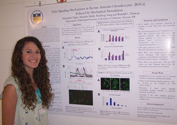 Early Signaling Mechanisms in Bovine Articular Chondrocytes Induced by Mechanical Stimulation. Samantha Nigro, Manisha Malik and Randall L. Duncan Department of Biological Sciences Osteoarthritis, a leading cause
of disability in the United States,
results in pain and swelling of the joints due to the loss of articular
cartilage that protects the bones of the joint. This loss reflects an
imbalance in the catabolic and anabolic activities of articular
chondrocytes, the cells of the cartilage. Because mechanical loading of
the joint can accelerate the degeneration of the articular cartilage,
the effects of mechanical stimuli on chondrogenesis was studied using
fluid shear and hypotonic swelling. We have shown that increased
intracellular Ca2+ levels ([Ca2+]i) and ATP release are initial events
in mechanical signaling in bone and postulate that similar signaling
occurs in chondrocytes. [Ca2+]i levels and ATP release were measured in
primary bovine articular chondrocytes (BAC) in response to hypotonic
swelling or fluid shear. We found that the [Ca2+]i response in BACs is
different from osteoblasts, exhibiting greater peak calcium levels and
spontaneous oscillations in [Ca2+]i. We also found that ATP was rapidly
released, but that this release was different when the cells were grown
on top of type II collagen or encased in the collagen. These responses
could be major factors in cartilage loss with prolonged usage incurred
in an aging individual. A better understanding of these early signals
will provide insight into the initial degradation of articular
cartilage during high impact loading and will potentially lead to an
understanding of cartilage repair. (supported by the Science and
Engineering Scholars Program and NIH/NIAMS R01 AR043222)
|
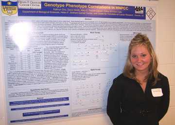 Genotype Phenotype Correlations in HNPCC Kathryn Orio, Mary C. Farach-Carson, and Zohra Ali-Khan Catts Helen F. Graham Cancer Center at Christiana Care Health System. Colorectal is the third most
common cancer and the second leading cause
of cancer-related death. Most colorectal cancers occur sporadically,
but 5-10% are hereditary due to a genetic predisposition. One of these
syndromes, hereditary non-polyposis colorectal cancer (HNPCC) is
characterized by an increased risk of colon cancer and many other types
of cancer. The pedigrees and test results for eleven families found in
the Familial Cancer Risk Assessment Program at the Helen F. Graham
Cancer Center who tested positive for HNPCC were collected and analyzed
for genotype phenotype correlations. Those positive for a mutation in
MLH1 (MutL homolog 1, colon cancer, nonpolyposis type 2 (E. coli)) were
found to have early onset colon cancer, an average diagnostic age for
endometrial cancer and absence of ovarian cancer. In contrast, those
with a nonsense alteration type typically presented with gastric
cancer. Those who tested positive for a mutation in MSH2 (mutS homolog
2, colon cancer, nonpolyposis type 1 (E. coli)) were found to have the
same early onset of colon cancer found in MLH1, but all families also
had either breast or ovarian. Those testing positive for MSH6 (mutS
homolog 6 (E. coli)) had an average age for colon cancer diagnosis
meaning that it was consistent with the reported age of 62 in HNPCC
individuals. This is a very small subsection of HNPCC families, so it
may be that these results cannot be extrapolated to general population.
Additional research will include more families with specific mutation
types to make statistically significant conclusions.
|
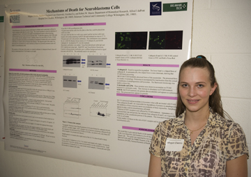 Mechanisms of Death for Neuroblastoma Cells Megan Owens, Matthew England, Guizhen Lu, Lisa Glazewski, and Robert W. Mason Department of Biomedical Research, Alfred I duPont Hospital for Children , Wilmington DE and Delaware Technical and Community College Neuroblastoma is the most
common soild tumor in children and is
particularly difficult to treat in patients older than two years of
age. The overall hypothesis is that the inhibition of cathepsins B and
L will result in the death of neuroblastoma tumors without harming
differentiated nervous tissue and non-neuronal cells. To meet this
objective, Some of the SK-N-SH were used as control (non FYAD treated)
and the others cells were treated with FYAD. FYAD induces cell death
and then the purification of lysosomes was confirmed by western blot
analysis. The objective of this study is to determine the mechanism by
which an inhibitor of two lysosomal proteases cause cell death in
neuroblastoma cells. This grant was supported by was supported by Grant
Number 2 P20 RR016472-08 under the INBRE Program of the National Center
for Research Resources (NCRR), National Institutes of Health (NIH).
|
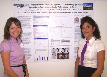 Prevalence of Non-Therapeutic Thoracotomy Operations for Indeterminate Pulmonary Nodules Yuliya Pepelyayeva, Sandy A. Almassou, Nancy Steward, Thomas L. Bauer. Section of Thoracic Surgery, Helen F. Graham Cancer Center, Christiana Care Health Services, Newark, Delaware The purpose of this research
was to determine the rate of
non-therapeutic surgeries for solitary pulmonary nodules. Researching
and viewing power charts, data bases, and medical records of the
patients provided information to evaluate the trends of the negative
surgeries for the past nine years. The sensitivity of CT scanning made
it possible to detect small pulmonary nodules. However, it is not
capable to accurately differentiate between benign and malignant
lesions, which expose patients to a risk of negative surgical
interventions. After the patients who underwent thoracic surgery were
identified, their operative and pathology reports were reviewed,
leaving patients who underwent resection for indeterminate solitary
pulmonary nodule(s) as the subject of the study. Non-therapeutic
surgeries were defined as a pathology reports demonstrating no
malignancy, fungal infection or other anomaly that would jeopardize the
patient’s health. The accessibility of PET scanning and the launch of
I-ELCAP study decreased the rate of non-therapeutic thoractomies from
23% (before 2002) to 9% (2003 till present). The average prevalence of
negative thoracotomies for the past five years at CCHS was determined
to be 7.4%. This grant was supported by was supported by Grant
Number 2 P20 RR016472-08 under the INBRE Program of the National Center
for Research Resources (NCRR), National Institutes of Health (NIH).
|
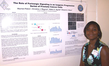 The role of purinergic signaling in an isogenic progression of prostate cancer cells Wachen Peters, Christine Maguire, Adam Aguair, and Robert A. Sikes Department of Biological Sciences Background: Purinergic
signaling stimulates many biological processes
such as cell proliferation, differentiation, and apoptosis. Two classes
of purinergic receptors, GPCR and S/T kinase, have been identified that
bind ATP and other nucleotides as ligands. ATP has an antitumor effect
on cancer in vivo. Thus clinical trials are being carried out to
determine that ATP can be used as a therapeutic agent for cancer. Here
we looked at the effect of ATP on an isogenic progression series of
prostate cancer (PCa) cell lines (LNCaP, C4 2, and C4 2B4). We
hypothesize that ATP stimulates neuroendocrine differentiation (NED)
and growth in PCa cells. NED occurs with relatively high frequency in
PCa and is correlated directly with poor prognosis. Methods: ATP,
0.001-1000μM, was added in log10 increments to PCa cell lines in vitro.
Morphology was examined by photomicroscopy; cell number was determined
using crystal violet staining; and, migration was examined using
scratch assays. Results: ATP increased the growth of PCa cells over
vehicle alone (LNCaP=2.7X, C4-2= 1.7X, C4-2B4= 1.4X) with peak
stimulation occurring at 1nM ATP. Morphology was largely unchanged in
response to ATP. Scratch assays are currently in progress.
Conclusions: Growth response to exogenous ATP decreases as PCa cells
become more androgen insensitive and metastatic. NED is not apparent
morphologically but biochemical analysis is required to confirm this
result.
Funding by DoD PCRP-W81XWH-06-1-0244
|
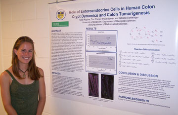 Role of Enteroendocrine Cells in Human Colon Crypt Dynamics and Colon Tumorigenesis Kelly A. Pippins, Tao Zhang, Bruce M. Boman, and Gilberto Schleiniger Department of Biological Sciences and Department of Mathematical Sciences Colon cancer arises from tissue
changes in normal colonic epithelium,
the structure of which consists of histologic subunits termed colonic
crypts that maintain an incredibly high level of organization during
the human lifespan. Slowly proliferating colonic stem cells normally
reside at the crypt base and are progenitors of all other cell types in
the crypt. Enteroendocrine cells — specialized endocrine cells of the
gastrointestinal tract that also have neuronal properties —are thought
to: i) be responsible for normally controlling the stem cell population
and ii) contribute to the development of tumors in the colon. We
hypothesize that tumorigenesis is coupled to abnormal cell cycle
proliferation and migration of enteroendocrine cells in the crypt. A
system of reaction-diffusion equations was designed to mathematically
model the crypt’s dynamic spatial-temporal organization involving eight
different cell types. The state of the system is predicted converge to
a steady-state distribution of cells, as found in a normal crypt.
Divergence from the normal steady state, in particular from the
proportion of different cell types and their distribution in normal
crypts, may predict development of a colon tumor. To obtain biologic
data for validation of model output, we did immunostaining of normal
and tumor tissue using antibodies against several different
neuroendocrine markers. Our results indicated that neuroendocrine cells
are abnormally distributed in colon tumor because positively-stained
cells were located in the upper level of tumor crypts, as compared to
being restricted to the lower level of normal crypt This finding
suggests that communication between neuroendocrine cells and stem cells
could be a key factor in tumor development. This project was funded by
the Howard Hughes Medical Institute.
|
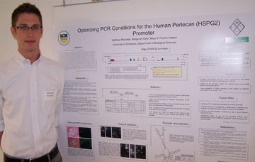 Optimizing PCR conditions for the Human Perlecan Promoter Matthew T. Richards, Benjamin Rohe, and Mary C. Farach Carson Department of Biological Sciences Perlecan, also called HSPG2, is
a heparan sulfate proteoglycan predominantly located in basement
membranes and the matrix surrounding endothelial, mesenchymal and
stromal cells. It is ubiquitously expressed in vascularized
tissue and the reactive stroma surrounding prostate cancer cell lines
produces high levels of the protein. Additional studies suggested
that perlecan plays a role in delivery of growth and angiogenic
factors, which aids survival and growth of metastatic tumors. The
overall goal of this project was to study the promoter in order to
understand the up-regulation of perlecan in the tumor reactive stroma
which occurs via transcriptional increases in perlecan
biosynthesis. Our first step was to analyze the promoter to find
conserved elements. The sequence for the human HSPG2 promoter was
found using public databases and compared to a published human sequence
(Iozzo et al., 1997) and a sequence for the mouse perlecan promoter
also found in online databases. Several transcription factor
binding sites of interest were identified for further study including
NFkappaB [-2410 to -2398], CREB ([-1797 to -1777] and [-709 to -689]),
Smad3 ([-1301 to -1293] and [-187 to -179]), Elk-1 [-1699 to -1679],
c-Jun ([-2453 to -2441] and [-2496 to -2476]) and TCF/LEF-1 ([-1521 to
-1505] and [-1247 to -1231]). Currently we are working to build a
promoter-reporter construct by isolating the promoter region using PCR
amplification. Our current strategy seeks to optimize the PCR
reaction with different sets of primers. The next step will be
creating a luciferase or RFP reporter construct in order to test the
effects of the identified pathways. This project was funded by
the Howard Hughes Medical Institute.
|
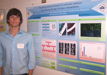 Targeting perlecan domain I and heparin binding growth factors to articular cartilage surfaces: A promising tool to repair knee osteoarthritis. Matthew D. Riscinti, David G. Tuke, Weidong Yang, Mary C. Farach-Carson, Xinqiao Jia, and Catherine B. Kirn-Safran Departments of Biological Sciences and Material Science Osteoarthritis (OA), a
degenerative joint disease, affects many people
over the age of 65 and a large portion of competitive athletes. Age,
mechanical stress, and genetics contribute to the disease. Currently
there are few effective therapeutic treatments. We are using use a
hyaluronic acid (HA) based scaffold delivery system in combination with
recombinant perlecan domain 1 (PlnDI) to permit the slow release of
heparin binding growth factors (HBGFs) into osteoarthritic knees. We
hypothesized that Synvisc®, a commercially available HA gel, would
decrease diffusion of injected PlnDI from the knee cavity. To test
this, we fluorescently labeled and injected PlnDI intra-articularly
into murine knees. The retention time of PlnDI was compared using
in-vivo imaging when injected alone or co-injected with Synvisc®.
In
both cases, PlnDI was undetectable in the knee cavity after 24 hrs,
therefore the Synvisc® failed to prevent PlnDI loss from the
injection
site. We next investigated PlnDI’s ability to diffuse into articular
cartilage of isolated femurs, tibias, and patellar explants by
incubating them with labeled PlnDI and nuclear dye. The PlnDI signal
did not overlap with the nuclear signal, but localized to a peripheral
region near the cartilage matrix. Current effort will develop a
papain-induced mouse model of OA for use in future studies to screen
the efficiency of different PlnDI-based repair complexes including
their ability to increase retention of HBGFs and for proper targeting
to OA articular surfaces. Funding for this project has been provided by
Charles Peter White Scholarship.
|
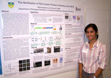 The Identification of PDZ Domain Proteins Interacting with JAM-B Ritika Samant and Ulhas P. Naik Department of Biological Sciences A family of Junctional Adhesion
Molecules (JAMs) consisting of JAM-A, JAM-B, and JAM-C has recently
been discovered. These transmembrane proteins are involved in
tight junctions, are often associated with PDZ domain-containing
proteins, help anchor transmembrane proteins to the cytoskeleton, and
hold together signaling complexes. JAM-A and JAM-B are known to
contain PDZ domain-binding motifs, and it has been shown that JAM-A
uses this motif to interact with cytoplasmic PDZ proteins.
However, interactions between JAM-B and PDZ domain-containing proteins
have not yet been identified. To study this, protein microarrays
can be used to identify protein-protein interactions between JAM-B and
PDZ domain-containing proteins. A cDNA construct encoding for a JAM-B
GST fusion protein with an HA-tag was first created using polymerase
chain reaction. The JAM-B fusion protein was expressed in IPTG
induced BL21 E. coli cell cultures, which were lysed using a French
press. Expression levels in induced cultures were observed with
Coomassie staining and Western blot analysis. The lysate was
incubated with glutathione sepharose beads to bind the fusion protein,
which was subsequently cleaved with thrombin, allowing the HA-tagged
protein to be purified. The purified HA-tagged protein will be
used to probe a protein microarray spotted with PDZ domain-containing
proteins and JAM-B – PDZ domain interactions will be detected
by chemiluminescence using an anti-HA primary antibody and an
HRP-conjugated secondary antibody. This identification of JAM-B
and its PDZ-domain containing binding partners will further our
understanding of signaling pathways and molecular interactions,
particularly pertaining to the formation and regulation of
intercellular pathways and tight junction integrity. Funding was
provided by the Beckman Foundation, and the NIH for Dr. Naik.
|
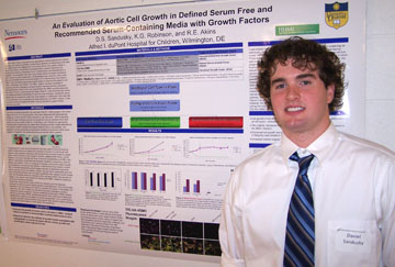 An Evaluation of Aortic Cell Growth in Defined Serum Free and Recommended Serum-Containing Media with Various Growth Factors Daniel S. Sandusky, K.G. Robinson, and R.E. Akins Alfred I. duPont Hospital for Children, Wilmington DE A viable route in tissue
engineering and regenerative medical
treatments for arterial diseases is to create synthetic, transplantable
stretches of arterial tissue that have the ability to grow and interact
with surrounding tissue in the patient. However, a serum free medium
must first be developed that is capable of growing and sustaining the
various cell types found in arterial tissue until transplantation is
possible. In my evaluation, the three major human arterial cell types
(endothelial cells, vascular smooth muscle cells, and fibroblasts) were
cultured in vitro in both their recommended serum-containing medium and
a defined serum free medium (SM3+) with and with out growth factors in
order to determine the extent of cell death, proliferation, and
viability. This SM3+ medium has been successful in sustaining cell life
in analogous cell types found in neonatal rat heart cultures for
extended periods of time, and theoretically should be able sustain cell
life in a human model. However, it was found that the overall cell
viability decreased substantially for the three cell types, causing the
cells to detach from the polystyrene plates they were seeded on.
Additionally, there was little or no proliferation and considerable
cell death observed in the SM3+ medium. This project was funded by
the Howard Hughes Medical Institute.
|
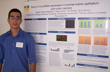 miRNA Profiling of Colonic Stemcell-like Cells and Colon Carcinoma Cells Sepehr Sedigh Haghighat, Tao Zhang, Bruce M. Boman, and Greg Gonye1 1Thomas Jefferson University Study of microRNA expression in
normal colonic epithelium and colon
cancers
Sepehr Sedigh Haghighat1, Greg Gonye2, Tao Zhang1, Bruce Boman1
1Department of Biologic Sciences, University of Delaware, Newark DE,
2Thomas Jefferson University, Philadelphia PA
Dysregulation of crypt cell proliferation and differentiation has been
implicated in the development of colorectal cancer (CRC). As part of
our investigation of regulatory factors involved in the stem cell
origin of CRC, we investigated the role that microRNAs (miRNAs) might
play in colon carcinogenesis. miRNAs are RNAs 20-24 nucleotides long
that were recently shown to modulate many cellular signaling pathways
through post-transcriptional regulation of messenger RNA (mRNA) levels
and thus protein synthesis. Our previous micorarray analysis (368 gene
chip) showed increased expression of 37 miRNAs in CRCs compared to
normal colonic epithelium. Here we further evaluated miRNA expression
in CRC versus purified colonic epithelium by quantitative PCR (QPCR).
Total RNA was immediately isolated from tissue by the TrizolTM method.
RNA was transcribed into first-strand complementary DNA (cDNA) through
reverse transcription (RT). cDNA was then amplified through QPCR.
Statistical analysis and plots of expression data for miRNAs were done
to find miRNA genes that are differentially expressed. We found, using
QPCR, five miRNAs to be differentially expressed in CRCs versus normal
colonic epithelium including mir-25 and mir-198. Identification of
miRNAs specifically expressed in normal colonic crypts and changes in
their expression in CRCs will provide important information to help
understand mechanisms of colon tumorigenesis.
|
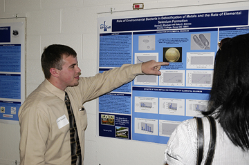 Role of Environmental Bacteria in Detoxification of Metals and the Rate of Elemental Selenium Formation Kevin E. Shuman and Keka C. Biswas Wesley College Selenium exists in the
environment in several ox-red states and one of
the unresolved features is the rate at which colloidal, elemental
selenium is produced from selenium oxy -anions. While few bacteria have
the capability of coupling the reduction of selenate or selenite to
elemental selenium, many aerobic bacteria have a glutathione-based
process of forming elemental selenium and several anaerobic bacteria
use metal reducing systems to produce elemental selenium. From this
study we have developed a novel colorimetric method for the measurement
of elemental selenium under several conditions including sulfidogenic
environments. Using environmental bacterial isolates, we have followed
the reduction of selenite to colloidal elemental selenium by pure
cultures and the method detects elemental selenium even if elemental
selenium is formed inside the cells. Our research addresses a method
for measuring colloidal red selenium in the presence of chemicals
typically associated with soil or water containing toxic compounds.
Using environmental bacterial isolates, we have followed the reduction
of selenite to colloidal elemental selenium by environmental isolates
and the method detects elemental selenium.
[This project described was supported by Delaware EPSCoR, through
National Science Foundation Grant EPS-0447610 and the State of
Delaware.]
|
 Laura Sloofman received the first place award for the best talk in the Sigma Xi competition. |
Effects of Diminished Protein Synthesis on Bone Anabolic Response to Load in RPL29-deficient Mice Laura G. Sloofman1, Christopher Price2, David Chen2, Xiaozhou Zhou2, John E. Novotny2, Liyun Wang2, and Catherine B. Kirn-Safran1 1Department of Biological Sciences and 2Department of Mechanical Engineering Ribosomal proteins (RPs) play
an important function in the maintenance
of a normal protein synthetic rate. Our group generated the first
viable mouse mutant model lacking an individual ribosomal protein
(RPL29). In these mutants, decreased rates of protein synthesis and
cell proliferation resulted in skeletal growth defects leading to short
adult stature. Recently, we demonstrated that the absence of RPL29
increases bone fragility due to poor tissue and extracellular matrix
(ECM) quality, suggesting an important link between efficient protein
production and skeletal tissue growth. We hypothesized that because of
impaired ability to synthesize large volumes of ECM proteins, RPL29
null bones will exhibit a reduced response to mechanical load as
compared to wild type controls. To establish a correlation between
defects in ECM protein biosynthesis and an impaired response to
mechanical stimuli, cyclic axial loading was performed in vivo on the
tibia of adult RPL29 null mice and age matched wild type controls of
the C57BL/6J (B6) background. The cortical and trabecular bone
microstructure of both the loaded and unloaded tibiae from each sample
was analyzed using micro-computed tomography (microCT). Three-point
bending tests on the loaded and unloaded tibiae will assess the extent
that cyclic loading improves the mechanical properties and fragility of
RPL29-deficient bones compared to controls. Studies of dynamic
histomorphometry will also be used to quantify the effect of mechanical
loading on bone formation capacity between the null mice and controls.
Altogether, these studies will establish the importance of high volume
protein synthesis for the regulation of bone formation during
adulthood. In the future, this information may be particularly useful
in treating diseases such as osteoporosis, in which bone
microarchitecture is altered due to an imbalance between mineral and
organic (collagen/non-collagenous proteins) phases. This project was
funded by
the Howard Hughes Medical Institute.
|
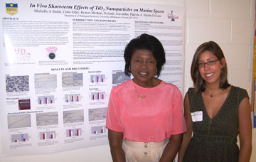 In Vivo Short-term Effects of TiO2 Nanoparticles on Murine Sperm Michelle A Smith, Chris Elder, Rowan Michael, Rolands Aravindan, Patricia A. Martin-DeLeon Department of Biological Sciences Titanium dioxide (TiO2)
nanoparticles are widely used and can be found
in sunscreens, paint and food coloring. These metallic compounds
readily donate electrons to create reactive oxygen species (ROS) and
induce oxidative stress. High levels of oxidative stress are toxic to
cells, particularly sperm which are devoid of repair enzymes and thus
may undergo deleterious effects. The objective of this project was to
examine the effects of TiO2 nanoparticle exposure during
spermiogenesis. Sexually mature male mice were injected
intraperitoneally with 100% anatase nano-TiO2 suspension at
0 mg/ml
(control), 0.625 mg/ml (low), and 1.25 mg/ml (high) for three
consecutive days. The mice were sacrificed after 24, 48 and 120 hours
from the final injection and caudal epididymal sperm were removed and
testis weights recorded. Sperm were analyzed for motility,
hyperactivated motility, retention of residual cytoplasm and the
ability to acrosome react. Testes were prepared for histological
examination, TEM microscopy and DNA analysis. The results showed a
significant (p<0.005) increase in the number of sperm retaining
excess cytoplasm, as well as a decrease in acrosome reaction rates and
motility with nano-TiO2 exposure. TEM microscopy revealed TiO2
nano
aggregates in fat tissue surrounding the testes. Testis histology was
abnormal and testicular DNA was tested for fragmentation. These results
indicate that TiO2 nanoparticles induce retention of excess
cytoplasm
on sperm by disrupting spermiogenesis and may lead to male
subfertility/infertility. Funding was provided by EPSCoR and the
Charles Peter White fellowship.
|
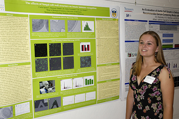 Relationship of Fungal Pathogen and Non-pathogen with Innate Immunity Carla A. Spence, Shannon Modla, Liz Adams, and Kirk Czymmek Delaware Biotechnology Institute Fungal infections are becoming
increasingly common, with the most
frequent fungal organism being Candida albicans. The molecular
mechanisms for this increase in pathogenicity of C. albicans, has yet
to be fully understood. For example, it still remains unknown as to why
certain microorganisms present a significant pathogenic risk to humans.
It has been previously hypothesized that fungal pathogens are able to
mask their -glucans within their cell wall to prevent recognition by
immune system cells; therefore, we hypothesize that the opportunistic
pathogen, C. albicans, conceals -glucans within its cell wall,
hindering recognition by pattern recognitions receptors (PRRs), such as
Dectin-1 and toll-like receptors (TLRs) expressed by cells of the
innate immune system. In this research we examined fungal cell wall
structure by Transmission Electron Microscopy (TEM) and macrophage
recognition of fungal pathogens by Atomic Force Microscopy (AFM). This
research revealed that C. albicans was found to have an average cell
wall thickness of 96nm, whilst Saccharomyces cerevisiae, a
non-pathogenic control, was found to have an average cell wall
thickness of 130nm. Immunolabeled sections imaged by TEM show that the
-glucans of C. albicans are clustered within the cell wall, rarely
being displayed on the surface. The importance of the -glucan-Dectin-1
interaction is further corroborated by AFM data showing a 28% reduction
in unbinding force when the β-glucans are blocked, and a 20% reduction
when Dectin-1 is blocked. In conclusion, the interaction between
Dectin-1 and β-glucan is important in innate recognition of pathogens,
and C. albicans is able to evade recognition by concealing β-glucans
within the cell wall.
|
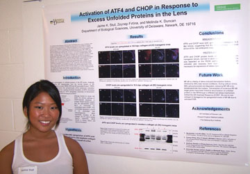 Activation of CREB-2/ATF4 and GADD153/CHOP in Response to Excess Unfolded Proteins in the Lens Jaime K. Stull, Zeynep Firtina, and Melinda K. Duncan Department of Biological Sciences The Unfolded Protein Response
(UPR) has been hypothesized to be a cause
of cataracts. UPR is a stress-induced autoregulatory mechanism that
arises from the accumulation of unfolded proteins in the endoplasmic
reticulum. It restores homeostasis in stressed cells by modifying gene
expression at both the transcriptional and translational levels or by
inducing apoptotis. UPR signaling pathways may be initiated by three ER
transmembrane proteins: IRE1, PERK, and ATF6. Our lab previously
confirmed IRE1 activation in mouse lenses that accumulate unfolded
collagen IV protein chains in the ER lumen. Therefore, we chose to
investigate another UPR pathway, the PERK pathway, in the collagen IV
transgenic mice. In response to ER stress, PERK phosphorylates eIF2α,
halting global ER protein translation but increasing the translation of
CREB2/ATF4. CREB2/ATF4 can then upregulate GADD153/CHOP, a
pro-apoptotic transcription factor. The mechanism behind cell death
occurring through CHOP-mediated apoptosis is not completely clear, but
is connected to the abnormal lens phenotypes seen in collagen IV
transgenic mice. Immunostaining of 16.5dpc lenses with ATF4 and CHOP
antibodies showed considerable upregulation in the transgenic lens
cells. Furthermore, ATF4 and CHOP protein levels in newborn lenses were
increased in transgenic mice, whereas adult transgenic lenses exhibited
normal levels of ATF4 and CHOP. This was expected, as the PERK pathway
is the first to be activated but then must be deactivated for
stress-specific gene induction. Collectively these data suggest that
the PERK pathway of the Unfolded Protein Response is active within
embryonic lens cells experiencing ER stress. Supported by Howard Hughes
Medical Institute and the National Eye Institute.
|
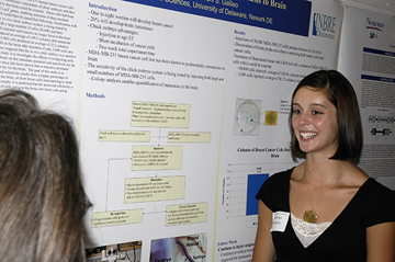 Quantitative Analysis of Breast Cancer Metastasis to the Brain Kathryn Teixeira and Deni S. Galileo Department of Biological Sciences Previous experiments have shown
that MDA-MB-231 human breast cancer cells could be injected into the
extra-embryonic vasculature of chick embryos and then cells which had
metastasized to the brain could be isolated. These cells had been
transfected with the neor and LacZ genes, enabling selection of drug
resistant colonies of cancer cells which could then be quantified
following visualization with X-Gal. The sensitivity of the in
vivo chick embryo system is being tested by initially injecting embryos
with a large number of cells (50,000) and then decreasing the number of
cells injected until tumors no longer form in the brain.
Injections of 50,000 cells produced an average of 199.5 colonies after
treatment with G418, and injections of 5,000 cells produced an average
of 32.2 colonies. Detection of breast cancer cells in the brain
after injection of only 5,000 cells into the extra-embryonic
vasculature suggests that the in vivo chick embryo is a sensitive
system. The next step of this study is to investigate the effects
of re-injecting cells which have been through the brain on the
metastatic potential and specificity for the brain. Experiments
with nude mice labs have shown that re-injection produced sublines with
enhanced selectivity for the brain. The goal of this study is to
determine whether the increased selectivity results from a higher
percentage of injected cells metastasizing to the brain, or from fewer
cells metastasizing to other organs. Based on preliminary
results, it is predicted that the increased selectivity is not due to a
higher percentage of cells going to the brain, but fewer cells going
elsewhere. This grant was supported by was supported by Grant
Number 2 P20 RR016472-08 under the INBRE Program of the National Center
for Research Resources (NCRR), National Institutes of Health (NIH).
|
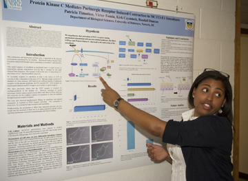 Protein Kinase C Mediates Purinergic Receptor Induced Contraction in MC3T3-E1 Osteoblasts. Patricia Timothee, Victor Fomin, Kirk Czymmek and Randall L. Duncan Department of Biological Sciences Osteoblasts respond to
mechanical load with a rapid release of ATP that, in turn, binds to two
classes of purinergic receptors (P2 X and P2Y). Our lab has reported
that P2X7 receptor activation is essential to mechanotransduction in
osteoblasts. We have recently observed that activation of this receptor
results in a rapid change in osteoblast morphology and induces cellular
contraction. We hypothesize that activation of P2X7 receptors during
mechanical stimulation activates two distinct pathways, the RhoA GTPase
and Protein Kinase C, that lead to the contraction of the osteoblast.
Here, we examined the changes in MC3T3-E1 preosteoblast
morphology and contraction using the Zeiss 5LIVE rapid confocal
microscope during activation of the P2X7 receptor and how these changes
were affected by inhibition of specific sites in the RhoA GTPase and
PKC pathways. BzATP, a known agonist of the P2X7 receptor, was
added to MC3T3-E1 cells and changes in cell area following BzATP
stimulation were quantitated using Differential Interphase Contrast
(DIC) microscopy. Addition of 0.5mM BzATP to MC3T3-E1 cells
resulted in a 42% reduction in cell area. Inhibition of PKC with
the non-specific inhibitor, GF109203X, attenuated the BzATP-induced
contraction with only an 11% reduction in cell area. We predicted
that activation of myosin light chain kinase, a modulator of
contraction and a downstream affector of RhoA, would have significant
effects on P2X7-induced contractions. However, inhibition of this
pathway failed to block BzATP-induced contraction. These studies
suggest that PKC interacts with purinergic signaling pathways to
increase the skeletal remodeling. (supported by INBRE2 P20)
|
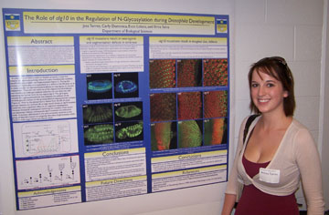 The Role of alg10 in N-glycosylation During Drosophila Development Jessica Torres, Carly Dominica, Evan Lebois, and Erica Selva Department of Biological Sciences Before a protein can properly
function in extracellular signaling, it
must first traverse the secretory pathway during which it undergoes
several
post-translational modifications. During
N-glycosylation, the alg10 gene
encodes for the glycosyltransferase responsible for the addition of the
terminal glucose residue onto the oligosaccharide-dolichol complex
prior to its en masse transfer to nascent
polypeptides. This terminal glucose is removed from the glycoprotein
previous
to it’s release from the luminal ER, so logic would dictate that an alg10 mutation would not significantly
disrupt function or development. However, an alg10
mutation is embryonic lethal and alg10 mutate embryos
show severe and pleiotropic defects indicating
its pivotal role in Drosophila
development. This study aimed to
characterize the alg10 gene during
development and identify the pathway(s) disrupted.
Embryonic antibody stainings revealed
segmentation and neurological defects in alg10
mutant embryos. In order to observe the
effect of alg10 mutations in adult
tissue, alg10 was removed from larval
imaginal disc tissue. The resultant eye
is reduced in size and disordered. Examination of molecular markers in
developing eye imaginal discs revealed a gain-of-function Sevenless
receptor
tyrosine kinase phenotype. This receptor tyrosine kinase pathway is
responsible
for the specification of the R7 photoreceptor. Our
data suggest N-glycosylation plays a significant role
in this
pathway and that the Sevenless pathway is a crucial target of alg10 function. Funding for
this project was provided by the
University of Delaware and the Jr. Life Sciences Scholars program.
|
 Down Regulation of TGF-β1 when treated with IGF-1 and the Effect on Prostate Cancer Elaina D. C. Welch and Carlton Cooper Department of Biological Sciences When caught early prostate
cancer (PCa) is treatable. Insulin-like
growth factor-1 (IGF-1) and transforming growth factor-Beta (TGF-Beta)
are cytokines that are both involved in the progression of cancer. TGF
is a tumor growth suppressor and IGF is a cancer growth promoter. It is
possible for IGF-1 to suppress the surface expression of TGF- Beta's
major receptor TR-II. Based on published findings it appears that IGF-1
down regulates TGF-Beta signaling. By using a progressive lineage of
PCa cells our objective is to explain the down regulation of TGF-Beta
by IGF-1, through TGF-Beta receptor (TR-II). As the disease progresses
TGF-Beta evolves from tumor suppressor to tumor promoter. TGF-Beta
usually decreases the likelihood of cell proliferation and IGF-1 could
be what causes TGF-Beta to mutate and possibly speed up the progression
of PCa and increase the risk of malignancy. Using FACS we sorted the
cells according to the intensity of fluorescence they acquired during
the process. The level of TR-II on the lineage of cells used: LNCap,
C4-2, and C4-2B4, was evaluated and compared to the controls which were
not treated with TR-II.
This project was funded by the HHMI NUCLEUS Program.
|