Abstracts from the Department of Biological Sciences
Undergraduate Summer Research Symposium August 10, 2005

Ordered alphabetically by student's last name


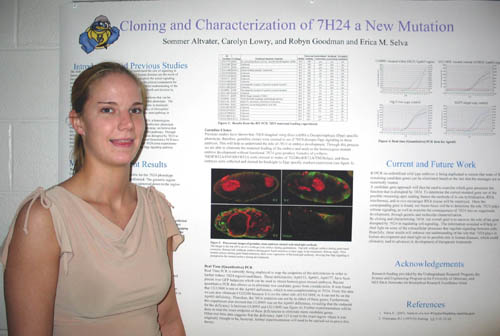
Cloning and Characterization of 7H24, a new mutation in Drosophila melanogaster

Sommer Altvater, Carolyn Lowry, and Erica Selva
Department of Biological Sciences

The focus of this laboratory is to investigate the extracellular interactions that influence signaling pathways between cells using Drosophila as a model.  Cell signaling is essential for correct development of all organisms. the study of development in Drosophila can help us understand the role of signaling in human development and human diseases because, many human diseases are the result of incorrect signaling, including cancer. The goal of my project is the cloning and characterization of 7H24, a homozygous recessive pupil lethal mutation that yields a lawn of denticles embryonic phenotypewhen the maternal contribution if removed. Based upon this phenotype, we believe that this mutation is involved in the Wingless (Wg) and Hedgehog (Hh) pathways. Through deficiency mapping we have narrowed down the region of the gene disrupted by 7H24 to the area between 62D2 and 63E5. RtPCR and quantitative PCR have  been used to try to further map the 7H24 mutation. Preliminary adult 7H24 clone experiments showed that 7H24 has a direct effect on the TGFB of the Dpp signaling pathway. Therefore, I am in the process of staining imaginal discs with anti-phosphoMAD antibody in order to further characterize this mutation during wing development. I am also in the process of generating germline clone embryos for staining in order to see if the Dpp specific markers are affected in these embryos because we see a Dpp phenotype in the wing discs. Funded by the Science and Engineering Scholars Program.


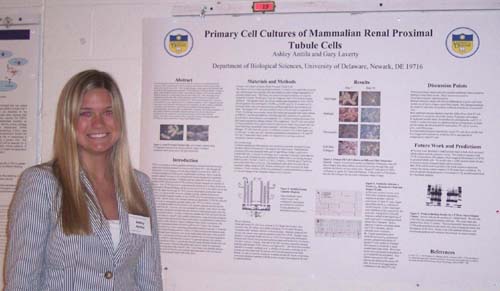
Primary Cell Cultures of Mammalian Renal Proximal Tubule Cells


Ashley Anttila and Gary Laverty
Department of Biological Sciences

Previous electrophysiological experiments in this lab revealed a novel  chloride transport system activated by parathyroid hormone in primary  cell cultures of chick renal proximal tubule cells (Laverty et al,  2003).   This chloride transport system seems to be associated with the  cystic fibrosis transmembrane regulator (CFTR) protein or CFTR-like  proteins.  In order to address if this transport system is unique to the  chick cell cultures, we have begun repeating identical experiments in  mammalian renal proximal tubule cells.  These electrophysiology  experiments must be done with similarly prepared confluent monolayers of primary cell cultures of mammalian renal proximal tubule cells.  Mouse proximal tubule (PT)  cells have been isolated by enzyme digestion  and Percoll gradient separation.  The PT fragments are then seeded onto  coated permeable filters and grown to confluence. In order to achieve  confluence on the filters, it was necessary to optimize growth conditions through changes in the extracellular matrix used on the filters, growth media composition, and enzyme preparation.  If these optimal conditions yield stable mammalian primary cell cultures, then identical experiments may be completed to reveal the physiological effects of this chloride channel.  This leads to the possibility of discovering more answers to the questions about CFTR and the deadly disease that it causes when mutation occurs.  Funded by Science and Engineering Scholars Program.


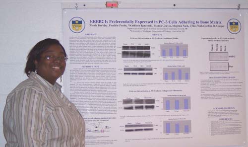
ERBB2 is Preferentially Expressed in PC-3 Cells Adhering to Bone Matrix


Nicole Barkley, *Kathleen Ignatoski, Freddie Pruitt, Magna Naik, Ulhas Naik, and Carlton R. Cooper
University of Delaware, Department of Biological Sciences, Newark, DE. *University of Michigan, Department of Urology, Ann Arbor, MI

Prostate cancer (PC) commonly metastasizes to the skeleton, where it becomes highly resistant to chemo-therapeutics. The components of the bone extracellular matrix microenvironment and other secreted factors have been implicated in PC cell proliferation. The purpose of this project was to determine the effects of PC-3 adhering to bone substrates on Akt activation. PC-3 cells plated on substrates composed of secreted factors from osteoblastic-like cells (SaOS2), bone stromal-like cells (HS-5), as well as collagen type-1, a major component of the bone matrix, and fibronectin. Western analysis was performed to resolve the phosphorylated Akt. We hypothesize that prostate cancer cells grown on substrate of secreted factors from osteoblast (SaOS2) and bone stromal (HS-5) cells will preferentially stimulate the activation of Akt. Also we hypothesize that PC-3 cells grown on type-1 collagen will preferentially stimulate Akt activation than on fibronectin. Western Blot analysis of Akt activation showed the greatest increase in PC-3 cells on SaOS-2 secreted substrate compared to control (plastic) and HS-5 conditioned media. In addition to this, we will present data showing the effect of prostate cancer cell adhesion to collagen type I and fibronectin on the activation of Akt. However, preliminary data suggest that secreted factors predominately from osteoblast cells enhance prostate cancer cell survival in the bone microenvironment and these factors may contribute to drug resistance. Funding by Charles Peter White Fellowship, HHMI, University of Delaware startup grant, and NIH K22 Career Transition Award


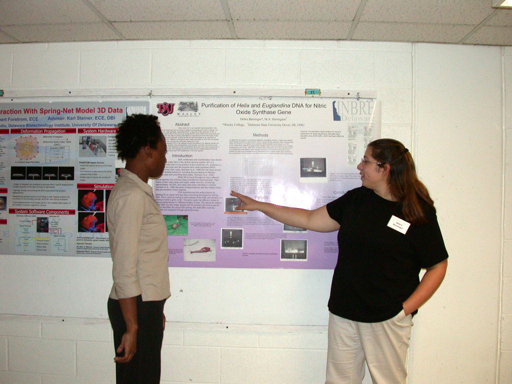
Purification of Euglandina and Helix DNA for Nitric Oxide Synthase Gene


Debra Barninger1 and  Melissa A. Harrington2
1Wesley College, Dover, DE 19901; 2Dept Biological Sci., Delaware State University, Dover, DE 19901

Nitric oxide, NO, is an important neurotransmitter in the ganglia of the snail brain and is involved in olfactory processing and control of eating. The enzyme nitric oxide synthase, abbreviated NOS, is required for the production of NO. A method of locating NOS in the snail is by cloning specific parts of DNA from Euglandina and Helix. To do this, two sets of primers were based on sequences from the NOS gene of the pond snail, Lymnaea stagnalis. Another primer set used was based on an alignment of mammalian NOS’s. Different combinations were made and tested on DNA from the garden snail, Helix aspersa, and the rosy wolf snail, Euglandina rosea. The samples underwent PCR in two different rounds and placed in 2% agarose solution for electrophoresis. Samples were photographed, and analyzed, and DNA removed from the gel. Samples and their respective primer combinations were sent off for gene sequencing. If results were produced, then samples would be cloned and the resulting sequence will be used to do gene knockout with RNA interference in the Helix species. Support contributed by: NIH NCRR INBRE grant 2P2ORRO16472-04 and NSF grants DBI-0320920 and IBN-0315551


Are Smoking Cessation Classes Beneficial or Ineffective Within a Lung Cancer Screening Program


Jamie Bartsch, Barbara Marconi, Angela Steele-Tilton, James Lally, and Thomas Bauer.
Christiana Care’s Helen F. Graham Cancer Center, Newark, DE 19716

The objective was to determine whether a professional smoking cessation coordinator was beneficial in increasing smoking cessation rates. Lung cancer is the leading cause of cancer deaths in the U.S. Forty nine million Americans smoke. Eighty five percent of lung cancer is because of smoking. Formal smoking cessation programs are established to increase quit rates. International Early Lung Cancer Action Project at Helen F. Graham Cancer Center (HFGCC) database was queried for all patients from April of 2003 to July of 2004. Patients were divided into 2 cohorts: cohort 1 was offered a formal smoking cessation program. Cohort 1 was divided into 3 groups: group A = personal contact with an invitation to the class, group B = a phone invitation to join class, and group C = written invitation. Cohort 2 patients were given information on smoking cessation. All 303 patients were called to answer questions on their current smoking status. Results found were: cohort 1 (192 patients), 35 quit smoking for an 18% quit rate, cohort 2 (111 patients) 19 quit smoking for a 17% quit rate. Increased class enrollment was noted in cohort 1 group A. Smoking cessation classes offered a 30% quit rate. In conclusion a formal smoking cessation program at the HFGCC was just as beneficial as an informal smoking cessation program. Individuals enrolled into the class simply represented a motivated segment of the smoker population. Further, prospective clinical trials are required to determine the best method for smoking cessation.


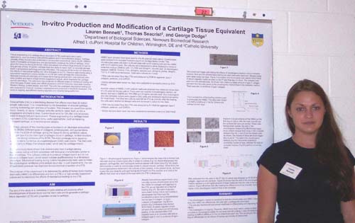
In-vitro Production and Modification of a Cartilage Tissue Equivalent


Lauren Bennett, Thomas Seacrist1, and George Dodge
Department of Biological Sciences, Nemours Biomedical Research, A.I. DuPont Hospital for Children, Wilmington, DE and 1Catholic University

Tissue engineering of a cartilage tissue equivalent (CTE) could have many useful applications, such as healing osteoarthritis patients and reconstructive surgery. Cartilage consists of few chondrocytes embedded in an abundant extracellular matrix (ECM). Different types of collagens, proteoglycans, and glycoproteins constitute the ECM of cartilage, which gives the tissue its strong yet tensile nature and allows it to endure large amounts of load for long periods of time. However, cartilage lacks the ability to repair itself, so damage acquired leads to progressive pathology and loss of function. The purpose of this study is to determine the effects of mechanical loading and other biologics on a CTE. Previous experiments using a specialized suspension culture resulted in a CTE with many cartilage-like characteristics. Neonatal porcine chondrocytes and human bone marrow-derived stem cells (stromal cells) were isolated and held in a high-density suspension culture, which maintains the cartilage specific phenotype. The cells were then subjected to different doses of mechanical loading or a matrix and biologically defined environment. Protein levels and mRNA expression of ECM molecules were determined by western blotting, protein assays, and PCR. Western blots of the cells subjected to the matrix environment show a rise in ECM proteins. The cells that underwent mechanical loading are also expected to reveal an increase in expression and production of the ECM molecules. This study is ongoing and different loading regimens are currently being performed. Funded by Milton Stetson Memorial .


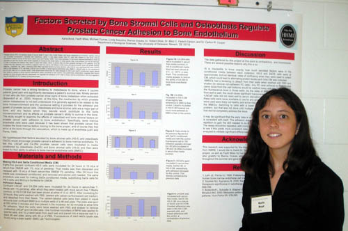
Factors Secreted by Bone Stromal Cells and Osteoblasts Regulate Prostate Cancer Adhesion to Bone Endothelium


Karla Boyd, Fayth Miles, Micheal Dumas, Linda Sequenia, Bianca Graves, Robert Sikes, Mary C. Farach-Carson, and Carlton R. Cooper
Department of Biological Sciences

Prostate cancer (PC) is a leading cause of cancer death in men in the United States, and bone metastasis is a common, painful, and as yet incurable complication. (Szostak and Kyprianou, 2000). It has been shown that PC cells first adhere to the bone marrow endothelium, before moving on to the bone matrix (Lehr and Pienta, 1998). This initial adhesion to the bone marrow endothelium must be regulated so that the cancer cells remain capable of moving on to the bone matrix. In other words, they must adhere, but not too strongly, in order to achieve the traction they need to reach the matrix, which is where they do their greatest damage to PC patients. It is yet unclear how this happens, but evidence suggests that it is due to the effects of the bone microenvironment, such as growth factors, integrins,  cytokines, etc. Here, I examine what effect secreted factors by bone stromal cells (HS-5) and osteoblast-like cells (SaOS) have on PC cell lines’ (LNCaP and C4-2B4) adhesion to bone marrow endothelial cells (BMEC). LNCaP cells do not metastasize but C4-2B4 cells do in a mouse model. Initial experiments show that those cells exposed to HS-5 and SaOS conditioned media were less adherent to bone marrow endothelium than control. This preliminary observation suggests that factors secreted by bone stromal cells and osteoblasts inhibit strong adhesion of PC cells to bone marrow endothelium, allowing them to move from that endothelium on to the bone matrix. Supported by the Howard Hughes Medical Institute 


Smoking Cessation:  An Opportunity to Have an Impact


Ashley Cephas and Thomas L. Bauer
Christiana Care’s Helen F. Graham Cancer Center, Newark, DE 19716

The time period at which a cancer diagnosis is made marks a significant point at which smoking cessation information can be offered to the patient.  This study assessed the smoking habits of forty-two patients (23 male, 19 female) who were seen in the Thoracic Unit of the Helen F. Graham Cancer Center and identified themselves as current smokers at their initial visit.  Of the 42 contacted patients, twenty-nine had quit smoking while thirteen admitted to still being current smokers.  Of the 29 patients who stopped smoking, 51% (15/29) cited a lung cancer diagnosis as the reason for quitting while 7 cited another cancer diagnosis or symptoms related to smoking.  This project was supported by the NIH NCRR INBRE grant to Delaware, grant number 2P20RR016472-04 .


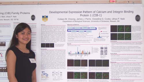
Developmental Expression Pattern of Calcium and Integrin-Binding Protein-1 (CIB-1)


Colleen M. Cheong, James J. Parris, Vesselina G. Cooke, and Ulhas P. Naik
Department of Biological Sciences

Calcium and integrin binding protein-1 (CIB-1) is known to have a key function in platelet activation through its interaction with integrin αIIbβ3.  Using Northern blots, it has also been found CIB-1 mRNA is present in a wide number of adult tissue including heart and kidney.  However, the location of CIB-1 in these tissues and the role of CIB-1 in development has yet to be studied.  In order to study the expression pattern of CIB-1, a series of immunofluorescence experiments were performed on various types of tissues.  First, the immunohistochemical staining protocol for the staining of CIB-1 was standardized.  Next, sections of heart and kidney tissue were stained and viewed using fluorescence microscopy.  From these experiments, it was found that CIB-1 protein is expressed in adult mouse heart and kidney tissues.  From the expression patterns, it appeaers that CIB-1 is expressed in the blood vessels of the heart and in the periphery of the glomeruli of the kidney.  Further experimentation is necessary to conclusively determine this expression pattern.  Additionally, timed pregnancies will be carried out and the embryos will be collected in various stages of development.  These embryos will be sectioned and stained to examine the contribution of CIB-1 in organ development.   Many types of tissues must be studied to have a greater understanding of the overall expression pattern of CIB-1.  This research was supported by the Howard Hughes Medical Institute. 


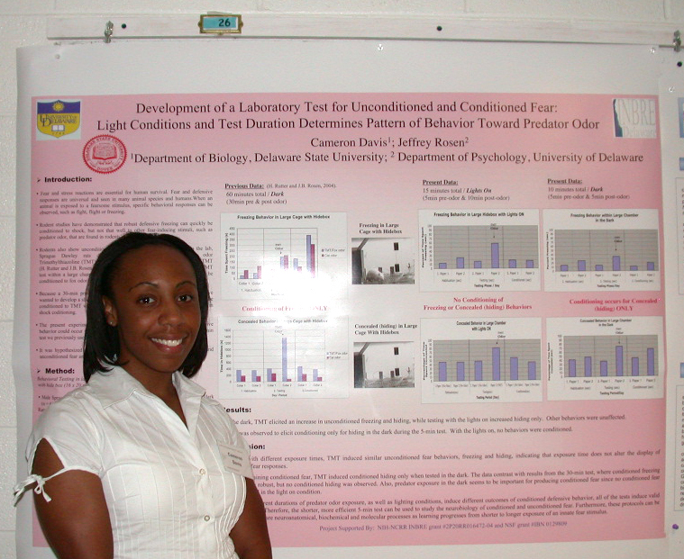
Development of a Laboratory Test for Unconditioned and Conditioned Fear: 

Light conditions and Test Duration Determines Pattern of Behavior toward Predator Odor

Cameron Davis1 and Jeffrey Rosen2
1Department of Biology, Delaware State University and 2Department of Psychology, University of Delaware

When an animal is exposed to learned and unlearned fearsome stimulus, specific behavioral responses are observed, such as fight, flight or freezing. Rodent studies find that robust defensive freezing can quickly be conditioned to shock, but not very well to other fear-inducing stimuli such as predator odor. In the lab, fox odor Trimethylthiazoline (TMT) induces unconditioned fear, but not conditioned fear in a small chamber.  However, in a large chamber with hidebox for 30 minutes, defensive behaviors can be conditioned to fox odor. Because a 30-min test is long and inefficient, we wanted to develop a shorter method to investigate conditioned and unconditioned fear while using predator odor. The present study tested whether defensive behavior in rats can also be conditioned to TMT when the time of testing is reduced to that similar used in shock conditioning. Animals were assessed within a large chamber containing a hidebox for a 10-min or 5-min duration. There were three days of testing: habituation, odor exposure (unconditioned), and conditioned retention. Testing the animals in the dark elicited an increase in defensive behavior (freezing, hiding), while testing using a lighted chamber increased hiding but decreased all other behaviors. TMT induced unconditioned freezing, but not conditioned freezing, whereas conditioned hiding increased. These results suggest that different durations of predator odor exposure, as well as lighting conditions, induce different outcomes of conditioned defensive behaviors.  The shorter more efficient test can be used to study the neurobiology of conditioned and unconditioned fear. Project Supported By: NIH-NCRR INBRE grant #2P20RR016472-04 and NSF grant #IBN 0129809


The Extralenticular Role of BetaB2-Crystallin

Kevin M. DuPrey and Melinda K. Duncan
Department of Biological Sciences

Crystallins are proteins responsible for proper lens function; yet the extralenticular role of the largest superfamily, beta/gamma-crystallins, remains unknown.  Previous studies have demonstrated that mice mutant for the betaB2-crystallin gene (Crybb2Phil) are sub-fertile compared to wild type mice due to a lack of sperm and egg production.  Morphometry of hematoxylin and eosin stained ovary and testis sections was performed to determine that the diameter of seminiferous tubules and ovarian follicles are significantly different at 1 week and 2 weeks of age probably due to the significant increase in apoptosis detected beginning between 2 and 3 weeks.  Immunohistochemical staining showed that betaB2-crystallin was localized to the developing acrosome of the spermatid, the corneal epithelium and endothelium, and the outer and inner plexiform layers of the retina. In order to confirm whether this infertility was due to the betaB2-crystallin mutation and! to assess possible functions of betaB2-crystallin in the retina, the mutation was moved from the original Swiss Webster genetic background which harbors a retinal degeneration mutation to the inbred C57Bl/6 background.  Initial studies on these mice determined that they did not have gonadal abnormalities and were not subfertile.  Studies on the possible function of betaB2-crystallin in the eye revealed that lenses from Crybb2Phil mice had defects in the actin cytoskeleton.  RT-PCR is being conducted to confirm the presence or absence of beta B2-crystallin in the cornea and retina of the C57B6 strain expressing the betaB2 mutation.  Source of funding: Howard Hughes Medical Institute and Charles Peter White Research Fellowships

Exploring O-GlcNAcylation and Phosphorylation in the Developing Chick Brain           

Andrew M. Farach and Deni S. Galileo
Department of Biological Sciences

Interest in post-translational modification of proteins by O-linked beta-N-acetylglucosamine (O-GlcNAc) is growing rapidly due to its continued implication in regulation of cellular processes. Since roles for O-GlcNAc in brain development have not yet been described, we chose to characterize the expression of O-GlcNAc in the developing chick brain to see if patterns existed in specific cell types, in subcellular processes such as axons, or in specific proteins. Based on the recurrent O-GlcNAcylation of highly phosphorylated proteins in vertebrate systems, it was hypothesized that phosphorylation and O-GlcNAcylation function reciprocally and neurofilament modification in neuronal axons was chosen to examine this; previous studies showed that the three subunits of the neurofilament trimer are extensively modified by phosphorylation and O-GlcNAcylation in mammals.  Preliminary studies indicate that a regulated developmental pattern to O-GlcNAcylation exists in the developing chick brain but a reciprocal relationship has not been conclusively identified. Immunohistochemical analyses in sections of progressive stages of development suggest upregulation of O-GlcNAc in the ependyma, tectobulbar neuron bodies, and glial processes, but not in axons. In contrast, double-label immunostaining of monolayer cultures made from dissociated E7 optic tecta suggests O-GlcNAcylation of most axons. Western blot analysis showed O-GlcNAc modification of a few discrete proteins throughout development, the most prominent weighing approximately 65kD. While these results indicate a developmental role for O-GlcNAc modification, further studies, including pharmacological inhibition in monolayer cultures and double-label immunostaining with radial glial markers, are needed to determine the relationship between O-GlcNAc, phosphorylation, and development. Funded in part by the Charles Peter White Fellowship.


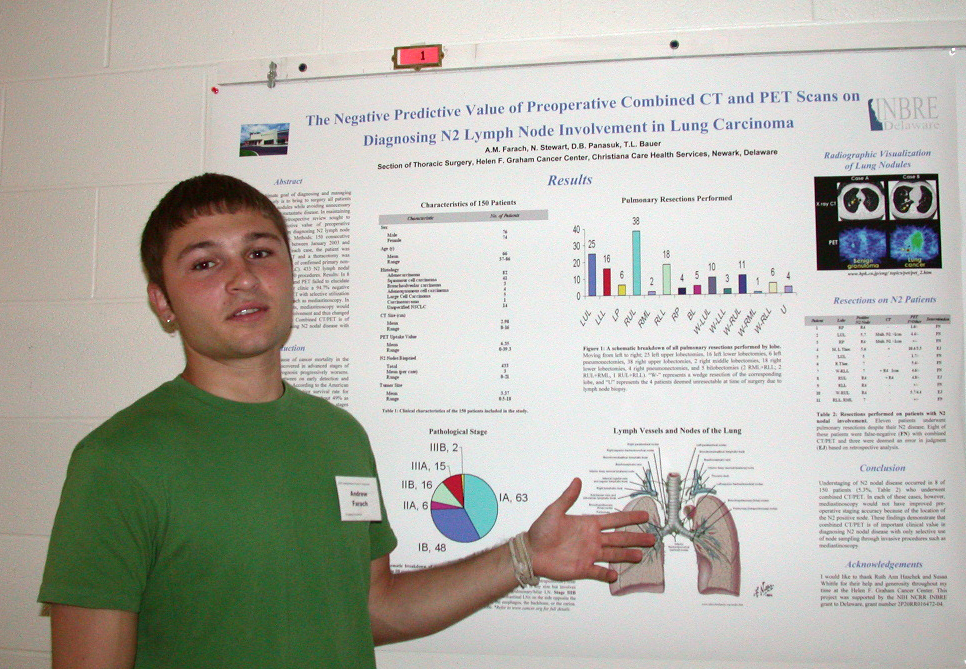
The Negative Predictive Value of Preoperative Combined CT and PET Scans

on Diagnosing N2 Lymph Node Involvement in Lung Carcinoma

Andrew M. Farach, Nancy Stewart, D. Bruce Panasuk, and Thomas L. Bauer
Section of Thoracic Surgery, Helen F. Graham Cancer Center, Christiana Care Health Services, Newark, Delaware

Background: The ultimate goal of diagnosing and managing nodal status preoperatively is to bring to surgery all patients with resectable malignant nodules while avoiding unnecessary thoracotomy on patients with metastatic disease. In maintaining this standard of care, this retrospective review sought to determine the negative predictive value of preoperative combined CT and PET scans on diagnosing N2 lymph node involvement in lung carcinoma. Methods: 150 consecutive pulmonary resection candidates between January 2003 and April 2005 were examined. In each case, the patient was preoperatively staged via CT/PET and a thoracotomy was performed for pulmonary resection of confirmed primary non-small cell lung carcinoma (NSCLC). 433 N2 lymph nodal stations were investigated in the 150 procedures. Results: In 8 out of 150 patients, combined CT and PET failed to elucidate N2 nodal involvement, giving our clinic a 94.7% negative predictive value to combined CT/PET with selective utilization of invasive diagnostic procedures such as mediastinoscopy. In each of these false-negative patients, mediastinoscopy would not have discovered lymph node involvement and thus changed preoperative staging.  Conclusion: Combined CT/PET is of important clinical value in diagnosing N2 nodal disease with selective use of mediastinoscopy. Funded in part by: NIH NCRR INBRE grant to Delaware, grant number 2P20RR016472-04


Haptic Interaction with Spring-Net Model 3D Data


Robert Forstrom
, Raymond Chen, and Karl Steiner
Delaware Biotechnology Institute

The spring-net deformable model of three-dimensial (3D) data aids in the accurate and real-life simulation of medical surgery.  This model's characteristics along with the haptic robot's force feedback allow for the 3D visualization and interaction processes to be highly believable when viewed by medically knowledgable personnel.  The use of this ability is hopefully to familiarize a surgeon with a specific patient before the actual surgery takes place.  Specific data sets can be acquired from a patient's CT, MRI or other volumetric scans.  This allows each type of data set with which a surgeon observes and interacts is patient-specific.  Using this method will allow the surgeon to be highly familiar with each patient.  Also, future versions of this project will allow particularly difficult surgeries to be practiced at will with precise feedback. This program was supported by the NIH NCRR INBRE grant to Delaware, grant number 2P20RR016472-04.


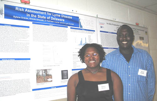
Risk Assessment for Lyme Disease in the State of Delaware


Xylene Graves, Arnold Omondi, Kathleen Curran, and Lynn Everett
Department of Biology, Wesley College, Dover, DE

According to the Centers for Disease Control, Delaware is a high risk state for Lyme disease. This disease is transmitted by the bite of Ixodes scapularis ticks infected with the bacterium Borrelia burgdorferi. Past vector ecology research in Delaware has shown that the number of cases of Lyme disease diagnosed in Kent and Sussex counties is quite large compared to the low rate of infected ticks. The first goal of our study is to determine tick density and infection rates in wildlife areas and state parks throughout Delaware because these regions are where most people come in contact with ticks. Ticks were collected by dragging. Only Ixodes scapularis nymphs were counted and used to calculate tick density. The nymphs were screened by PCR and gel electrophoresis for the presence of B. burgdorferi. Thus far, we have determined infection rates for the following for state parks: Killens Pond 2.0%, Trap Pond 8.6%, and Brandywine (Nature Trail) 14.3% and for Blackiston Wildlife Area 14.3%. Infection was confirmed by amplifying a region of the B. burgdorferi outer surface protein A (ospA) gene, 4 variations of which have been identified in nearby states. Our second goal is to identify genetic variants of ospA in Delaware ticks. PCR products from the nymphs found to be positive for B. burgdorferi were sequenced, and ten of the samples yielded usable sequence information. Analysis of the sequences indicated that each nymph harbored a single variant, nine of which were identified as variant 1 and one as variant 4.


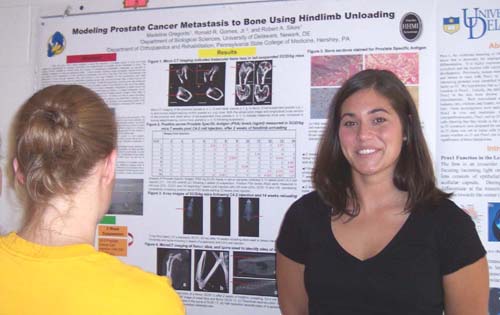
Modeling Prostate Cancer Metastasis to Bone Using Hindlimb Unloading


Madeline Gregorits and Robert Sikes
Department of Biological Sciences

There are only a few models available to study the interaction of prostate cancer in bone and these models are inappropriate for studying colonization and establishment of prostate cancer metastases in bone. The existing models available to directly study the process of spontaneous osteoblastic metastasis, as opposed to osteolytic metastasis to bone are extremely limited.  Current models, including mouse xenografts, require more than six months to form metastases in bone, producing metastases only 20% of the time (Wu, Sikes et al, 1998; Thalmann, Sikes et al, 2000). We have hypothesized that differences in bone turnover between mice and humans is the main limitation for the spontaneous and rapid colonization of marrow forming bone by osteoblastic prostate cancer cells, as bone turnover rates in adult mice do not mimic the higher, continuous turnover rates observed in humans. To test this hypothesis, bone turnover was enhanced in the hindlimbs of SCID/bg mice by housing them in a ground-based model of microgravity for two and three weeks, termed hindlimb suspension, followed by intracardial injection of a highly metastatic, osteoblastic prostate cancer cell line (C4-2). SCID/bg mice were allowed to recover under normal load bearing conditions. To detect metastases, serum samples were collected weekly during the reloading period and analyzed for Prostate Specific Antigen (PSA). After 14 weeks of reloading, the SCID/bg mice were imaged by X-ray and MicroCT. All tibias and femurs were harvested, imaged by MicroCT, paraffin-embedded, and sectioned. Hematoxylin-eosin staining was performed routinely for all sections. Following 14 days of suspension and intracardiac C4-2 injection, 2/8 (25%) SCID/bg mice expressed positive PSA levels 7 weeks post injection with an additional 2/8 (25%) SCID/bg mice expressing consistently increasing PSA levels 12 weeks post injection for a total of 50% PSA positive animals. Future work will involve PSA staining of the paraffin-embedded bone sections to detect the presence of cancerous cells in the unloaded hindlimbs. Funding provided by The Howard Hughes Medical Institute, Biological Sciences Start-Up Funds, University of Delaware Research Foundation, and University of Delaware Undergraduate Research Program.


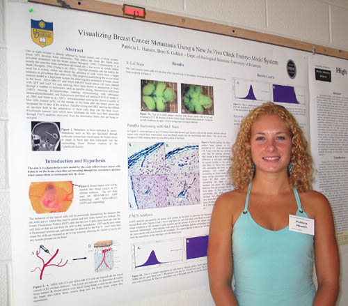
Visualizing Breast Cancer Metastasis Using a New In Vivo Chick Embryo Model System


Patricia L. Hansen and Deni S. Galileo
Department of Biological Sciences

Of the one in eight women directly affected by breast cancer, 20% of them will develop brain metastases and most of them will die shortly after diagnosis. This project is establishing the in vivo chick embryo model as a legitimate system for observing this metastasis of breast cancer in the brain.  Human MDA-MB-231 and MDA-MB-435 breast cancer cell lines have been infected with vectors containing the LacZ and GFP (Green Fluorescent Protein) genes for tracking purposes.  The LacZ gene turns blue when exposed to a β-Galactosidase solution, and GFP shines green under fluorescent light. These cell lines have shown to metastasize to brain through a number of techniques, such as paraffin sectioning, hemotoxylin and eosin (H&E) staining done by the Agriculture Department, β-Galactosidase staining, cryosectioning with subsequent immunohistochemistry, and fluorescence activated cell sorting (FACS).  β-Galactosidase staining has shown many blue cells on the outside of the brain after the cancer has incubated for 10 days in the embryo, and paraffin sectioning has offered an up-close look at the penetration of those cells deep into the brain tissue. GFP expressing MDA-MB-435 cells which have extravasated into the brain have been quantified through FACS analysis, recovered from the dissociated brains and are being re-cultured.  Through these methods, we hope to verify and then use this new model for the study of how breast cancer cells home in on the brain when they are traveling through the vasculature, and what causes them to extravasate into the tissue. Funded by the HHMI Program.


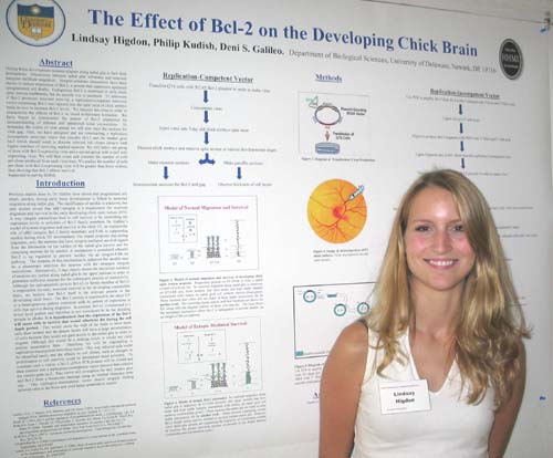
The Role of Bcl-2 in the Development of the Chick Optic Tectum


Lindsay Higdon, Philip Kudish, and Deni Galileo
Department of Biological Sciences

During brain development neurons migrate along radial glia to their final destinations.  Interactions between radial glial substrates and neuronal integrins facilitate migration.  Integrin-substrate interactions have been shown to induce expression of Bcl-2, a protein that suppresses apoptosis (programmed cell death).  Endogenous Bcl-2 is expressed in early chick optic tectum (midbrain), but its specific role is uncertain.  To determine if Bcl-2 promotes neuronal survival, a replication-competent retroviral vector expressing Bcl-2 was injected into the optic tecta of chick embryo brain in vivo to increase Bcl-2 levels.  We injected this virus in order to characterize the effects of Bcl-2 on tectal architecture formation.  We have begun to characterize the pattern of Bcl-2 expression by immunostaining of infected and uninfected tectal cryosections.  To determine the extent of viral spread we will also stain the sections for viral gag. Also, we have designed and are constructing a replication incompetent retroviral vector that encodes Bcl-2 and the marker gene lacZ which should result in discrete infected cell clones (arrays) with higher numbers of surviving marked neurons. We will infect one group of tecta with Bcl-2-expressing virus and a second group with a lacZ only expressing virus. We will then count and compare the number of cells per clone produced from each virus type. We predict the number of cells per clone with Bcl-2-expressing virus will be greater than those without, thus showing that Bcl-2 affects survival. Supported in part by HHMI.


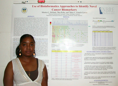
Use of Bioinformatics Approaches to Identify Novel Cancer Biomarkers

 
Monica L. Holland1, Ben Rohe2, and Mary C. Farach-Carson2
1Delaware Technical and Community College, Wilmington, Delaware
2Department of Biological Sciences, University of Delaware, Newark, Delaware

An urgent need exists to identify new biomarkers that will detect patients at risk for development of colon cancer. The initial purpose of this research was to use a bioinformatics approach to discover novel biomarkers associated with cell transformation in colon cells. ERp57/GRP58/1,25-D3 MARRS is a redox sensitive protein found in intestinal epithelial cells that facilitates ion transport in response to vitamin D hormone. Disruption in the function of this protein interferes with differentiation of colonic cells and may augment tumorigenesis. This research served to identify a family of genes (cluster) that are co-regulated with 1,25D3-MARRS in the mouse genome expression map. Quantitative analysis of transcriptional co-expression is a tool that has been used to investigate regulatory networks and to predict the functions of new genes found by genome sequencing. To form the mouse gene prediction database (http://mgpd.med.utoronto.ca/), microarray expression data was created for approximately 40,000 known or predicted mRNAs in 55 mouse tissues. Data obtained from analysis of custom-built oligonucleotide arrays was used to show that quantitative transcriptional co-expression is a substantial predictor of gene function and regulation of expression. The mouse prediction database, MGPD, was used in this work for the initial analysis of genes likely to be co-regulated with 1,25D3-MARRS with the assumption that the regulation of expression of mouse and human genes are similar. Selected genes were chosen for further analysis from the MGPD cluster based upon their similarity of expression, predicted function, and presence in colon. Specific functional response elements were searched for in the predicted 5’ promoter sequences (1000 bp upstream of transcriptional start site) of the selected genes. Each gene was scored based on the number of specific elements of interest that were found.  Antibodies to 1,25D3-MARRS and to the chosen gene products will be used to perform immunofluorescent staining to determine 1) if they are present in colon cells (IEC-6 or IEC-18 cell line); 2) if they also are present in colon tumor specimens; and 3) if they are co-expressed with 1,25D3-MARRS.  In the long term, it is hoped that this protein cluster approach will prove useful for identification of new candidate biomarkers for colon cancer. This work was supported by the NIH NCRR INBRE grant to Delaware, grant number 2P20RR016472-04 and by a grant from NIAMS AR48554.


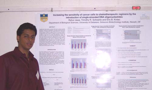
Increasing the Sensitivity of Cancer Cells to Chemotherapeutic Regimens

by the Introduction of Single-stranded DNA Oligonucleotides

Rahul Jawa, Timothy R. Schwartz and Eric B. Kmiec
Department of Biological Sciences and the Delaware Biotechnology Institute

Targeted gene repair utilizes a synthetic DNA vector to direct nucleotide alterations in specific DNA sequences. It has been determined that the transfection of single-stranded DNA oligonucleotides into mammalian cells can cause a delay in S-phase of the cell cycle by inducing a damage response and inhibiting DNA replication. This inhibition leads to a majority of the cells to stall in S-phase, potentially increasing the sensitivity of these cells to specific chemotherapeutic drugs. We examined the effect of a number of single-stranded oligonucleotides in DLD-1 and HCT116 (human colon adenocarcinoma) cells, to inhibit progression of cells from S-phase, and evaluated the sensitizing effect of the oligonucleotides on the cells by introducing doses of the chemotherapeutic drugs 5-fluorouracil (5-FU) and cisplatin. Cell viability was assessed via an MTT cytotoxicity assay. Our results indicate that a 74-mer all DNA oligonucleotide of mixed content (32% G-rich, 27% T-rich, 55% GC-rich ) and a 20-mer oligonucleotide containing all-T or all-G bases delayed cells in S phase or G1 respectively, and further resulted in significant sensitization of the cells to 5-FU and cisplatin, reducing viability with increasing doses of the drugs.


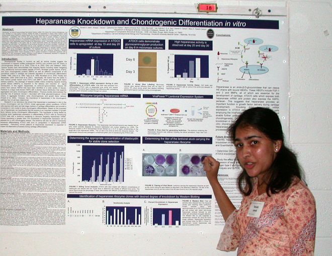
Heparanase Knockdown and Chondrogenic Differentiation in vitro


Sonali Joshi1, Anissa J. Brown1, Catherine Kirn-Safran1, Madhuchhanda Roy2, Dario Marchetti2, Daniel D. Carson1, Mary C. Farach-Carson1
1Department of Biological Sciences, University of Delaware, Newark, DE 19716
2Department of Comparative Biomedical Sciences – SVM, LSU-Baton Rouge, Baton Rouge, LA, 70803

Heparanase, an endoglucuronidase that cleaves heparan sulfate (HS) chains from various proteoglycans, has been identified in a wide variety of tissues. Despite the ability of heparanase to release active heparan sulfate binding growth factors (HBGFs) in various tissues, the role that heparanase plays in chondrogenesis has not been determined.  Quantitative PCR (Q-PCR) and activity assays were used to monitor changes in heparanase expression and activity during in vitro chondrogenic differentiation of mouse ATDC5 cells.  Q-PCR showed that heparanase transcript expression is transiently increased 60-fold in maturing chondrocytes. Heparanase activity assays show robust heparanase activity throughout the differentiation period.  Surprisingly, during the mid-point of chondrogenic differentiation in this system (day 15), overall heparanase activity was decreased although protein expression was still evident. The mechanism for this functional decrease has not been elucidated. Collectively, our data indicate that chondrogenic differentiation may be, in part, regulated by changes in heparanase expression and activity.  Based on these findings we hypothesize that the enzyme heparanase is an important mediator of growth factor delivery during chondrogenic differentiation. This hypothesis was tested by knocking down heparanase expression in differentiating ATDC5 cells by transducing ATDC5 cells with a lenti-virus containing a ribozyme targeting heparanase mRNA. Clones expressing a greater than 70% knockdown in heparanase expression will be selected for in vitro chondrogenic assays. Perturbing normal patterns of heparanase expression is expected to impact HS-dependent processes during cartilage differentiation.   This study was funded by HHMI and NIH.


The Role of Junctional Adhesion Molecule A (JAM-A) in the Corneal Epithelium

Liang-I Kang, Yan Wang, Vesselina G. Cooke, Ulhas P. Naik, and Melinda K. Duncan
Department of Biological Sciences

Junctional Adhesion Molecule-A (JAM-A) is a ~32 kDa protein that has been implicated in a variety of roles in the body, including platelet activation and adhesion, leukocyte migration, and the structural integrity of endothelial and epithelial cell layers.  Recently, our lab has begun to characterize JAM-A function in the eye since its expression was upregulated in the lens of Pax6 transgenic mice.  The presence of JAM-A, JAM-B, and JAM-C mRNA in the adult wildtype lens and cornea was confirmed through Real Time RT-PCR, with no significant level of compensatory expression of JAM-B and -C in JAM-A -/- tissue observed.  Using immunohistochemistry, JAM-A protein was found in the blood vessels of the developing eye as early as 12.5 dpc (days post conception) and in the corneal epithelium by 13.5 dpc.  PLAP (placenta alkaline phosphatase) reporter gene activity was detected in the JAM-A heterozygous and homozygous cornea, indicating the insertion of the gene-trap reporter construct in these mice, and immunohistochemistry showing the absence of JAM-A staining in knockout cornea validated both JAM-A antibody specificity and the knockout genotype.  While the eye appears to develop normally in JAM-A knockout mice, older animals have subtle abnormalities in the corneal epithelium.  We hypothesize that JAM-A has a role in the maintenance of the cornea and future functionality studies will include corneal wound healing assays. Supported by the Beckman Scholars Program.


The Role of Spot 14 in Cholesterol’s Influence on Fatty Acid Synthesis in Adipocytes


James Kelleher, John David, and David Usher
Department of Biological Sciences

A functioning organism is maintained only by the intricate chemical balance within the body.  Along with storing triglycerides, fat cells, or adipocytes, play an important role in the homeostasis of both glucose and fatty acids.  Spot 14 is a transcription factor, that in the liver, acts on various lipogenic genes.  It is also found in adipocytes where it is believed to have a similar function.  Spot 14’s expression is thought to be controlled from various factors, perhaps indicating it as a control point in the lipogenesis pathway.  In this study, 3T3-L1 adipocytes were differentiated, then analyzed under different treatments.  Some cells were treated with β-cyclodextrin (CD), a method of cholesterol depletion.  Another line was treated with pioglitizone, a drug that increases insulin-sensitivity.  The expression of Spot 14 and its hypothesized target genes—ACLY, ME, and PCK1—were quantified over a time course of the different treatments.  The mRNA of RBP4, Glut4, and GK, all involved in the transport and phosphorylation of glucose, along with glucose sensors USF1/USF2 and NR2F2, were also analyzed.  The information gathered from this study should lead to a better understanding of the effect of cholesterol on fatty acid synthesis, and the involvement of Spot 14 in the process. This research was funded in part by the Howard Hughes Medical Institute Undergraduate Science Education program.

Histotechnology: Technical Procedures

Joanne M. Kramer and Robert A. Sikes
Laboratory for Cancer Ontogeny & Therapeutics, Department of Biological Sciences

Histology is the branch of science that deals with microscopic cell identification, structure, and organization into various body tissues.  The histology technician prepares tissue specimens (both human and animal), for the pathologist to examine for diagnostic, research, or teaching purposes.  From fixation to the staining of tissue specimens, it is important that each step in the process be executed properly to ensure a high quality microscope slide.  A poorly prepared slide is difficult for the pathologist to read and cannot be utilized for patient diagnoses.  The tissue samples presented here are of prostate and bone, extracted from mice.  These specimens are used for prostate cancer research.  Aside from the prostate tissue itself, the bone study is vital to this project, as this is the site of prostate cancer metastases.

Currently a student in the histotechnology program at Delaware Technical and Community College, the INBRE grant has provided me the opportunity for a tremendous head start in acquiring the practical skills necessary to become a licensed histotechnician.Dr. Robert A. Sikes had a backlog of fixed tissues that needed to be placed onto slides for microscopic examination as part of his prostate cancer research.  With his assistance and that of many others affiliated with this project, I was able to prepare approximately 2000 slides.  This opportunity has allowed me to work as a histotechnician before my clinical internship requirement.  Funded by NIH NCRR INBRE grant to Delaware, grant number 2P20RR016472-04.


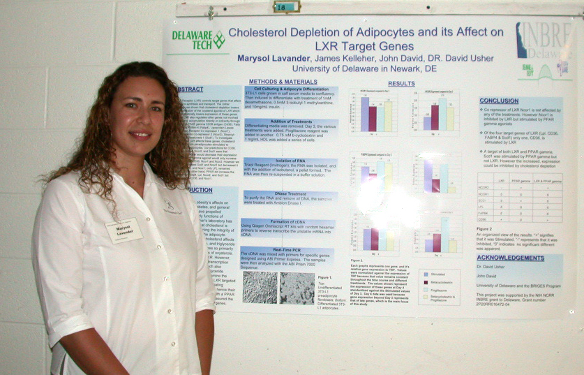
Cholesterol Depletion of Adipocytes and its Affect on LXR Target Genes

Marysol Lavander, John David, and David Usher
Department of Biological Sciences

Liver X Receptor (LXR) controls target genes that affect cholesterol synthesis and transport. The Usher laboratory has shown that cholesterol depletion lowers the concentration of the oxysterol agonist of LXR which in turn dramatically lowers expression of these genes.  However LXR also regulates other genes not involved in cholesterol accumulation directly or indirectly through regulation of PPAR gamma CD36 antigen (Cd36), Fatty Acid Binding Protein 4 (Fabp4), Lipoprotein Lipase (Lpl), Nuclear Receptor Co-repressor 1 (Ncor1), Nuclear Receptor Co-repressor 2 (Ncor2), Stearoyl-Coenzyme A Desaturase 1 (Scd1). To investigate whether or not LXR affects these genes, cholesterol was depleted from peradipocytes stimulated to differentiate to adipocytes. Our predictions for CD36, Fabp4, Lpl, Ncor1, Ncor2, and Scd1 were that cholesterol depletion would decrease their expression and that a PPAR gamma agonist would only increase the expression of CD36, NCOR1 and NCOR2. However we found LXR increased Scd1 and Ncor2 but decreased it for Fabp4, CSD36, Ncor1, LPL remained constant. On the other hand, PPAR did increase the expression for Fabp4, Lpl, Ncor2, and Scd1 but decreased it for CD36, and Ncor1. Funded by the NIH Bridges Program.


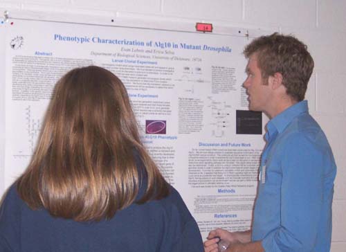
Phenotypic Characterization of Alg10 in Developing Drosophila


Evan P. Lebois and Erica Selva
Department of Biological Sciences

In this study we begin to phenotypically characterize Alg10, a gene encoding a glucosyltransferase that functions in N-linked glycosylation of nascent polypeptides in the ER, by use of genetic experiments conducted at various developmental stages of the Drosophila life cycle.  There are several reasons as to why further investigation into this mutation in glycosylation is very worthwhile.  Since many N-glycosylated proteins are targeted to the cell surface or for secretion, further study will lead to an increased understanding of how the extracellular environment influences cell signaling.  Also, because defects in glycosylation have been shown to play critical roles in human development, both in muscular dystrophy formation as well as in congenital disorders of glycosylation, our study of Alg10 will serve as an ideal model to better understand the role glycosylation plays in organismal development.  Clonal analysis in the developing wing yields a phenotype in adult flies where wings appear both shorter and rounder than wild type wings, implicating the involvement of glycosylation.  The embryonic mutant phenotype of Alg10 is a severely disrupted denticle banding pattern and results in a ventral open embryo.  In addition to the genetic experiments I have prepared hairpin RNAi constructs that will be expressed throughout development by using the UAS-Gal4 system in order to temporally and spatially characterize the Alg10 phenotype in developing Drosophila.  Constructs against Wingless (Wg) and Porcupine (Porc) have been synthesized as well as an Alg10-cDNA rescue construct to aid in this approach. Source of Funding: Charles Peter White Scholar Award


Microarray Analysis in Mammalian Systems Using Oligonucleotides for Gene Editing
Arjun K. Manrai1, Luciana Ferrara2, Hetal Parekh-Olmedo2, and Eric B. Kmiec2
1Department of Physics, Harvard College and  2Department of Biological Sciences and Biotechnology Institute, University of Delaware

Targeted Gene Repair is the process in which a modified single-stranded oligonucleotide directs the exchange of a single mutated DNA base.  This correction is site-specific, utilizing the cell’s endogenous repair mechanisms.  Results have shown that upon correction induced by the oligonucleotide, many corrected cells cease active replication.  Recent protein-level work has suggested that a large proportion of corrected cells enter into cellular senescence and hence, the proportion of corrected cells declines as noncorrected cells continue to divide.  Using DLD-1 cells stably integrated with a mutant eGFP gene, we perform the gene repair reaction and run microarray analysis using cell-cycle specific oligonucleotide microarrays to capture the transcriptional profile of gene-repaired cells.  We compare targeted cells with control samples at 8, 24, and 48 hours to obtain results representing the evolution of the transcriptional profile over time.  After mathematically correcting for background and normalizing the data, we confirm validity by verifying agreement with Bayesian (BAM) and Significance Analysis (SAM) techniques.  From these microarray data, we then construct a pathway and propose a regulatory network that accounts for the observed protein-level data and helps to explain the induction of targeted cells into senescence.  This is the first study to obtain and analyze transcriptional profiles of targeted DLD-1 cells and we hope that, in addition to providing information about the observed regulatory pathways of targeted cells in vitro, this work provides valuable insight for future in vivo gene repair applications. This project was supported by NIH NCRR INBRE grant to Delaware, grant number 2P20RR016472-04, and NIH grant RO1 CA89325.


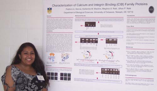
Characterization of Calcium and Integrin Binding (CIB) Family Proteins


Mini Manrai, Katherine M. Maddox, Meghna U. Naik, and Ulhas P. Naik
Department of Biological Sciences

It is known that CIB1 is involved in platelet spreading and adhesion via interaction with the αIIbβ3 integrin as seen in cell adhesion and spreading assays.  The functions of three novel CIB family proteins known as CIB2, CIB3, and CIB4 in comparison to the function of CIB1 in cell migration and cell adhesion are unknown.  Using Hind III and Sac II restriction sites, CIB proteins were subcloned into the pEGFP N1 and pEGFP C3 vectors in order to create GFP-CIB1, GFP-CIB2, and GFP-CIB4 fusion protein constructs for both the N and C terminals of GFP.  These constructs will be transfected into Chinese Hamster Ovary (CHO) cells in order to study the localization of each CIB family protein and the possible competition during cell localization between the CIB family proteins when co-expressed.  This summer project was funded by the Howard Hughes Medical Institute Summer Undergraduate Research Scholarship.


The Role of Autoinducer-2 Interspecies Communication in Escherichia coli and Helicobacter pylori Biofilm Development


Christina Nichols, M. Thompson, and Diane S. Herson
Department of Biological Sciences

During a process known as quorum sensing, bacteria produce molecular signals called autoinducers (AIs) to communicate. A specific group of quorum sensing molecules collectively referred to as autoinducer-2 (AI-2) have recently been discovered as part of a signal relay to stimulate bioluminescence in the marine bacterium Vibrio harveyi. Recent studies have implicated bacterial quorum sensing via AI-2 as a form of global communication during biofilm formation. We hypothesize that AI-2 serves as a form of intercellular communication during mixed species biofilm development for Helicobacter pylori and Escherichia coli. The E. coli strain DH5α is known to have a frameshift mutation preventing it from producing AI-2 and stimulate bioluminescence in the marine bacterium V.  harveyi. A luxS mutant strain of H. pylori and E. coli DH5α will be used to evaluate the role of AI-2 in coordinating the development of protective biofilm communities. Preliminary data with monospecies H. pylori biofilms has shown that, compared to a wildtype strain, the luxS mutant H. pylori produces relatively more dense biofilms. An hourly biofilm assay with E. coli has similarly shown that DH5α produces relatively more dense biofilms compared to a wildtype E. coliH. pylori to attach to existing E. coli biofilms. strain. Microscopic analysis of mixed biofilms will be performed over time to evaluate the ability of


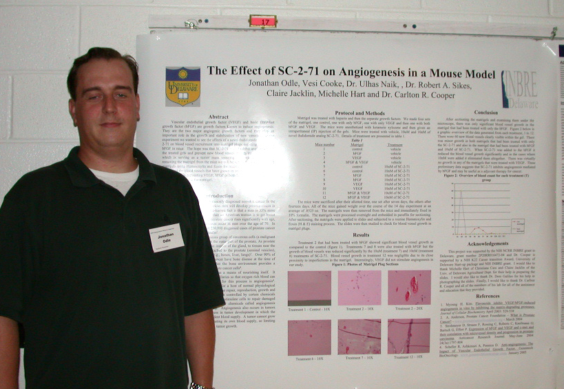
The Effect of SC-2-71 on Angiogenesis in a Mouse Model


Jonathan Odle, Vesi Cooke, Ulhas Naik, , Robert A. Sikes, Claire Jacklin, Michelle Hart, and Carlton R. Cooper
Department of Biological Sciences

Vascular endothelial growth factor (VEGF) and basic fibroblast growth factor (bFGF) are growth factors known to induce angiogenesis.  They are the two major angiogenic growth factors and they play an important role in the growth and stabilization of new vessels .  In our experiment we wanted to see the affects of a novel thalidomide analog SC-2-71 on blood vessel recruitment into matrigel plugs secreting VEGF and bFGF in mice.  The hope was that SC-2-71 would inhibit angiogenesis in the treated gels and prevent new blood vessel growth into the matrigel, which is serving as a tumor mass secreting antigenic factor.  After removing the matrigel from the mice we will be sectioning and staining the matrigels using hematoxylin and eosin for microscope viewing.  We will then count the blood vessels that have grown in order to see if the SC-2-71 was effective in preventing VEGF, bFGF or both from recruiting any blood vessel growth within the matrigels.


The Role of JAM-A in Breast Cancer


James J. Parris, Katherine M. Maddox, Sonali Raje, and Ulhas P. Naik
Department of Biological Sciences

Breast cancer is the second most common type of cancer among women: over 2 million women in the US currently have breast cancer. In order to develop effective treatments for this disease, the molecular mechanisms underlying cancer growth and metastasis must be investigated. Our lab studies Junctional Adhesion Molecule-A (JAM-A, also known as JAM-1), a transmembrane protein localized at tight junctions (TJs) and involved in angiogenesis, leukocyte transmigration, and platelet adhesion. Recently, we have observed that the less invasive T47D breast cancer cell line expresses higher levels of JAM-A than the more invasive MDA-MB-231 (231) cell line. This, in addition to the role of JAM-A in cell adhesion and migration, have led us to the hypothesis that JAM-A is an important factor in the growth and metastasis of cancer. We predict that increasing the level of JAM-A will prevent invasion due to increased cell adhesion. Alternatively, it is possible that increasing JAM-A will promote metastasis to bone because an increase in adhesion to endothelium will facilitate this process. To test this hypothesis, highly metastatic 231 human cancer cells will be engineered to overexpress murine JAM-A (mJAM-A). A construct was generated by cloning mJAM-A cDNA into a pcDNA3.1(+) expression vector. This construct, along with an empty control vector, will be transfected into 231 cells to overexpress JAM-A. Subcutaneous injection of cells into immunocompromised mice will be used to study tumor growth, and intracardiac injection will be used to determine the effect of JAM-A on metastasis. Funding was generously provided by HHMI.


A Role of Acute Phase Protein in Regulation of Lipid Metabolism or Transport in Adipocytes


Maulika Patel1, John David2, and David Usher2.
1Delaware Technical & Community College and 2Department of Biological Sciences, University of Delaware, and

In response to pathogen ingestion by macrophages, several cytokines include interleukin-6 (IL-6) & tumor necrosis factor alpha (TNF-ά) are secreted which causes the liver to synthesize and secrete the proteins into the blood.  The plasma concentrations of these acute phase proteins increase (or decrease) by 25% or more during certain inflammatory disorders.  These proteins include C-reactive protein, complement protein, serum amyloid protein, mannan binding lectin and fibrinogen.  They function to mediate destruction or limit the spread of a pathogen.  Adipocytes also make acute phase proteins C-reactor protein (CRP), Orosomucoid (ORM1), and Angipoietin-like4 (ANGPTL-4), indicating that some acute phase proteins have functions other than simply to limit the spread of pathogens.  To determine if CRP, ORM1, & ANGPTL-4 play a role in the regulation of lipid metabolism or transport, experiments were done on these gene under condition of cholesterol depletion.  Specifically differentiating 3T3L1 peradipocytes were treated with cyclodextrin (CD), preglutozol (a PPARg against) and CD + preglutozol and then the level of mRNA in the cells was determined at various times by quantitative PCR.


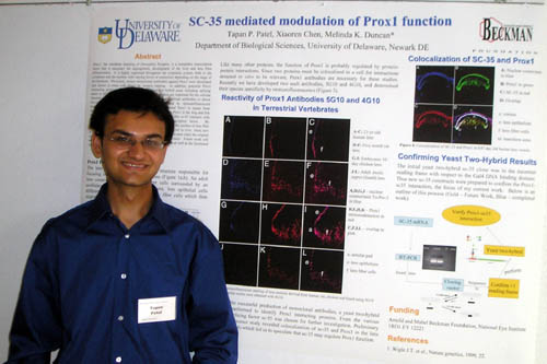
SC-35 Mediated Modulation of Prox1 Function


Tapan P. Patel, Xiaoren Chen, Melinda K. Duncan
Department of Biological Sciences

Prox-1, the vertebrate homolog of Drosophila Prospero, is a homeobox transcription factor that is necessary for angiogenesis, development of the liver and lens fiber differentiation.  It is highly expressed throughout the lymphatic system, both in the cytoplasm and the nucleus with varying levels of expression depending on the stage of development.  Potential Prox1 interacting proteins were identified by the yeast-two hybrid assay including splicing factor sc-35.  We hypothesize that sc-35 Prox1 interactions are important for the cellular function of Prox1. Next, investigations were initiated into the possibility that sc-35 interacts with Prox1 in vivo and modulates its function as a transcription factor.  By immunofluorescence, Prox1 and sc-35 protein are colocalized in the nucleus of lens fiber cells showing that they reside in the same location in the same cell in vivo.  Next, new sc-35 constructs were prepared for use in the yeast-two hybrid system since the original sc-35 clone was not in frame with the Gal4 DNA binding domain.  Future work will assess whether sc-35 and Prox1 can interact in vivo and in vitro as well as the functional significance of these interactions.


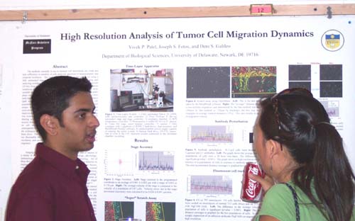
High Resolution Analysis of Tumor Cell Migration Dynami
cs


Vivek P. Patel, Joseph S. Fotos, and Deni S. Galileo
Department of Biological Sciences

The methods currently in use to measure cell movements are crude and lack sufficiency in numbers of cells analyzed, precision of measurements, and temporal resolution.  The study of cell motility is greatly enhanced using a fully automated time-lapse microscopy system capable of collecting and analyzing motility data at closely spaced time points (5 min) for long periods (20 hrs) from multiple areas of interest under several different experimental conditions simultaneously.  This system is designed to be significantly more versatile and less costly than commercial systems and can collect data under phase contrast or widefield fluorescent illumination concurrently.  Data were analyzed using motility parameters of velocity, total accumulated distance, and directionality for individual cells or for averaged cell populations.  Quantitation of “wound healing” assays revealed unique motility dynamics of drug-treated, antibody-treated, or adhesion molecule-transfected cells with high resolution and, thus, is a vast improvement over current methods.  In addition, GFP fluorescent labeling was evaluated for its usefulness in time-lapse studies.  Importantly, GFP labeling is not toxic under certain time-lapse conditions as shown by its toxicity kinetics.  Ultimately, fluorescent cells were tracked while migrating on cell monolayers expressing ectopic adhesion molecules, which consequently resulted in altered migration velocities.  Future work will utilize the techniques developed in this project to study glioma motility on brain slices.  The current work reveals cell motility behavior not discernable by currently used methods and increases the application, resolution, and accuracy of existing assays – enhancing the study of cell migration.  Funded in part by the McNair Scholars Program.

Large T Antigen Hexamer Assembly During SV 40 DNA Replication

Joseph Petfield, David Manna, and Daniel T. Simmons
Department of Biological Sciences

Simian virus 40 (SV40) is used as a model for eukaryotic DNA replication, transcription, and malignant transformation.  SV40 DNA encodes a multifunctional protein called large T-antigen, which is the only viral protein required for DNA replication, as the host cell provides all other replication machinery.  In order to initiate DNA replication, large T antigen forms a double hexameric structure over the viral origin in the presence of ATP.  This double hexamer serves to melt and structurally distort the DNA, as well as to recruit other necessary cellular proteins.  Five large T antigen mutants are currently being generated featuring single-residue, conservative point mutations of amino acids hypothesized to participate in the monomer-monomer interactions of the double hexameric structure.  Oligomerization assays are being performed to assess the ability of the mutants to form hexamers as compared to wild type T antigen.  Preliminary data show that mutants are less effective than wild-type T antigen in forming hexamers.  Future tests will compare the mutants’ capability to form double hexamers, as well as their ATP binding and helicase activities.  This research is funded by the National Cancer Institute and the Howard Hughes Medical Institute. 

Evidence for Hyal3 as a Murine Reproductive Hyaluronidase

Kristen Reese and Patricia Martin-DeLeon
Department of Biological Science

The extracellular matrix is rich in hyaluronic acid (HA), a repeated unit of disaccharides, which is degraded by enzymes termed hyaluronidases.  Specific hyaluronidases (hyases) on sperm are encoded by genes on human chromosome 7q/3p, and are essential in the fertilization process since they assist in the penetration of an unfertilized egg. SPAM1/Spam1 is the most characterized sperm hyaluronidase, and is localized to the acrosome cap of mature sperm.  However, it has been recently discovered that sperm from mice lacking Spam1 are still able to fertilize an egg (Baba et al. 2002).  Therefore, it is predicted that the related hyase genes contribute functionally to fertilization. Further characterization of the somatic and reproductive hyases related to Spam1 will provide an understanding of the contribution each hyaluronidase makes to the fertilization process.  HYAL3/Hyal3, located on human chromosome 3p21/mouse 9F1, has 67.1% similarity to SPAM1/Spam1, which is equal to or greater than other reproductive hyases’ similarities to Spam1, yet remains highly uncharacterized. It is important to note that humans do not have functional hyaluronidases located on the 3p21 chromosome. Therefore, it is possible that Hyal3 could act as a reproductive hyaluronidase in addition to or in the absence of a functional Spam1 protein.  It is the goal of this study to perform bioinformatic and functional studies to determine the characteristics, cell type expression, intracellular location, and function of Hyal3 in the mouse model.  Preliminary data suggests that Hyal3 contains three hyaluronidase domains present in reproductive hyases. Immunocytochemistry on mature caudal sperm indicate the presence of Hyal3 on the sperm head.  Western Dot Blot analysis also detects Hyal3 in the female uterine luminal fluid (ULF).  Taken together, these results indicate that Hyal3 has similar characteristics to the reproductive hyaluronidase Spam1, and may be involved in the fertilization process. Research Funded by HHMI and NIH

 
Bone Marrow Engraftment Monitoring

Adeline Richez1, Lynnelle W. Thorpe2, Deb Stabley3 and Katia Sol-Church3
1 Universite de Poitiers, France;  2University of Delaware, Newark DE;  3duPont Hospital For Children, Wilmington DE

This project is based on the development of new diagnostic tools for bone marrow transplantation monitoring.  Allogeneic bone marrow transplantation (BMT) is a well-established procedure conducted at the A. I. duPont Hospital for Children for the treatment of a variety of hematologic malignancies.  Typically, malignant cells are eradicated from the host by irradiation and/or high dose chemotherapy.  While killing the malignant cells this therapy also destroys the host's normal bone marrow cells, thus infusion of bone marrow from a healthy donor is required to restore hematological and immune functions. The key to success for BMT is the reconstitution of patient's bone marrow with the donor stem cells.   This full chimerism is consistent with full engraftment in the clinical setting and disease remission.  However, persistence of host hematopoietic cells, which results in a mixture of donor and recipient cells (mixed chimerism) are often associated with poor prognosis and disease relapse. The goal of this study was to compare two techniques using biomarkers to determine which one is the most sensitive to monitor the hematopoietic chimerism. The biomarkers tested were the Single Nucleotide Polymorphism (SNPs) analyzed by pyrosequencing, versus the Short Tandem Repeats (STR) analyzed by capillary electrophoresis. The technique using STR as biomarkers proved to be the most reliable and sensitive method for the detection of low level of chimerism after BMT.

 
The Effect of G-rich Oligonucleotides on the Inhibition of Huntingtin Protein Aggregation

Michael Skogen, Hetal Parekh-Olmedo, Eric B. Kmiec
Department of Biological Sciences, and Delaware Biotechnology Institute

Huntington’s disease is a neurodegenerative disorder caused by an expansion of polyglutamine repeats in the first exon of the Huntington gene, located on chromosome 4.  These polyglutamine tracts range from 40 to 122 glutamines and form inclusion bodies or aggregates.  The mutant protein, htt, has the acquired ability to assume an altered configuration allowing it to misfold and accumulate abnormal polypeptides within the nucleus.  The inclusion formation of mutant huntingtin leads to the death of specific neuronal populations in patients with Huntington’s disease leading to neurotoxicity.  In our results, poly-guanine oligonucleotides, or G-rich ODNs, have been shown to reduce the rate of aggregation of the huntingtin protein in vitro, as well as showing a structure dependent response in their ability to retard htt aggregation.  These G-rich oligonucleotides have been reported to form G-quartet secondary structures that may be the active component in repressing the aggregation of the htt protein.  In the present study, an in vitro biochemical filter retardation analysis was performed in addition to utilizing the human embryonic kidney cell line (HEK293T) to test a variety of G-rich ODNs and their affect on the inhibition of the htt aggregation.  We have seen that an oligonucleotide consisting of 20 guanine nucleotide repeats exhibits the greatest response in reducing protein aggregation when introduced both into cells expressing the mutant huntingtin protein and in a biochemical screen utilizing a truncated htt protein.  These discoveries will permit future work to be pursued in examining the effect of G-rich ODNs in vivo as well as leading to novel compounds for the treatment of HD.


The Role of Junctional Adhesion Molecule-A Homodimerization in Angiogenesis


Wen Allen Tseng and Ulhas P. Naik
Department of Biological Sciences

Junctional adhesion molecule-A (JAM-A or JAM-1) is found at the tight junctions of endothelial and epithelial cells.  Our lab has shown that JAM-A is involved in basic fibroblast growth factor (bFGF)-induced angiogenesis.  It has also been demonstrated that JAM-A is capable of cis-homodimerization. This study intends to test the hypothesis that cis-homodimerization of JAM-A is necessary for JAM-A-mediated angiogenic activities of endothelial cells to occur.  Site-directed mutagenesis was used to create constructs coding for mutant JAM-A proteins with substitutions of residues within the homodimer interface.  These constructs were transiently transfected into Chinese hamster ovary cells to determine whether these mutants have an impaired ability to form homodimers.  The extent of homodimerization will be determined by Western blotting after treating the transfected cells with an extracellular cross-linker.  After these assays are completed, these constructs will be stably transfected into human umbilical vein endothelial cells (HUVECs), which will be assayed to determine their ability to perform activities involved in angiogenesis as compared to mock-transfected and wild-type JAM-A-overexpressing HUVECs. This research is supported by the Arnold and Mabel Beckman Foundation.


Links: Summer 2005 Undergraduate Research Symposium, Symposium Abstracts from other Colleges and Departments,
Undergraduate Research Summer Enrichment ProgramUnversity of Delaware Undergraduate Research Program, Howard Hughes Undergraduate Program.
Created  4 August 2005. Last up dated 3 August 2005 by Hal White
Copyright 2005, University of Delaware