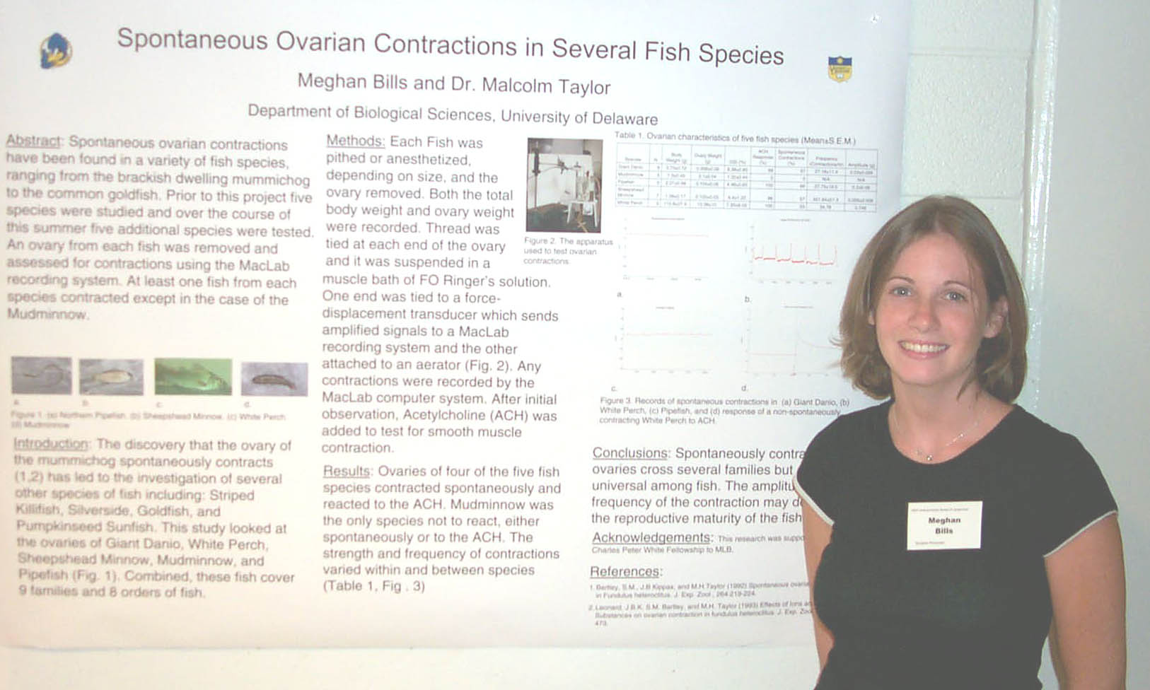
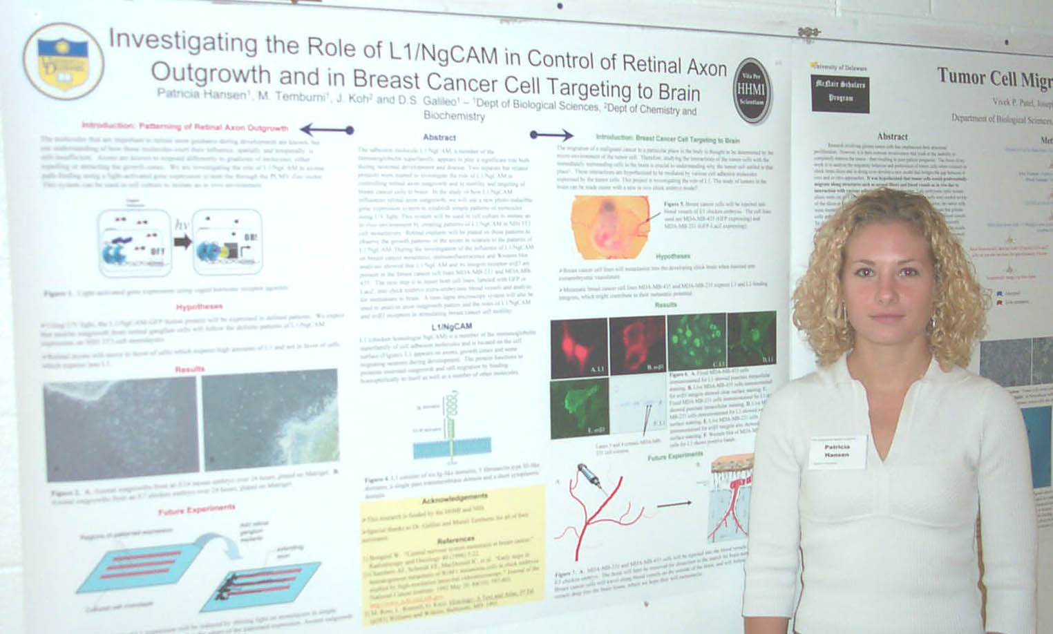
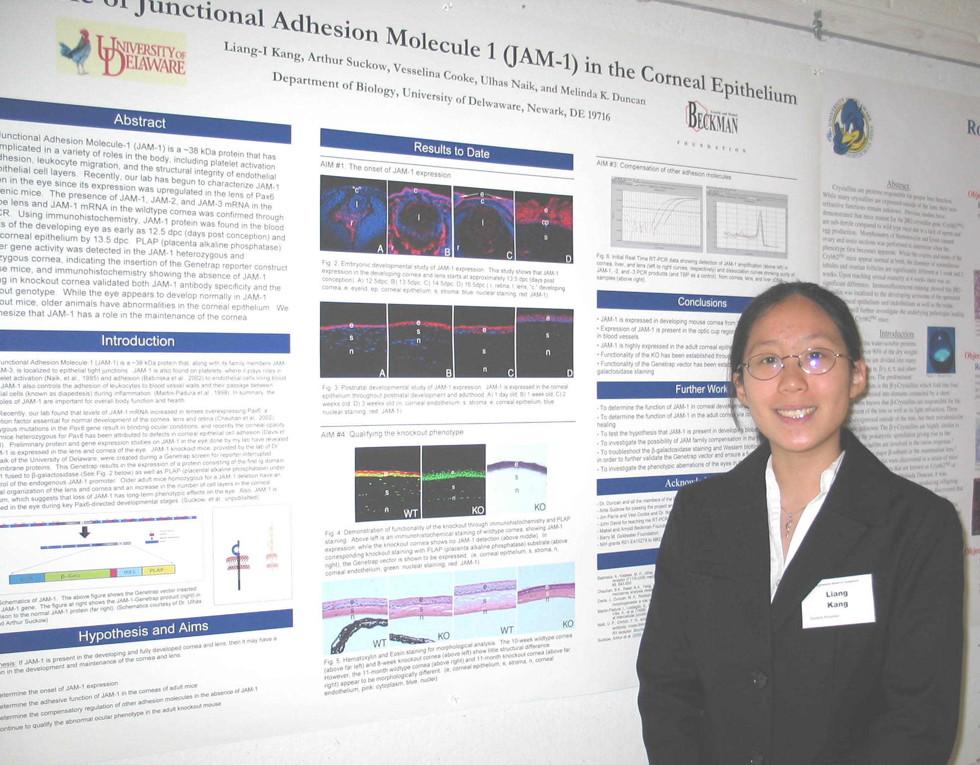
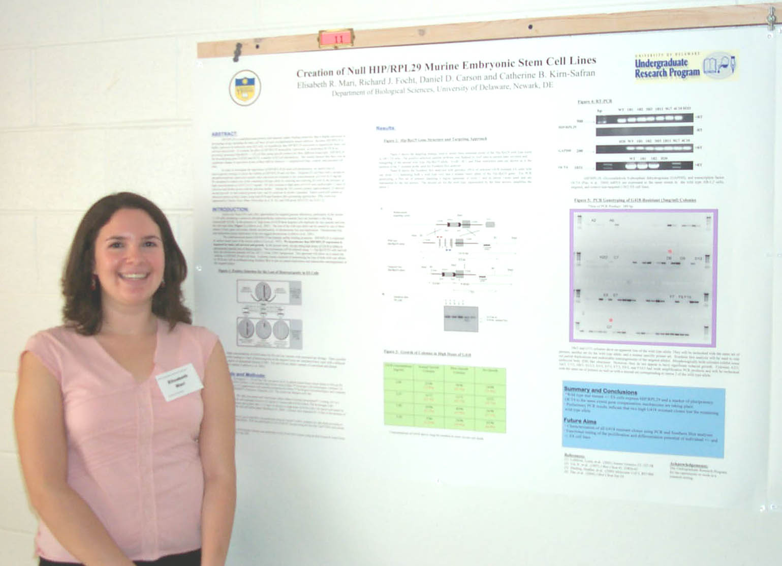
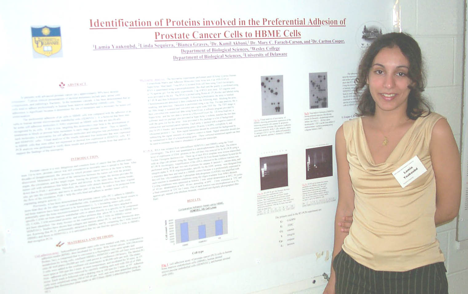
Abstracts from the Department of Biological Sciences
Undergraduate Summer Research Symposium August 11, 2004

Ordered alphabetically by student's last name


The expression of CD44 in bone marrow endothelial cells:
a target for bone metastasizing prostate cancer cells expressing hyaluronan.

Michael Arienmughare1, Cliff Piondexter2, Linda Sequeria2Robert A. Sikes2, George Dodge3, & Carlton R. Cooper2
1Department of Biology, Lincoln University, Lincoln, PA
2Department of Biological Sciences, University of Delaware, Newark, DE
3A. I Dupont Children Hospital, Wilmington, DE


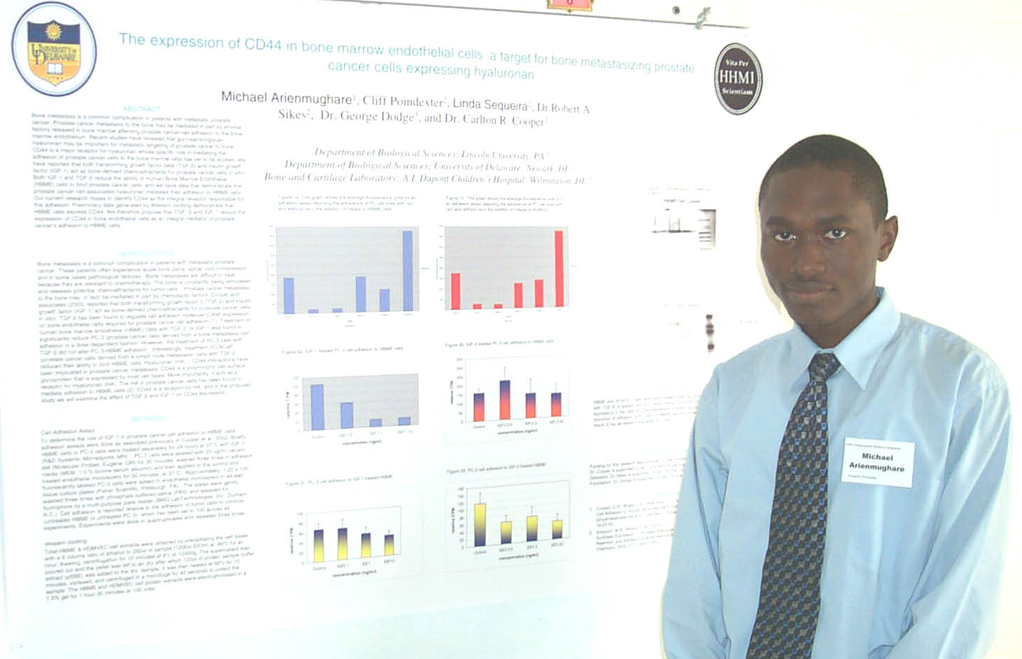
Bone metastasis is a common complication in patients with metastatic prostate cancer.    Prostate cancer metastasis to the bone may be mediated in part by stromal factors released in bone marrow affecting prostate cancer cell adhesion to the bone-marrow endothelium.  Recent studies have revealed that glycosaminoglycan hyaluronan may be important for metastatic targeting of prostate cancer to bone.  CD44 is a major receptor for hyaluronan whose specific role in mediating the adhesion of prostate cancer cell to the bone-marrow cells has yet to be studied.  We have reported that both transforming growth factor beta (TGF-β) and insulin growth factor (IGF-1) act as bone-derived chemoattractants for prostate cancer cells in vitro.  Both IGF-1 and TGF-β reduce the ability of Human Bone Marrow Endothelial (HBME) cells to bind prostate cancer cells, and we have data that demonstrate that prostate cancer cell-associated hyaluronan mediates their adhesion to HBME cells.  Our current research hopes to identify CD44 as the integral receptor responsible for this adhesion. Preliminary data generated by Western blotting demonstrate that HBME cells express CD44. We therefore propose that TGF- β and IGF-1 reduce the expression of CD44 in bone endothelial cells as an integral mediator of prostate cancer’s adhesion to HBME cells.

Supported in part by the HHMI Program.

Spontaneous Ovarian Contractions in Several Fish Species.
Meghan Bills and Malcolm Taylor, Department of Biological Sciences


The discovery that the ovary of Fundulus heteroclitus spontaneously contracts has led to the investigation of several other species of fish including: Striped Killifish, Silverside, Goldfish, and Pumpkinseed Sunfish. I was able to investigate five additional fish species: Giant Danio, Sheepshead Minnow, White Perch, Mudminnow, and Pipefish. In order to measure ovarian contractions, the fish were pithed or anesthetized, depending on size, weighed, and the ovaries removed and weighed. A string was tied to each end of the ovary and the ovary suspended in a muscle bath of FO Ringer’s solution. One end was attached to a force displacement transducer which sends amplified signals to a MacLab recording system and the other to an aerator. The tension could be adjusted and any movement was recorded by the MacLab system. After initial observation, Acetylcholine (ACH), a standard stimulus for smooth muscle, was added to create a concentration of 10-5M. Excluding Mudminnow all of the species spontaneously contracted and reacted to the ACH. The strength and frequency of the contractions is different for each species and appears at different times of reproductive maturity. The function of these contractions is not entirely understood but they may serve to mix the oocytes and ovarian fluids.

Supported by a Charles Peter White Fellowship.

Effects of Chemokine Treatment on Cancer Cell Behavior
Amanda Bly and Robert. A. Sikes
Department of Biological Sciences, University of Delaware


Prostate cancer (PCa) occurs at a high frequency. Overall there is a 1 in 6 lifetime chance of developing PCa in North American males. Survival rates of patients with PCa are good if the disease is localized; however, 45-50% of all PCas eventually metastasize to bone. Upon autopsy, more than 80% of patients diagnosed with localized PCa have developed micrometastases.  The LNCaP (androgen sensitive, weakly tumorigenic) and C4-2 (androgen independent, aggressive) are lineage-related PCa cell lines that represent two stages in the progression of PCa. C4-2 cells can spontaneously metastasize to bone, while LNCaP cells cannot.  Both will grow when implanted into bone marrow; and, LNCaP and C4-2 cell lines adhere similarly to bone marrow endothelial cells, the first cell that the metastatic cancer cell will contact when entering the bone. This implies that the primary difference in metastatic capability is due to inherent differences in the invasive abilities of the tumor cell. Chemokines and their receptors play a role in lymphocyte migration and chemotaxis as well as angiogenesis. Among these are IL-8 and CXCR12 (SDF-1). C4-2 cells have been known to secrete high levels of IL-8, which may provide a clue to their increased invasive potential. Chemokine receptors have been found on both cell types. These cell lines are to be tested for changes in adhesion and invasion following treatment with chemokines, IL-8 and CXCL12, to ascertain their effects on cell motility.

Funding for this project has been provided by the HHMI Science and Engineering Summer Research Scholarship.


Mechanism of Action of SV40 T Antigen Helicase
Josh R. Brooks; Junfang Jiaou, and Daniel Simmons, Department of Biological Sciences


T-Antigen is a DNA helicase which assembles over the simian virus 40 origin and functions in unwinding of double stranded DNA into two complementary single strands.  Normal T-antigen can load onto artificial replication forks from either a double stranded or single stranded region.  A mutant T Antigen (462-GA) with a mutation in the helicase domain is defective in double-stranded DNA binding and subsequently is not able to load onto a double-stranded end.  The aim of this project is to characterize the ability of this mutant T Antigen to propel itself along a 3’ single-stranded end.  We also wish to test how WT and Mutant T-Antigen load onto the DNA by testing Y forks with various chemical groups over the 3’ single stranded tail of the Y forks.  So far we have seen that the mutant T-antigen is not very efficient at unwinding helicase substrates.  Wild type T-antigen has shown to be a much better helicase on a variety of substrates. 

Funding was provided by a Charles Peter White Scholarship.


Polycystin-2 Expression in Osteocytes
Ashley Cephas and Norman J. Karin, Department of Biological Sciences

Autosomal Dominant Polycystic Kidney Disease is a genetic disorder caused by mutations in either the PKD1 or PKD2 gene, and in some cases both1.  The function of polycystin-1, the protein product of PKD1 has not yet been determined.  However, polycystin-2, the protein product of PKD2, has been shown to be a mechanosensitive calcium channel in kidney cells2.  The transmembrane protein is thought to act in association with polycystin-1 at crucial stages of bone development. We postulate that polycystins are mechanosensors in osteocytes. The goal of this project is to generate osteocytes with decreased expression of polycystin-2. RNAi constructs were inserted into osteocytes obtained from the cell line MLO-Y4.  Of the 12 clones obtained from the insert, five were screened using the western blot technique. None of the clones screened showed a significant decrease in the polycystin-2 expression. We are now screening the remaining clones.

Supported in part by a Charles Peter White scholarship.


Role of betaB2-Crystallin in Fertility
Kevin M. DuPrey and Melinda K. Duncan
Department of Biological Sciences


Crystallins are proteins responsible for proper lens function.  While many crystallins are expressed outside of the lens, their non-refractive functions remain unknown.  Previous studies have demonstrated mice mutant for the beta B2-crystallin gene (Crybb2Phil) are sub-fertile compared to wild type mice due to a lack of sperm and egg production.  Morphometry of hemotoxylin and eosin stained ovary and testis sections was performed to determine when the phenotype first becomes apparent.  While the ovaries and testes of the Crybb2Phil mice appear normal at birth, the diameter of seminiferous tubules and ovarian follicles are significantly different at 1 week and 2 weeks. Upon reaching sexual maturity at 4 weeks there was no significant difference.  Immunohistochemical staining showed that beta B2-crystallin was localized to the developing acrosome of the spermatid and the corneal epithelium and endothelium as well as the retina.  Future work will further investigate the underlying pathologies leading to infertility in Crybb2Phil mice.

Source of funding: The Charles Peter White Scholars Program


Investigation of the Expression of Murine Hyal5
in the Male Reproductive Tract, Accessory Organs, and Kidney

Mesha Eaton, Hong Zhang, and Patricia Martin-DeLeon, Department of Biological Sciences


Hyaluronidases are enzymes involved in the degradation and remodeling of the extracellular matrix. A group of closely linked hyaluronidase gene residing on human7q31/ mouse 6A2 chromosome contains a well studied, highly conserved protein, SPAM1 that has important roles in mammalian fertilization including the dissolution of the ECM around cumulus cells of the oocyte. This is necessary for sperm penetration of the oocyte. Recently a study revealed that fertility was unaffected in Spam1 Knockout mice, and suggested that there was a mouse-specific hyaluronidase, Hyal5, which encodes Hyal5, with similar functions to Spam1 (Baba et. al., 2002). The goal of this study was to determine if Hyal5 is similar to Spam1 in regards to its mRNA and protein expression in the male reproductive tract, the accessory organs, (prostate and seminal vesicles) and kidney. An mRNA expression analysis was performed using RT-PCR on the testis and three regions of the epididymis, caput, corpus, and cauda, (which were rendered sperm-free after excessive washing) and the kidney. Although preliminary, the results suggest the presence of Hyal5 in the cauda and the kidney. Protein expression in the testis, epididymis, kidney, and prostate was analyzed by Western Blot. The results suggest the presence of Hyal5 protein in the epididymis, kidney cortex, kidney medulla, and prostate. The overlapping functions of Spam1 and Hyal5 underscore the importance of the roles they play in the reproductive process and the kidney.

Supported in part by the Beckman Scholars Program.


Time Lapse Microscopy and Motion Analysis of Glioma Tumor Cells.
Joseph S. Fotos, Vivek P. Patel, and Deni S. Galileo, Department of Biological Sciences.

The aggressive invasiveness associated with some gliomas may be due to expression and proteolytic cleavage (shedding) of specific membrane molecules. Toward this end, a detailed motion analysis of fluorescently labeled tumor cells was performed using a sophisticated time lapse microscopy system. This system is fully automated and is capable of recording time lapse images of live cells in phase contrast and fluorescence.  Tumor cells were fluorescently labeled with either DiO (a vital fluorescent dye) or Q Tracker (a new type of intracellular staining). In addition, GFP expression was used to determine the most suitable method of labeling (i.e. maximum detectable fluorescence, cell viability, and minimum bleaching). MetaMorph software was used to track labeled cells and analyze cell motility parameters such as velocity, total distance traveled, and the angular direction of movements.  Ultimately, fluorescently labeled glioma cells will be placed on monolayers of normal 3T3 cells, as well as monolayers of 3T3 cells that express a specific adhesion molecule on their surface that is implicated in stimulating glioma motility.  We hypothesize that tumor cell lines will show significantly increased motile activity on 3T3 monolayers with surface expression of specific adhesion molecules then on plain 3T3 monolayers.

Funded by NIH and HHMI


Synthetic and natural inhibitors of angiogenic activity in endothelial cells
Janelle Green1, Angelo Evans1, Nikhil Patel1, Robert A. Sikes1, Merita McCluney2,  Benjie Blair2,  Milton Brown3, and Carlton R. Cooper1
1 Department of Biological Sciences, University of Delaware
2 Department of Biology, Jacksonville State University, Jacksonville, Alabama,
3 Department of Chemistry, University of Virginia-Charlottesville, Charlottesville, Virginia


Angiogenesis has been implicated in the progression and metastasis of cancer.  This process of forming new blood vessels from pre-existing vessels is mediated by a disruption in the balance of endogenous proangiogenic and antiangiogenic factors.  Natural and synthetic compounds are being evaluated for potential angiogenic inhibition.  A novel compound thalidomide-derivative, SC-2-71, was tested on Human Bone Marrow Endothelial cells (HBME) and Human Dermal Microvascular Endothelial Cells (HDMVEC). In addition, bacterial by-products that created a clear zone against Pseudomonas Aeruginosa were selected to study their effects on HDMVECs.  We hypothesize that these organic and synthetic compounds may be useful in the treatment of metastasis, particularly bone metastasis by reducing angiogenic activity of HBME cells. A growth assay demonstrated that SC-2-71 significantly inhibited the growth of both HBME and HDMVEC, suggesting that this compound is a potent angiogenic inhibitor. In addition, bacterial by-products inhibit the growth of HDMVEC.  SC-2-71 significantly inhibit endothelial tubular formation in the nanomolar range, while bacterial by-product did not demonstrate an effect. Taken together, this study concludes that synthetic and natural inhibitors of angiogenesis down-regulate specific angiogenic activities (i.e, endothelial proliferation and migration) in a concentration dependent manner.

JG supported by the BRIN Program. CRC is supported by a NIH transition award and NIH-INBRE Award; RAS is supported by a NIH award and Department of Defense Award; and MB is supported by a NIH award.


Fecal Coliform Regrowth or Reactivation in Biosolids
Sean Gillow1, Yinan Qi2, Brady Redmond1, Todd McVoy1, Steve Dentel2 and Diane S. Herson1
1Department ofBiological Sciences and 2Department of Civil and Environmental Engineering


A goal of wastewater treatment (WWT) is to decrease the levels of disease causing organisms such as bacteria, viruses, protozoa, and helminth ova in domestic sewage.  These organisms are not always present in sewage and it is not feasible to assay for all of them.  Instead fecal coliforms, which are always present in human waste, serve as indicators of possible pathogen contamination.  If treatment is successful, the resulting residue, called biosolids, can be land-applied and used as a soil conditioner or fertilizer.  Recently some wastewater treatment plants (WWTPs) have reported an increase in fecal coliforms when reference enumeration numbers (pre-dewatering) are compared to numbers obtained after the dewatering step.  We have received funding from the Water Environment Research Foundation (WERF) to verify this observation by assaying pre- and post-dewatered biosolids.  Our results confirmed that only post-dewatered samples from some WWTPs exhibited a significant regrowth or reactivation of fecal coliforms either during the dewatering step or after dewatering and subsequent incubation.  This increase is a health concern since it is possible that pathogens, such as Salmonella also increase.  To study this, we are currently initiating studies to detect Salmonella.  In these we will develop protocols using molecular techniques and compare these with standard methods currently in use.  The classical method uses selective enrichment, as well as biochemical and immunological techniques to detect Salmonella.  Molecular analysis will include use of the polymerase chain reaction (PCR) and immunological techniques.  Fecal coliform assays will be done by EPA Method 1680 on pre- and post-dewatered biosolids.  

Maximal Induction of Brain Creatine Kinase by AP2 also Requires Factor NF-Y
Brian Gladnick and George Molloy, Department of Biological Sciences


Astrocytes perform a variety of critical functions in the brain.  Most of these functions are highly energy-demanding and will occur too slowly unless ATP is regenerated.  An important enzyme for regenerating ATP is brain creatine kinase (CKB).  I have investigated how nuclear factors AP2α and NF-Y induce transcription of CKB in U87-MG glioblastoma, an attractive model system for astrocytes.  AP2α binds to the GCCAATGGG element located at –50bp in the proximal CKB promoter.  Interestingly, NF-Y has been shown to bind CCAAT sequences, one of which is located at –50bp in the CKB promoter within the –50bp AP2α element.  Transfections show that AP2α induces transcription of CKB by 7-10-fold.  However, the ability of AP2α to induce CKB was dramatically reduced when factor NF-Y could not function.  This supports the model whereby both AP2α and NF-Y associate with the –50bp GCCAATGGG element and induce CKB transcription by stabilizing the binding of factor TFIID to the (-28bp) TTAA box and RNA Polymerase II.  In agreement, increasing the levels of NF-Y increased the ability of AP2α to induce CKB by 2-fold.  The results provide new information on how factors AP2α and NF-Y function to regulate CKB, ensuring that ATP can be rapidly regenerated.

Supported in part by HHMI.

Effects of Tail Suspension on CB. 17 SCID/beige Mice
Madeline Gregorits, Ronald R. Gomes, and Robert A. Sikes, Department of Biological Sciences


The rodent tail suspension model developed in the mid-1970’s is widely used to study the musculoskeletal adaptation to microgravity.  In this model, rodents are suspended by their tails such that the hindlimbs fail to contact the floor.  Under these conditions, the non-weight-bearing hindlimbs rapidly undergo remodeling, characterized by a loss of skeletal muscle and bone mass.  Best characterized using rats; few studies have adapted tail suspension to mice. Thus, the major goals of this study were:  1) Determine if the CB. 17 SCID/beige mice can adapt to the stress induced by tail suspension; 2) Determine if two weeks of tail suspension will induce a significant loss of trabecular bone in non-weight bearing hindlimbs. To test our hypothesis, five CB. 17 SCID/beige mice were tail suspended for two weeks. Mice were monitored regularly for changes in body weight, and daily for eating, drinking, and grooming.  Blood collected on days 0, 7, and 14 was analyzed for serum cortisol, a marker of stress.  On day 14 the animals were sacrificed and the heart, adrenal glands, gastrocnemius, femurs, and tibias harvested.  A significant decrease in body weights of the tail-suspended mice was observed on day 4 and remained ~10% below controls.  Expressed relative to body weight, gastrocnemius mass and femur dry weights were significantly reduced in tail-suspended mice. Differences in heart, adrenal, and tibia masses were not observed.  These findings suggest the CB. 17 SCID/beige mice adapt to fourteen days of suspension and produce changes in the unloaded hindlimbs classically associated with this model. 

Funding provided by the Howard Hughes Medical Institute, University of Delaware Research Foundation, and Biological Sciences Start-up Funds.


Investigating the Role of L1/NgCAM in control of retinal axon outgrowth and in breast cancer cell targeting to brain.
Patricia Hansen1, Murali Temburni1, John Koh2, Deni S Galileo1,
1Dept of Biological Science, 2Dept of Chemistry and Biochemistry


The adhesion molecule L1/NgCAM, a member of the immunoglobulin superfamily, appears to play a significant role both during neuronal development and disease. Two separate but related projects were started to investigate the role of L1/NgCAM in controlling retinal axon outgrowth and in motility and targeting of breast cancer cells to brain.  In the study of how L1/NgCAM influences retinal axon outgrowth, we will use a new photo-inducible gene expression system to establish simple patterns of molecules using UV light. This system will be used in cell culture to imitate an in vivo environment by creating patterns of L1/NgCAM in NIH 3T3 cell monolayers.  Retinal explants will be plated on these patterns to observe the growth patterns of the axons in relation to the patterns of L1/NgCAM. During the investigation of the influence of L1/NgCAM on breast cancer metastasis, immunofluorescence and Western blot analyses showed that L1/NgCAM and its integrin receptor αvβ3 are present in the breast cancer cell lines MDA-MB-231 and MDA-MB-435.  The next step is to inject both cell lines, labeled with GFP or LacZ, into chick embryo extra-embryonic blood vessels and analyze for metastases to brain.  A time-lapse microscopy system will also be used to analyze axon outgrowth patters and the roles of L1/NgCAM and αvβ3 receptors in stimulating breast cancer cell motility. 

Funded by HHMI and NIH.


Geographic Variation in Random Genetic Markers in the Blue Mussel, Mytilus edulis
Nicole Hitchen and John McDonald, Department of Biological Sciences


Several allozyme loci exhibit large differences in allele frequency over relatively short geographic distances in the blue mussel, Mytilus edulis. To determine whether this results from natural selection or random drift, geographic variation of non-coding nuclear DNA polymorphisms is being surveyed. Random DNA fragments are amplified using random primers under low stringency conditions, then sequenced. Specific primers for each fragment are designed, the fragment is sequenced in several individuals, and a restriction site polymorphism is identified. One fragment has been surveyed for one restriction site polymorphism in fifty individuals from six locations ranging from Gloucester, Massachusetts to Oregon Inlet, North Carolina. It is anticipated that we will have nine more fragments surveyed in the next several months. The DNA polymorphism examined here shows that the random marker is geographically uniform in frequency. This is consistent with the idea that migration keeps neutral polymorphisms uniform in frequency, while selection causes the geographic variation in allozyme allele frequencies. If some of the restriction site polymorphisms show geographic variation, further research will consist of fine-scale geographic and temporal surveys in areas of differentiation to see if groups of mussels are partially reproductively isolated and are therefore starting to evolve into different species. If all of the restriction site polymorphisms exhibit geographic uniformity in the eastern United States, the survey could be extended to European and Southern Hemisphere populations of Mytilus edulis.

Studies of calcium channel subunit assembly and association with AHNAK:
Regulation by vitamin D hormone
Andrea Joseph, Kamil Akanbi, and Mary C. Farach-Carson, Department of Biological Sciences


The voltage dependent calcium channel is a multimeric protein consisting of several subunits including, alpha 1,alpha 2 delta, beta, and gamma.  However, the gamma subunit is not expressed in osteoblastic cells. The alpha 1c and the beta subunit are structurally linked. In addition, an unusually large protein named Ahnak whose exact function is unknown has been found to be directly associated to the beta subunit as well. Previous studies in our laboratory has shown that 1, 25 dihydroxyvitamin D3 down regulated the expression of the alpha 1c subunit of the voltage gated calcium channels in osteoblastic cell lines. In the present study we treated MC3T3-E1 cells with 1,25 dihydroxyvitamin D3, to determine if alpha 1c subunit of the voltage gated calcium channels and its associated beta subunit and Ahnak expressions are coordinately regulated. Our results indicate decrease in the expression of AHNAK in response to the 1,25 dihydroxyvitamin D3 treatment.

Funded in part by the Undergraduate Research program and the HHMI NUCLEUS Program.

Perlecan Knockdown and Chondrogenic Differentiation in Vitro
Sonali S. Joshi, R.R. Gomes Jr.,  D.D. Carson, and M.C. Farach-Carson Department of Biological Sciences


Perlecan, a 470 kDa heparan sulfate proteoglycan, plays an important role in chondrogenic differentiation in vivo and in vitro. Cells grown on surfaces coated with perlecan domain I, aggregate and form condensations which undergo chondrogenic differentiation. Moreover, condensations treated with Bone Morphogenic Protein-2 (BMP-2) undergo chondrogenic maturation. The role of endogenous perlecan expression in these processes is unknown. This study tested the hypothesis that endogenous perlecan expression is required for aggregation, chondrogenic differentiation and maturation of C3H10T1/2 cells in vitro. To test this hypothesis, a hammer headed ribozyme, targeting the domain I coding region of perlecan mRNA, was stably transfected into C3H10T1/2 fibroblasts. Dot blot analysis of conditioned media identified two clones exhibiting a 60-70% knockdown in perlecan secretion. These clones were characterized by growth curve analysis and their poliferative response to BMP-2. Finally, clones were tested for their ability to aggregate, maintain condensations and undergo chondrogenic maturation in response to BMP-2 treatment.  Knockdown clones demonstrated a decreased poliferative response following treatment with BMP-2. Interestingly, condensations from knockdown clones, expressed reduced levels of collagen type X following BMP-2 treatment.  Collectively these findings suggest that endogenous perlecan secretion is important for condensation formation, chondrogenic differentiation and maturation of C3H10T1/2 cells in vitro.

This study was funded by HHMI Undergraduate Research Award to S.S.J, NIH-NRSA (IF32 AG20078) to R.R.G and NIH (ROIDE 13542) to D.D.C.


The Role of Junctional Adhesion Molecule 1 (JAM-1) in the Corneal Epithelium
Liang Kang, Vesselina Cooke, Ulhas Naik, and Melinda K. Duncan, Department of Biological Sciences


Junctional Adhesion Molecule-1 (JAM-1) is a ~38 kDa protein that has been implicated in a variety of roles in the body, including platelet activation and adhesion, leukocyte migration, and the structural integrity of endothelial and epithelial cell layers.  Recently, our lab has begun to characterize JAM-1 function in the eye since its expression was upregulated in the lens of Pax6 transgenic mice.  The presence of JAM-1, JAM-2, and JAM-3 mRNA in the wildtype lens and JAM-1 mRNA in the wildtype cornea was confirmed through RT-PCR.  Using immunohistochemistry, JAM-1 protein was found in the blood vessels of the developing eye as early as 12.5 dpc (days post conception) and in the corneal epithelium by 13.5 dpc.  PLAP (placenta alkaline phosphatase) reporter gene activity was detected in the JAM-1 heterozygous and homozygous cornea, indicating the insertion of the Genetrap reporter construct in these mice, and immunohistochemistry showing the absence of JAM-1 staining in knockout cornea validated both JAM-1 antibody specificity and the knockout genotype.  While the eye appears to develop normally in JAM-1 knockout mice, older animals have abnormalities in the corneal epithelium.  We hypothesize that JAM-1 has a role in the maintenance of the cornea.

Supported by a Beckman Scholarship and NIH grants R01-EY15279 (MKD) and R01-HL63960 (UN).

Ca2+ Effects on Ahnak Protein Expression
Erin E. Kenaley, Kamil A. Akanbi, and Mary C. Farach-Carson, Department of Biological Sciences


Ahnak is a 700 kDa protein that is expressed in a variety of cells. The precise function of Ahnak remains a mystery. Ahnak has been proposed to be involved in actin cytoskeleton rearrangement in some cell types and in calcium homeostasis. Ahnak cellular location is influenced by intracellular calcium levels.  In this study, we assessed the effect of Ca2+ on the expression of Ahnak in 3T3-L1 preadipocytes.  RT-PCR and Western blot analyses revealed that Ahnak is present in the 3T3-L1 preadipocyte cell line. Immunostaining of 3T3-L1 cells with Ahnak specific antibodies showed that Ahnak is localized in the cytoplasm and along the plasma membrane. Immunostaining of proliferating 3T3-L1 cells (in exponential growth phase) cultured in varying levels of extracellular Ca2+ showed that Ahnak protein expression may increase with increasing extracellular Ca2+ levels.  When cytoplasmic Ca2+ levels were raised with thapsigargin or Ca2+ ionophore Ahnak protein expression also appeared to increase in cells treated with these agents compared to control. These data suggest that Ca2+ is an important regulator of Ahnak expression in 3T3-L1 cells.

This work was partially supported by a grant from the Howard Hughes Medical Institute (to EK) and by grants from the NIDCR (to MCFC and KA).


Identification and Biochemical Characterization of 7E4, a Mutation in Drosophila
Evan Lebois and Erica Selva, Department of Biological Sciences


The focus of this project is the cloning and characterization of 7E4, an ethylmethane sulfonate (EMS) mutation that disrupts the Wingless (Wg) signaling pathway.  Based upon preliminary phenotypic characterization, 7E4 is believed to encode a protein vital for Wg signal transduction, either via posttranslational enzymatic modification, or as a component of the secretory pathway.  Deficiency mapping has led to the narrowing of our field of genetic candidates responsible for encoding the gene disrupted by 7E4 and imparted focus upon ALG10, a critical enzyme in the N-linked glycosylation process.  PCR has been used to amplify ALG10 from 7E4 mutants in order to ultimately determine by sequence analysis if the genetic lesion that results in the 7E4 mutant phenotype is present in ALG10.  I am also persuing an alternative in vivo approach to identify the gene disrupted by the 7E4 mutation.  Molecular techniques are being utilized to create RNA intereference (RNAi) constructs designed to specifically target our best candidates within the delimited 7E4 region – ALG10 and GlcN – as well as known components of the Wg pathway.  The intent of targeting these components is in order to unambiguously verify that we indeed have the correct genetic candidate by recapitulation of the 7E4 mutant phenotype.

Source of funding: University of Delaware Research Foundation.


Mapping and Cloning of a New Mutation in Hedgehog and Dpp Signaling
Carolyn Lowry, Robyn Goodman, Erik Welf,  Shreya Thombre, and Erica Selva, PhD.
Department of Biological Sciences, University of Delaware


The focus of our research is to investigate the extracellular interactions that influence signaling pathways between cells. Using Drosophila melanogaster as a model, homozygous lethal mutations have been generated that can be categorized into signaling pathways based on their embryonic cuticle phenotype. The mutation being studied in this project is 7H24, which was found to be linked to Hedgehog (Hh) and/or Dpp signaling via phenotypic analysis in the wing disc and embryo. The goal of my project is to map 7H24 to its corresponding gene through various genetic mapping techniques, then to determine the consequences of 7H24 loss on development through genetic and molecular characterization. The signaling mechanisms within the fly are remarkably similar to those in mammals.  Therefore, the information gained by studying the signal pathways of Drosophila melanogaster can be directly applied to our understanding of the same pathway in humans. By gaining a better understanding of the extracellular components that regulate cellular growth and division via modulation of signaling that occurs between cells, we hope to provide information that could ultimately be used to develop better therapeutics for diseases that can be caused by abnormal signaling and pathway regulation, such as cancer.

Creation of Null HIP/RPL29 Murine Embryonic Stem Cell Lines
Elisabeth R. Mari, Richard J. Focht, Daniel D. Carson, and Catherine B. Kirn-Safran,
Department of Biological Sciences


    HIP/RPL29 is a multifunctional protein with heparan sulfate binding properties that is highly expressed in developing tissues including the inner cell mass of early preimplantation mouse embryos.  Because HIP/RPL29 is highly expressed in embryonic stem (ES) cells, we hypothesize that HIP/RPL29 expression is required for basic cell survival and growth.  To evaluate the effect of HIP/RPL29 monoallelic expression, we performed RT-PCR on previously generated Hip/Rpl29 +/- ES cell lines using specific primers for three different transcripts: HIP/RPL29, the housekeeping gene GAPDH and OCT4, a marker of ES cell pluripotency.  Our results showed that there was no significant change in expression of any of these mRNAs between +/- targeted cell lines, control, and parental cell lines.    
    In order to investigate the importance of HIP/RPL29 for stem cell pluripotency, we used a loss of heterozygosity strategy to assess the viability of HIP/RPL29 null cell lines.  Targeted ES cell lines with a neomycin phosphotransferase expression cassette (neo) insertion are resistant to low concentrations of G418 (0.25 mg/ml).   We attempted to induce loss of the remaining wild type allele by culturing neo-carrying ES cells in the presence of high concentrations of G418 (1.75-3.5 mg/ml).  ES cells resistant to high doses of G418 were picked after 12 days of selection and further grown with the selection marker.   Among the 192 colonies picked, approximately 60 showed normal growth, 80 had reduced growth rates, and 52 could not be further expanded.  Future work will consist of characterization of these clones using both PCR and Southern Blot genotyping approaches.

This work was supported by Charles Pete White Fellowship [to E. R. M.] and NIH grant HD25235 [to D.D.C.]).


Expression Analysis of Hyalp1, a Murine Reproductive Hyaluronidase
Kim Miller, Hong Zhang, and Patricia A. Martin-DeLeon, Department of Biological Sciences


The cumulus cells surrounding the unfertilized egg have an extracellular matrix rich in hyaluronic acid.  The digestion of hyaluronic acid and subsequent penetration of the cumulus cells, required for fertilization, is aided by enzymes called ‘reproductive hyaluronidases’.  Located on mouse chromosome 6, the gene family contains Hyalp1, whose encoded protein remains almost fully uncharacterized, in addition to Hyal4, Spam1, and Hyal5.  Recently, it was discovered that sperm from Spam1 knockout mice were able to penetrate the cumulus and successfully fertilize an egg, indicating that other proteins involved in this process can functionally compensate for a lack of Spam1.  The aim of this study is to determine both the expression patterns of Hyalp1 in the testis and its possible role in murine fertilization.

 
Detection of Salmonella spp. in Puerto Rican Drinking Water
Christina D. Nichols1, Graciela Ramirez-Toro2, Kate Verville3 and Diane S. Herson1
Department of Biological Sciences1, University of Delaware, Newark DE
CECIA2, InterAmerican University of Puerto Rico, San German, PR
Department of Biological Sciences3, Washington College, Chestertown MD


The CDC estimates that every year there are 1.4 million cases of Salmonellosis in the U.S. resulting in 1,000 deaths1. Salmonella infection can be introduced through contaminated food or inadequately treated water. We are working in collaboration with the InterAmerican University of Puerto Rico and Washington College to analyze rural Puerto Rican drinking systems to determine the quality of source and distributed waters. We hypothesize intermittent treatment with tablet chlorination is not sufficiently eliminating the health threat posed by this pathogen. Our qualitative procedure is designed to verify the presence/absence of culturable Salmonella species. To detect Salmonella spp., samples were selectively enriched, concentrated by Immunomagnetic Separation (IMS), and typical colonies were verified with biochemical and serological assays. Tests for coliform indicator organisms were also performed in Puerto Rico. Our joint data will help establish the efficacy of current water treatment in selected small water systems serving fewer than 3,300 residents. Preliminary results indicate approximately 33% of the distributed drinking water tested contained viable Salmonella, while 67% of source water was positive with the possibility of passing through sporadically treated systems. Our findings suggest that more rigorous water treatment procedures are needed in some of these remote rural locations, and current tablet chlorination systems are not effectively removing pathogens. Funding from the Science and Engineering Scholars Program and CECIA.


Downward Regulated Genes and Adipocyte Differentiation
Chinedu Nworu, Amanda Peters, John David, Stephanie Yudkovitz, and David C. Usher ,  
Department of Biological Sciences.


Gene expression during adipocyte differentiation in 3T3-L1 cells was studied by SAGE (Serial Analysis of Gene Expression). SAGE results revealed four highly down-regulated genes, namely, delta-like homolog (Drosophila) (Pref-1), THY1 (Thy1 cell surfacing antigen), Fibronectin 1 (Fn1), and S100 calcium binding protein A4 (S100A4). These down-regulated genes may prove to be very important to adipogenesis because a number of them are responsible for signal transduction and the determination of cell linage. With real time PCR, we verified the SAGE results, and in addition, elucidated their gene expression pattern. 3T3-L1 cells are progenitor cells that are committed to differentiate from a fibroblast to a mature adipocyte. To further confirm the importance of these genes we used 10T ½ cells. These murine pluripotent stem cells posses the ability to differentiate to chondrocytes, myocytes and osteocytes as well as adipocytes. Real time PCR was also used to determine whether those specific genes that are down-regulated in 3T3-L1 cells are expressed the same way in pluripotent stem cells that were placed under the same 3T3-L1 conditions that were used for adipocyte differentiation.


 Supported in part and funded by the HHMI Undergraduate Science Education Program and McNair Scholarship.


Assembly of the DNA unwinding complex on Simian Virus 40 (SV40) DNA
Oyekanmi J. Oyeyemi and Daniel Simmons
Department of Biological Sciences


The replication of SV40 DNA with the aid of T antigen is an indeed complex one involving the presence and correct order of other proteins i.e. replication protein A (RPA), topoisomerase Ι (Topo Ι), polymerase α/ primase (pol/prim) as well as a suitable replication environment. These series of experiment sought to determine the order of recruitment of these proteins in the formation of a replication complex by using a Glutathione Sepharose Transferase (GST) tagged T-antigen. DNA binding assays were done with this specially tagged T-antigen such that whatever was bound to the T-antigen at the origin DNA would bind to Glutathione Sepharose beads which would then be quantitated through western blot. Graphical representation of amount of protein bound showed that at an optimum amount of Topo Ι, RPA seemed to bind to T-antigen regardless of the presence or absence of DNA. However, for other concentrations of Topo Ι, there seemed to be more RPA binding in the presence of DNA than in its absence. In a second study, several forms of mutant T-antigen were used to find out how well they bound SV40 origin DNA and whether Topo Ι and RPA were recruited to the initiation complex.

OJO supported by HHMI Undergraduate Science Education Program.

Tumor Cell Migration on Brain Slices
Vivek P. Patel, Joseph S. Fotos, and Deni S. Galileo, Department of Biological Sciences.


Research involving glioma tumor cells has emphasized their abnormal proliferation.  However, it is their extreme invasiveness that leads to the inability to completely remove the tumor – thus resulting in poor patient prognosis.  The focus of my work is to analyze the migratory behavior and preferences of tumor cells when cultured on chick brain slices and in doing so to develop a new model that bridges the gap between in vivo and in vitro approaches.  It was hypothesized that tumor cells would preferentially migrate along structures such as axonal fibers and blood vessels as in vivo due to interaction with various adhesion and ECM molecules.  Late embryonic optic tectum slices were cut on a vibrating tissue slicer.  Rat C6/lacZ7 glioma cells were seeded on top of the slices at various percentages and cultured overnight.  After fixation, the tumor cells were stained with X-gal to allow for detection.  Preliminary results indicate that glioma cells indeed associated themselves with structures such as axonal fibers and blood vessels.  To directly observe their migratory activity on slices, tumor cells will be fluorescently labeled with QTracker and sequentially imaged using time-lapse microscopy.  The results will undoubtedly present further questions regarding the molecular mechanisms involved and the advances made at understanding the invasiveness of glioma cells may eventually help to develop new treatments for this lethal disease. 

The project was funded by the HHMI and the McNair Scholarship.


JAM-1 is Expressed during Vasculogenesis In Vivo
James J. Parris, Vessilina G. Cooke, Patrick B. Kelly, Melinda K. Duncan, Ulhas P. Naik,
Department of Biological Sciences

Cell adhesion molecules of the Ig superfamily play an important role in embryonic development. We have recently shown that JAM-1, a member of this family, is involved in endothelial cell adhesion and migration during angiogenesis. This suggested that JAM-1 may also play a role in vasculogenesis; however, the embryonic expression of JAM-1 was not known. Embryonic mice heterozygous for a JAM-1-β-geo genetrap were stained for β-geo activity. JAM-1 gene activity was broadly detected as early as 8.5 days post coitum (dpc) and at sites of neovascularization in both intersomitic blood vessels and cranial vasculature by 9.5 dpc. This was confirmed by colocalization of both β-geo activity and endogenous JAM-1 protein with PECAM-1, a known blood vessel endothelial cell marker. JAM-1 gene activity was also found in many epithelial tissues including the otic and olfactory systems at the placodal stage of their development. Additionally, expression in the epithelial components of the developing lung and kidney was detected at 11.5 dpc during their formation via branching morphogenesis. Thus, JAM-1 may have a role during vasculogenesis and epithelial tissue development.

Funding fromR01HL63960 to UPN; R01E4015279 to MKD; JJP was an HHMI and Dean's scholar.


Cholesterol Efflux and Adipocyte Differentiation
Amanda Peters, Jennifer Risser, John David, and David Usher
Department of Biological Sciences


Adipocytes are known to be important regulators of fatty acid homeostasis.   The Serial Analysis of Gene Expression (SAGE) library of 3T3-L1 adipocytes, constructed in our laboratory, has implicated cholesterol in having an important role in this process.  As determined by RT-PCR, the relative expressions of several genes involved in cholesterol transport, Cav, SREBP-1c, Abca1, and SR-B1, were found to be highly upregulated during 3T3-L1 differentiation.  To test the dependence of adipocytes differentiation on cholesterol in forming their phenotypic lipid droplet, cholesterol efflux was induced in cultured 3T3-L1 adipocytes on day 3 of differentiation, ensuring that “early” genes, such as transcription factors, -cyclodextrin and HDL towere allowed time for upregulation.  The addition of  the growth medium provided a tool for cholesterol removal from 3T3-L1 cells without toxicity.  Oil red-O staining of mature 3T3-L1 cells after experimental treatment revealed that cholesterol efflux during the late phase of differentiation greatly hindered lipid droplet formation in adipocytes.  This suggests that cholesterol is necessary for lipid droplet development in adipocytes.  Expression patterns of “late” genes, including Cav, SREBP-1c, Abca1, SR-B1, Adipsin, DGAT2, CD36, PPAR, and Fabp4, were then compared to those under conditions of cholesterol efflux. 

Funded by a grant from the Howard Hughes Medical Institute.


Analysis of sex chromosomes in the American Eel
Steve Pine and  Malcolm Taylor, Department of Biological Sciences


The American Eel has been shown to have different sex ratios in estuaries on the East Coast of the United States.  Because of this, many people believe that the American Eel has environmental sex determination.  This study will try to determine if the difference in sex ratios is the result of differences in genetic sex, or environmental influences on phenotypic sex.  To determine the genetic sex lymphocytes are grown for up to 6 days in culture with mitogens such as PHA-W and Lipopolysaccharide (LPS).  Cell division is then stopped during metaphase of mitosis with colchicine (Colcemid).  Chromosomal pairing of the sex chromosomes will be determined and a genetic sex for each eel will be compared to the sex of the gonads.  Trial cultures have been successful in causing cell division and proliferation but we have not yet succeeded in obtaining metaphase chromosomes. 

Supported in part by a Charles Peter White Scholarship.


Mutational Analysis of Junctional Adhesion Molecule-1 (JAM-1)
Swati S. Pradhan, Vessilina G. Cooke, Meghna U. Naik, and Ulhas P. Naik, Department of Biological Sciences


Angiogenesis, a condition that occurs in diseases like cancer, rheumatoid arthritis, psoriasis, etc., takes place when new blood vessels feed diseased tissues and destroy normal tissues.  We have previously shown that JAM-1, a member of the immunoglobulin superfamily is involved in endothelial cell adhesion and migration leading to angiogenesis.  JAM-1 localizes at the tight junctions of epithelial and endothelial cells and is involved in the regulation of junctional integrity and permeability.  JAM-1 consists of an extracellular domain, a single transmembrane domain, and a cytoplasmic tail. The cytoplasmic tail contains a PDZ domain-binding motif, which is important for JAM-1 binding to the cytoplasmic tight junction associated protein ZO-1.  We have generated several mutations within the JAM-1 protein to correlate the structural components of JAM-1 with its functions.  Preliminary results with deletions of the first and second Ig domains showed inhibition of JAM-1 adhesion on fibronectin.  The presence of both deletion mutants and wild-type JAM-1, on the cell surface, was confirmed by flow cytometry.  Immunofluorescence experiments showed that the deletion of PDZ domain-binding motif and the point mutation from serine to alanine do not appear to have an effect on the localization of JAM-1 protein, while the mutation of tyrosine to phenylalanine inhibits the localization of JAM-1 to tight junctions.  It can be thus concluded that the two Ig domains and the tyrosine residue in the sequence of JAM-1 are important to the adhesive properties and the localization of JAM-1. 

Funded by the Ronald E. McNair Program

The Characterization of Hyal3 in the Murine Testicular Cells
Kristen Reese and Patricia A. Martin-DeLeon, Department of Biological Sciences


Hyaluronan, a simple repeated unit of disaccharides, is a major component of the extracellular matrix.  Hyaluronan’s deposit in the extracellular matrix leads to an expansion of the tissue, allowing for facilitated cell migration during embryonic development, cellular proliferation and differentiation, and has a structural role in connective tissue.  The hyaluronan in the extracellular matrix is broken down by enzymes, termed hyaluronidases.  Multiple hyaluronidase genes have been identified, but the exact physiological role each plays is not yet clear.  To date, all that is known about Hyal3, one of the three murine somatic hyaluronidases located on mouse chromosome 9, is that it is most highly expressed in the testis and is most active at an acidic pH.  It is the goal of this study to determine the stage in development that Hyal3 is first expressed in the testis, the specific cell type of expression, as well as the intracellular location of the protein.  Findings from this study will allow the determination of whether Hyal3 participates in the process of fertilization.

Supported in part by the HHMI Undergraduate Science Education Program

Apoptosis Inhibition:  Cell Rescue and Subsequent Gene Repair Efficiencies
Paige Selvy 1, Luciana Ferrara 2, and Eric B. Kmiec 2,
1Mount Holyoke College, 2Department of Biological Sciences University of Delaware


The most promising alternative to gene therapy is a new method termed targeted gene repair.  This approach seeks to correct native genetic material by utilizing the cell’s own gene repair machinery, thereby maintaining the regulatory factors as well as the gene expression patterns within a cell.  This process entails two general steps:  DNA pairing, which refers to the hybridizing of a single-stranded oligonucleotide with almost complete complementarity to the target gene, except for one base, and homologous recombination repair, which is upregulated through a cascade of proteins in order to repair the gene.  Although this approach has proved effective in cell-based assays, the efficiencies of gene correction are still low and variability is seen in different labs.  Delivery of the oligonucleotide via electroporation, or the oligonucleotide itself may induce cells to undergo apoptosis following correction, as observed through FACS analyses.  Rescue of these cells from apoptosis could significantly increase correction efficiency by allowing corrected cells to survive.  Apoptosis inhibitors were used to target different points of the cell death signaling pathway in two cell lines, where these were incubated either alone or with DNA-damaging drug treatments to observe the effect of cell rescue on overall gene repair frequencies. 

Support and Funding provided by the NIH through the BRIN internship program.


The Effect of Feeding JAM-1 Null Mice a High Fat Diet on Adipogenesis
Laura Shankman, Vesi Cooke, Deepika Vuppalanchi, and  Ulhas Naik, Department of Biological Sciences


Diets high in fat have been shown to induce adipocyte formation and are correlated with heart diseases such as atherosclerosis. Adipogenesis, the formation of adiopocytes, is believed to require angiogenesis. Our lab is currently researching on Junction Adhesion Molecule 1 (JAM-1), a member of the Ig superfamily shown to be involved in endothelial cell adhesion and migration which leads to angiogenesis. Although many factors lead to atherosclerosis, when angiogenesis is reduced the amount of triglycerides in the bloodstream increases. This alters the physical characteristics of low density lipoproteins (LDL) so that they become poorly recognized by LDL receptors and more likely to enter the intima of the arteries. Reducing angiogenesis should result in fewer adiopocytes, higher levels of LDL in the blood plasma and more atherosclerotic plaques. By analyzing the fat pads, lipoprotein profiles and histological characteristics of JAM-1 null mice and wild type mice fed a high fat diet we can interpret the effects of angiogenesis on adipogenesis.

The work that was performed was funded by Science and Engineering Scholars Program.


Do Hyal5 and Spam1 Have Overlapping Functions?
Stacy Shertok, Hong Zhang, and Patricia A. Martin-DeLeon


This study was conducted to characterize the expression pattern of the rodent-specific Hyal5 in the mouse testis, as a means of determining if the protein has redundant or overlapping functions with Spam1. Hyaluronidase activity in the testis was measured using Hyaluronic Acid Substrate Gel Electrophoresis (HASGE).  The enzyme phosphatidylinositol-specific phospholipase C (PI-PLC) was used to determine if Hyal5 is glycosyl-phosphatidylinositol (GPI)-linked on sperm. To  determine the role of Hyal5 in cumulus penetration and in early steps of fertilization, an in vitro assay was performed.  Analysis of the Hyal5 cDNA sequence from mice carrying either of two Robertsonian translocations on chromosome 6, Rb(6.16) and Rb(6.15), reveals multiple point mutations, base insertions and base deletions, suggesting that the protein may play a role in the transmission ratio distortion seen in the progeny of the mice.
 This study was supported by grants from the Howard Hughes Medical Institute and National Institutes of Health.

The Cloning and Characterization of the 8J16 Mutation
Andrea Short, Robyn Goodman, and Erica Selva, Department of Biological Sciences


Embryonic development in organisms is very dependent upon signal transduction pathways in the cell.  The purpose of this research project is to study the extracellular signaling components of signaling pathways using Drosophila melanogaster as a model.   Several homozygous lethal ethylmethane sulfonate (EMS) mutations have been created which are known to be mutations in signaling components required for normal embryonic development. The EMS mutations being studied in this project are 8J16, and a possible second allele, 9E6.  Based on the embryonic “lawn of denticles” phenotype seen in 8J16, it is believed that it is involved in either the Hedgehog (Hh) or Wingless (Wg) signaling pathway.  Using various gene mapping techniques, the immediate goal of our project is to map and clone the 8J16 mutation.  Once this is accomplished, we will be able to study the effects that the loss of 8J16 has on development using different genetic and molecular techniques.  Signaling pathways in Drosophila are very similar to those found in humans.  Specifically, it is known that abnormal signaling in Hh and Wg pathways in humans is involved in diseases such as cancer.  We hope that in the future we will be able to use the knowledge we gain from this study to identify targets for treatments of such diseases.

This project is funded by the Howard Hughes Medical Institute.


Activation of Signaling Pathways in IEC-6 cells by 1,25-dihydroxyvitamin D3.
Jane Ullah1, Benjamin Rohe2, Susan E. Safford1, and Mary C. Farach-Carson2
2
Department of Biological Sciences, University of Delaware:
1Department of Biological Sciences, Lincoln University


    1,25 dihydroxyvitamin D3 [1,25(OH)2D3] elicits both a genomic and rapid non-genomic membrane response in target cells. It has been shown that addition of 1, 25(OH)2D3 to target cells, including the intestinal cell line IEC-6, leads to rapid activation of protein kinase C (PKC).  Within milliseconds to minutes after exposure of target cells to 1,25(OH)2D3, the events initiated in response to the binding of the vitamin D ligand to its putative plasma membrane receptor include a rapid elevation in intracellular Ca2+, G-protein coupled activation of protein kinase C and activation of calcium channels. In this study, IEC-6 cells were stimulated with the active metabolite of vitamin D, proteins were extracted, and PKC was partially purified using batch chromatography with DEAE cellulose (DE-52).  My goal was to adapt a commercially available protein kinase assay in order to detect the activity of PKC in our IEC-6 cell line before and after hormone treatment.
    The PepTag® Assays utilize fluorescent peptide substrates that are highly specific for select protein kinases. Phosphorylation of the peptide by the activation of PKC is expected to alter the peptide’s net charge from positive to negative, thus only the phosphorylated peptide is expected to bind the DEAE resin. This change allows the phosphorylated and nonphosphorylated substrate to be separated using an agarose gel. We hope to use the Promega PepTag® Protein Kinase Assay system to determine if 1,25(OH)2D3 causes the activation of PKC in IEC-6 cells. Development of this assay will facilitate our efforts to determine if 1,25(OH)2D3 activates PKC in intestinal cells under a variety of conditions.

Funding for this study was provided by the Howard Hughes Medical Institute (HHMI) and additional support was obtained from NIH grant (S11 AR48554) to Susan E. Safford.


Assembly of Initiation and Elongation Complexes during SV40 DNA Synthesis
Allison Wojcik, Rebekah Parsons, and Daniel Simmons, Department of Biological Sciences


Simian virus 40 is used as a model for researching mechanisms of DNA replication, transcription, and malignant transformation.  The monopolymerase system of initiation uses T antigen, DNA polymerase /primase, RPA and topoisomerase I.  Efficient elongation of growing strands requires the proteins Replication Factor C (RF-C), Proliferating Cell Nuclear Antigen (PCNA), and Polymerase delta (pol ).   RF-C catalyzes the loading of PCNA onto the DNA template by using ATP hydrolysis.  This complex tethers pol , which is needed for elongation of DNA synthesis.  DNA binding assays are being performed to determine the order of binding of elongation factors and how their presence affects the binding or retention of the initiation proteins.  Biotinylated DNA is incubated with the initiation proteins and various combinations of the elongation factors.  Results show that the elongation factors have little effect on the binding of T antigen.  However, both RF-C and pol  can individually decrease the binding of Topo I.  Furthermore, RPA binding increases in the presence of increasing amounts of pol .  Finally, PCNA binding to the elongation complex is dependent on RF-C or pol . These results indicate that replication of SV40 DNA is dependent on the formation of dynamic initiation and elongation complexes.

Supported by the HHMI Undergraduate Science Education Program

Identification of proteins involved in the preferential adhesion of prostate cancer cells to HBME cells
Lamia Yaakoubd, Linda Sequiera, Bianca Graves, and Carlton Cooper, Department of Biological Sciences


In patients with advanced prostate cancer (pc), approximately 90% have skeletal metastases.  Typical clinical presentations of skeletal metastasis include pain, spinal cord compression, and pathologic fractures.  In the metastatic cascade, it has been demonstrated that pc cells tend to adhere preferentially to human bone marrow endothelial (HBME) cells.  This adherence is significant because it determines the site of metastasis and is necessary for tumor cell extravasation.

The preferential adhesion of pc cells to HBME cells was compared with the adhesion of pc cells to human dermal microvascular endothelial cells (HDMEVC).  It is believed that there may be some cell adhesion molecules (CAMs) expressed on HBME cells that are specifically recognized by pc cells.  If this is true, targeting this molecule(s) may help in the development of treatments to block or prevent bone metastasis in early-stage prostate cancer.  Thus, to identify such molecules, a microarray for cell adhesion molecules and integrins was performed on HBME and HDMVEC cells.  Visual analysis of the results indicated four molecules that were expressed in HBME cells that were either not expressed or showed low expression in HDMVEC cells.  RT-PCR analysis was performed to verify these results and preliminary results from that analysis do support the findings of the microarray.
   
Start-up funds provided by the University of Delaware and the BRIN program.


Gene Expression during Adipocyte Differentiation
Stephanie Yudkovitz, John David, Amanda Peters, Chinedu Nworu, and David C. Usher,
Department of Biological Sciences


Orosomucoid 1, Angiopoietin-like 4 and Haptoglobin are acute phase proteins that are secreted by adipocytes.  Based on previous experimentation and research, such as the SAGE Library (Serial Analysis of Gene Expression), it has been shown that these secreted proteins are upregulated in adipocytes, when compared to preadipocytes.  Their secretion is increased after an inflammatory event by the liver.  Their production is believed to be controlled by inflammatory cytokines. 3T3-L1 cells were differentiated and Real Time PCR was used to show the gene expression of the acute phase proteins studied.  It was found that several acute phase proteins, such as Orm1, Angptl4 and Hp, were upregulated during differentiation.  Time course assays were performed on these acute phase proteins on days 0, 3, 4, 5 and 8.

Supported in part by a Charles Peter White Fellowship.


Links: Summer 2004 Undergraduate Research Symposium, Symposium Abstracts from other Colleges and Departments,
Undergraduate Research Summer Enrichment ProgramUnversity of Delaware Undergraduate Research Program, Howard Hughes Undergraduate Program.
Created  30 July 2004. Last up dated  13 August 2004 by Hal White
Copyright 2004, University of Delaware