 |
REAP AWARDS AT THE AMERICAN SOCIETY FOR BIOCHEMISTRY AND MOLECULAR BIOLOGY (ASBMB) UNDERGRADUATE POSTER COMPETITION DURING THE EXPERIMENTAL BIOLOGY MEETINGS IN SAN DIEGO, APRIL 11-15, 2003 |
 |
|
REAP AWARDS AT THE AMERICAN SOCIETY FOR BIOCHEMISTRY AND MOLECULAR BIOLOGY (ASBMB) UNDERGRADUATE POSTER COMPETITION DURING THE EXPERIMENTAL BIOLOGY MEETINGS IN SAN DIEGO, APRIL 11-15, 2003 |
|
Some attractions like bonobos
at the San Diego Zoo and nearby Balboa Park were part of the trip to the
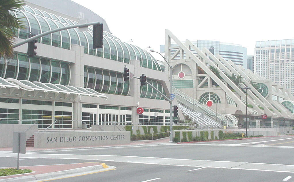
Experimental Biology Meetings, but most of the time was spent in the San
Diego Conference Center that seemed a mile long.
 |
 |
 |
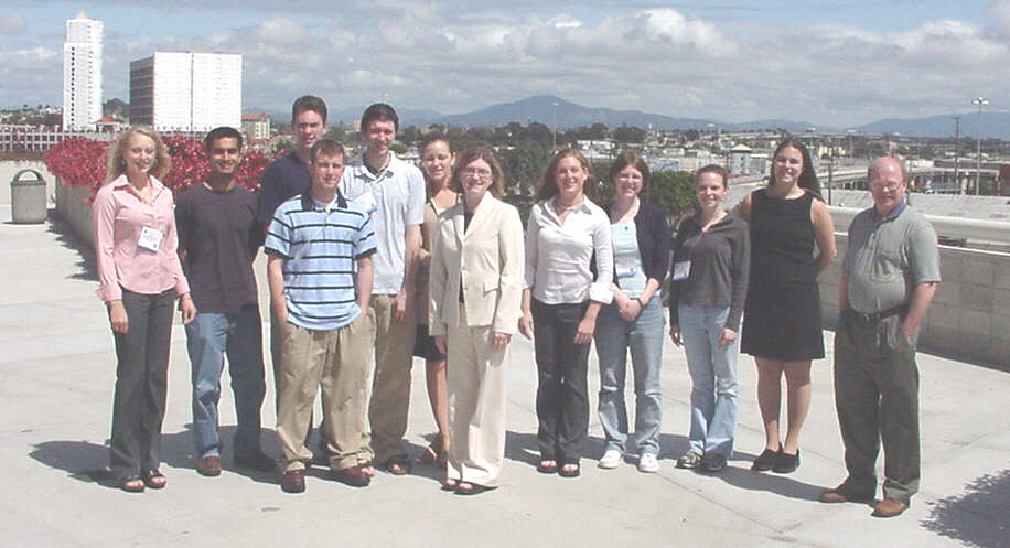
The University of Delaware Group after the Awards Ceremony for the ASBMB Undergraduate Poster Competition
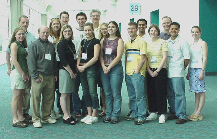 |
Danielle Skorupa, Prof. David Usher, Brandy Heckman, Jennifer Risser, Alaina Brown, Kenny Stapleford, Bevan Kirley, Erwin Puente, Karyn Oberman. Back Row from left to right:
|
The University of Delaware group included three faculty and thirteen undergraduates.
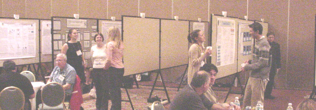
The ASBMB Undergraduate Poster Competition at the Marriott, Sunday 13 April |
 |
Sajid Noor receives the top award of from Peter Parker, representing The Biochemical Journal |

Alaina Brown, Sajid Noor, Erwin Puente, and Artie Suchow, First Place Winners in the Undergraduate Poster Competition. These four students accounted for half of the awards presented. |
Based on their abstracts, Stephanie Miller and Kenny Stapleford presented invited talks in the regular scientific sessions. This was the first time Delaware students were recognized in this way |
|
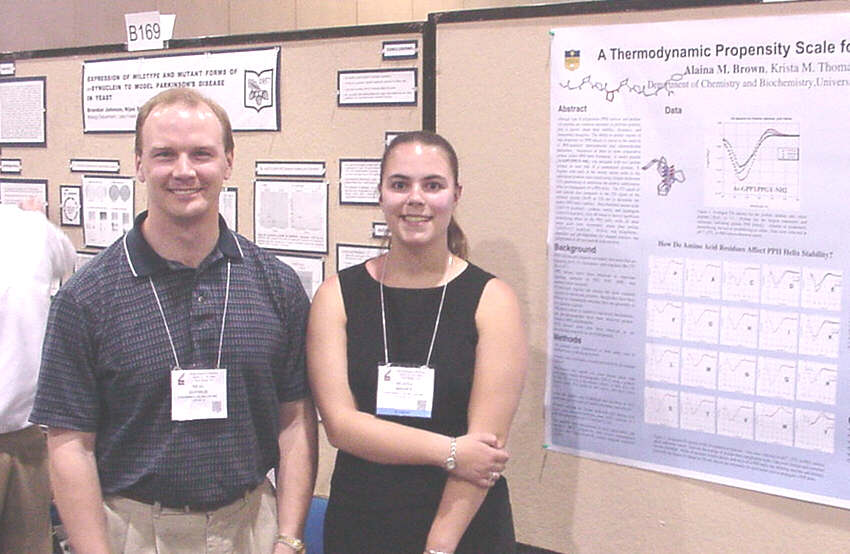
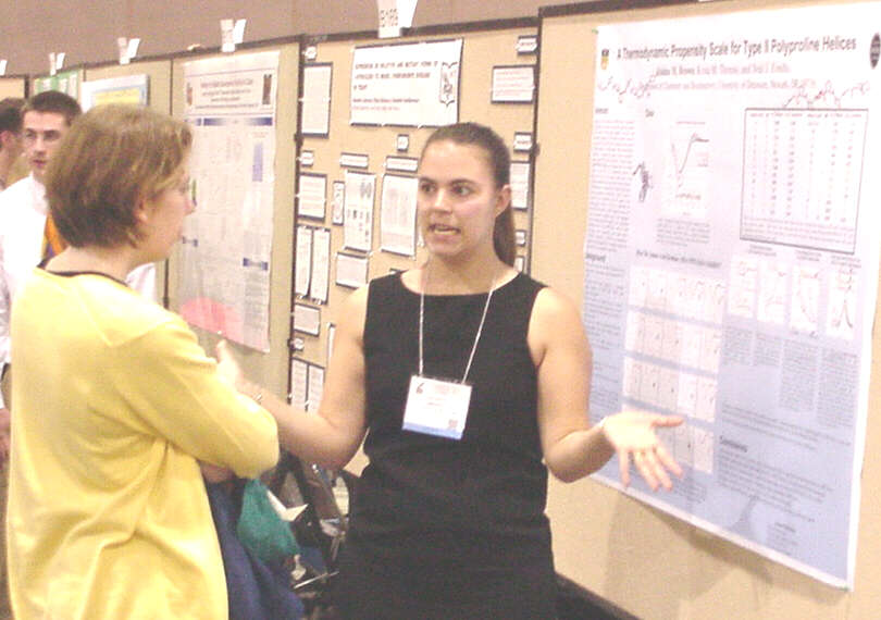
for Type II Polyproline Helices Alaina
M. Brown, Krista M. Thomas, and Neal
J. Zondlo
|
Although type II polyproline (PPII) helices and proline-rich peptides are common structures in globular proteins, little is known about their stability, dynamics, and fundamental energetics. The ability to predict regions of high propensity for PPII helices is crucial to the analysis of PPII-mediated intermolecular and intramolecular interactions. Sequences of three or more consecutive prolines induce PPII helix formation. A model peptide (Ac-GPPXPPGY-NH2) was designed with two proline residues on each side of a randomized position, X. Peptides with each of the twenty amino acids in the randomized position were tested using circular dichroism (CD) spectroscopy to determine the relative stabilization effect on propagation of a PPII helix. The CD signal of each peptide was compared to the CD signal of the reference peptide (X=P) at 229 nm to determine the relative PPII helix stability. Beta-branched amino acids (??G?1.6 kcal/mol), cysteine, serine, and asparagine (??G?1.0 kcal/mol), were all found to have a significant destabilizing effect on the PPII helix, while all other amino acids were moderately worse than proline (??G?0.3-0.7 kcal/mol). Helicity was temperature-dependant and pH-dependant for charged residues, but independent of salt and peptide concentration. This research was funded by a grant from the Howard Hughes Medical Institute and start-up funds from the University of Delaware for Dr. Zondlo. |
|
and Mutation Detection Without the Use of PCR Amplification or DNA Sequencing Brandy Heckman, Michael
Rice, Michael Usher, Eric
Kmiec
|
Escherichia coli RecA protein has been shown to catalyze the pairing of single strand DNA and double-stranded DNA molecules. The mechanism of this pairing proceeds initially through the formation of a stable nucleoprotein filament composed of RecA and a single-stranded DNA followed by assimilation into a double-stranded homologous target. The joint molecule takes the form of a triplex composed of two paired strands and a displaced strand known as a displacement (D) loop. Addition of a second single stranded molecule, complementary to the displaced strand, results in the formation of a four strand complement-stabilized double displacement (D) loop. Double D-loops can form in chromosomes, viral templates, artificial chromosomes, or plasmids and are more stable than D-loops. This complex is resistant to dissociation when the joint molecule formed has perfect complimentarity. In sharp contrast, the presence of a single mismatched base pair in one of the molecules confers a level of instability that induces dissociation. The disparate stability between these two reaction conditions enables the use of this approach to discriminate between highly homologous genomes (etc.) bearing single base heterology. Thus, we present, for the first time a protocol that can be used to identify SNPs or mutations in large DNA fragments without the need for PCR amplification or DNA sequencing. |
|
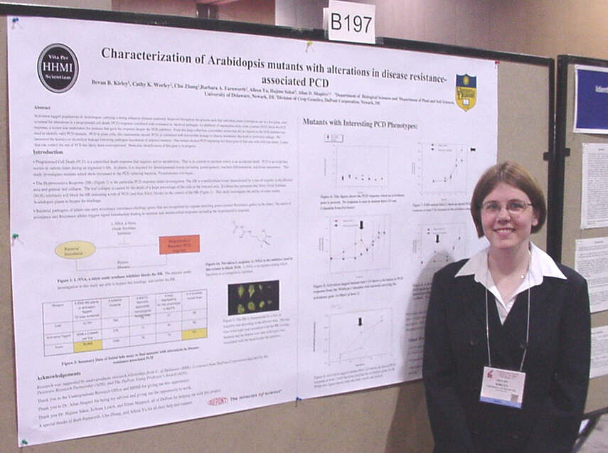
with Alterations in Disease Resistance-associated PCD Bevan
B. Kirley1, Barbara A. Farnworth2, Chu Zhang2,
|
Activation tagged populations of Arabidopsis, carrying a strong enhancer element randomly dispersed throughout the genome such that individual plants overexpress one or a few genes, were screened for alterations in a programmed cell death (PCD) response correlated with resistance to bacterial pathogen. As inhibitors of mammalian nitric oxide synthase (NOS) block this PCD response, a screen was undertaken for mutants that gave the response despite the NOS inhibitor. From this large collection, a secondary screen that did not depend on the NOS inhibitor was used to identify valid PCD mutants. PCD in plant cells, like mammalian oncotic PCD, is correlated with irreversible damage to plasma membranes that leads to electrolyte leakage. We measured the kinetics of electrolyte leakage following pathogen inoculation of selected mutants. One mutant showed PCD beginning two hours prior to that seen with wild type plants. A gene that can control the rate of PCD has likely been overexpressed. Molecular identification of this gene is in progress. Research was supported by undergraduate research fellowships from U. of Delaware (BBK) and a contract from DuPont Corporation matched by the Delaware Research Partnership (ADS). |
|
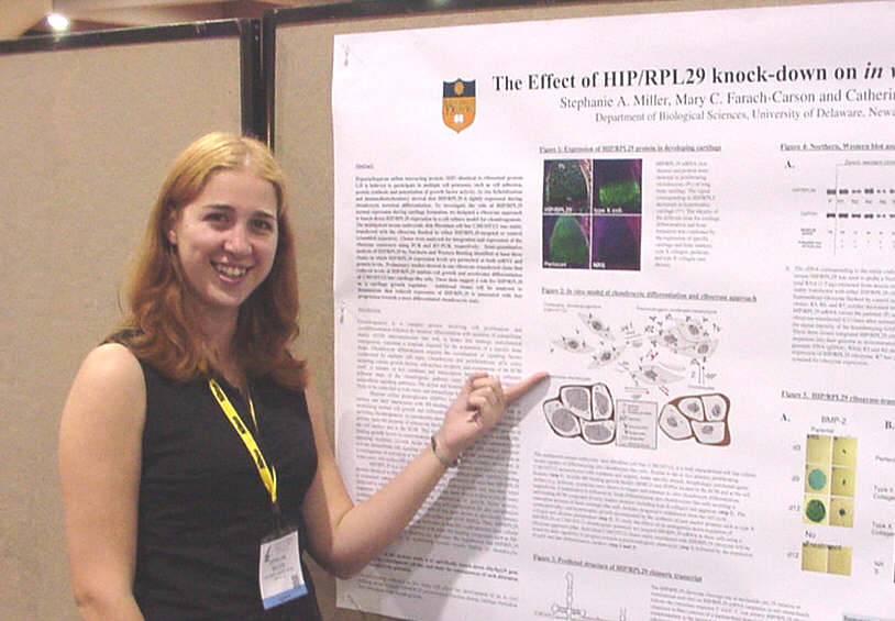
on "in vitro" Chondrocyte Differentiation Stephanie Miller,
Mary
C. Farach-Carson, and
|
Heparin/heparan sulfate interacting protein (HIP) identical to ribosomal protein L29 is believed to participate in multiple cell processes, such as cell adhesion, protein synthesis and potentiation of growth factor activity. In situ hybridization and immunohistochemistry showed that HIP/RPL29 is tightly expressed during chondrocyte terminal differentiation. To investigate the role of HIP/RPL29 normal expression during cartilage formation, we designed a ribozyme approach to knock-down HIP/RPL29 expression in a cell culture model for chondrogenesis. The multipotent mouse embryonic skin fibroblast cell line C3H/10T1/2 was stably transfected with the ribozyme flanked by either HIP/RPL29-targeted or control scrambled sequences. Clones were analyzed for integration and expression of the ribozyme constructs using PCR and RT-PCR, respectively. Semi-quantitative analysis of HIP/RPL29 by Northern and Western Blotting identified at least three clones in which HIP/RPL29 expression levels are perturbed at both mRNA and protein levels. Preliminary studies showed in one ribozyme-transfected clone that reduced levels of HIP/RPL29 inhibits cell growth and accelerates differentiation of C3H/10T1/2 into cartilage-like cells. These data suggest a role for HIP/RPL29 as a cartilage growth regulator. Additional clones will be analyzed to demonstrate that reduced expression of HIP/RPL29 is associated with fast progression towards a more differentiated chondrocytic state. |
|
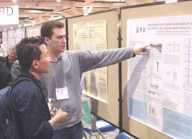
to Discriminate Between OPN Charge Forms in Biological Fluids Ian
Musselman, R. Al-Shami, D.
Carson, M.C.
Farach-Carson
|
Osteopontin (OPN) is a major non-collagenous phosphoprotein located in the bone extracellular matrix. OPN also has been found in the luminal surfaces of different glandular tissues and in many biological fluids. It is a secreted, highly acidic protein that binds to hydroxyapatite and Ca2+ in the context of mineralization and can support cell attachment/migration. Its amino acid sequence contains a conserved Gly-Arg-Gly-Asp-Ser (GRGDS) sequence, which allows it to bind effectively to the integrins. Two charge forms of OPN differing in their extent of phosphorylation have been identified in osteoblasts upon treatment with 1?, 25-dihydroxyvitamin D3. OPN-1 is the highly phosphorylated protein (pI 4.6) while OPN-2 is the less phosphorylated form (pI 5.1). It is thought that the extent of phosphorylation affects the ability of OPN to regulate crystal formation in solution. A current research focus is to establish a methodology for studying the expression of these OPN forms in human biological fluids. Examples include urine, human milk, cerebral spinal fluid, and secretions of human cell lines. Using the SELDI ProteinChip® technology, a mass spectrometry based technique, it is possible to determine a relationship between the levels of each charge form of OPN. These data then will be confirmed using Western blotting in conjunction with isoelectric focusing and 1-D and 2-D SDS-PAGE. (Supported by NIH grant HD25235 to DDC) |
|
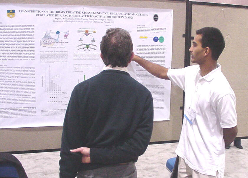
Creatine Kinase Gene in Glioblastoma Cells is Regulated by a Factor Related to Activator Protein 2 (AP2) Sajid
A. Noor, Dianna Willis, Yanping Zhang, and George
R. Molloy
|
Astrocytes maintain proper
synapse connections between neurons in the brain by regulating uptake of
extracellular glutamate and its conversion to glutamine, both of which
are energy-demanding events. Cyclic AMP (cAMP) regulates extension
of the astrocyte plasma membrane to generate processes which (i) facilitate
glutamate uptake and (ii) maintain the efficiency and number of neuronal
synapses. Our lab has shown forskolin-mediated elevations in cAMP
in U87 glioblastoma cells increased transcription of brain creatine kinase
(CKB) gene despite the absence of a cAMP response element in the CKB promoter.
CKB is essential for regenerating ATP in glial and neuronal cells.
However, the 200 bp proximal CKB promoter has four AP2 elements and transfection
experiments show that transcription of CKB in U87 cells is induced by transcription
factor AP2?, which is activated by cAMP. Mutagenesis has shown induction
of CKB by AP2? is principally mediated through the AP2 element located
at -55bp in the promoter. Nuclear extracts from U87 contain a protein
that binds to a consensus AP2 element as well as the region in CKB containing
the AP2 elements. However, this factor does not appear to be any
of the four previously-defined AP2 family members (AP2?, ?, ?, or ?) but
rather a new AP2-related factor. This may establish a possible connection
between the uptake of glutamate and its conversion to glutamine and increase
expression of CKB.
(Support provided by HHMI Undergraduate Biological Sciences Education Program, American Heart Association, and National Multiple Sclerosis Society Pilot Project) |
|
Karyn
G Oberman, Timothy R Schwartz, John M David,
|
Autosomal dominant polycystic kidney disease (ADPKD) is a common genetic disorder in humans caused by mutations in one of two genes, PKD1 or PKD2. The physiological function of the gene products, polycystin-1 and polycystin-2, is not clear, however polycystin-2 is a cation channel with a high permeability to Ca2+. RT-PCR revealed that mRNA encoding both polycystins is expressed in mouse MC3T3-E1 pre-osteoblastic cells. This finding may be related to the skeletal deformities seen in PKD1-null mice [Boulter et al., PNAS 98:12174 (2001)]. Sequence analysis revealed that the pre-osteoblast polycystin-2 mRNA is a novel, alternatively spliced variant of the PKD2 gene product expressed in kidney: a 108-base region encoding amino acids 117-152 is deleted in the pre-osteoblast transcript. Osteoblasts and adipocytes arise from a common mesenchymal precursor cell. Serial Analysis of Gene Expression (SAGE) analysis revealed expression of polycystin-1 mRNA in mouse 3T3 L1 pre-adipocytes, but expression was turned off as the cells differentiated into mature adipocytes. We currently are constructing stable MC3T3-E1 cell lines in which individual polycystin levels are knocked down using antisense technology. Funded in part by the Howard Hughes Medical Institute. |
|
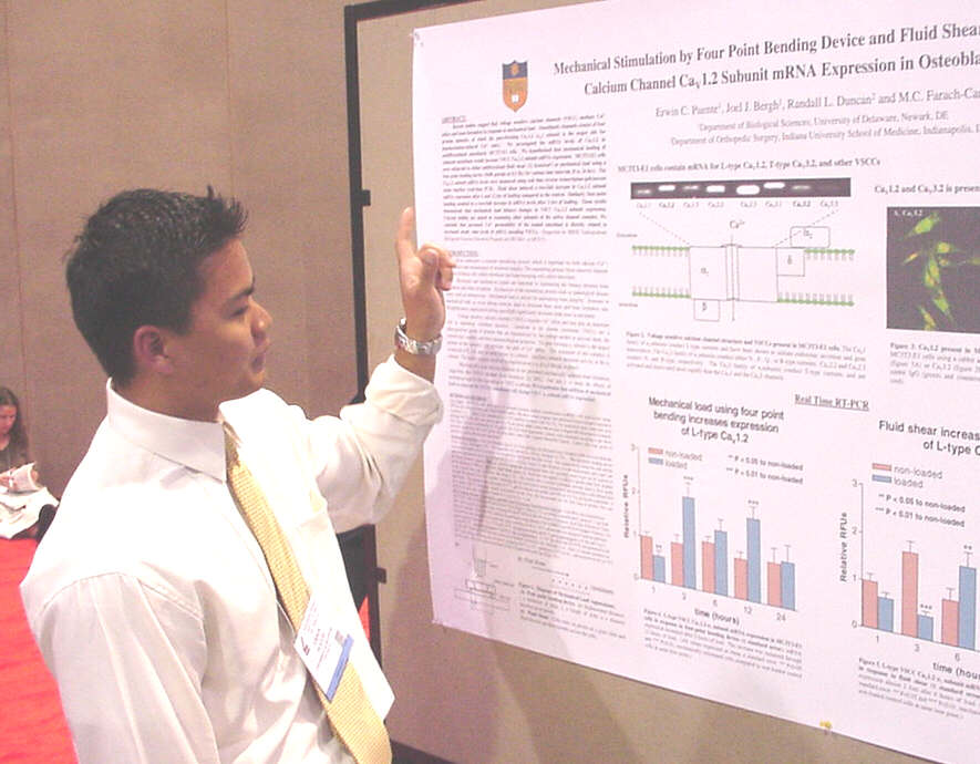
Bending Device and Fluid Shear Increase Voltage Sensitive Calcium Channel Cav1.2 Subunit mRNA Expression in Osteoblastic MC3T3-E1 Cells Erwin
C. Puente, Joel J. Bergh, Randall L. Duncan, and
|
Recent studies suggest that voltage sensitive calcium channels (VSCC) mediate Ca2+ influx and bone formation in response to mechanical load. Osteoblastic channels consist of four protein subunits, of which the pore-forming Cav1.2 (?1c) subunit is the major site for depolarization-induced Ca2+ entry. We investigated the mRNA levels of Cav1.2 in undifferentiated osteoblastic MC3T3-E1 cells. We hypothesized that mechanical loading of cultured osteoblasts would increase VSCC Cav1.2 subunit mRNA expression. MC3T3-E1 cells were subjected to either unidirectional fluid shear (12 dynes/cm2) or mechanical load using a four-point bending device (5640 µstrain at 0.5 Hz) for various time intervals (0 to 24 hrs). The Cav1.2 subunit mRNA levels were measured using real time reverse transcriptase-polymerase chain reaction. Fluid shear induced a three-fold increase in Cav1.2 subunit mRNA expression after 6 and 12 hrs of loading compared to the control. Similarly, four-point bending resulted in a three-fold increase in mRNA levels after 8 and 12 hrs of loading. These results demonstrate that mechanical load induces changes in VSCC Cav1.2 subunit expression. Current studies are aimed at examining other subunits of the active channel complex. We conclude that increased Ca2+ permeability of the loaded osteoblast is directly related to increased steady state levels of mRNA encoding VSCCs. (Supported by DE12641, to MCF-C). |
|
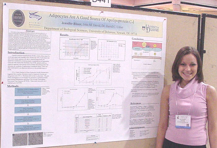
of Apolipoprotein C-I Jennifer
Lynn Risser, John M. David, and David
C. Usher
|
Serial analysis of gene expression (SAGE) used to obtain gene expression profiles of 3T3 L1 adipocytes and preadipocytes suggested that apolipoproteins A-I, A-IV, C-I, D and E were being expressed. To determine if these genes were being expressed differentially, Real-Time RT-PCR was performed. Differentiation of 3T3 L1 preadipocytes was induced by the addition of dexamethasone, 3-isobutyl-1-methylxanthine and insulin. Trizol was then used to extract RNA from cells harvested daily for two weeks. An ABI Prism 7700 with SYBR-green detection was used to quantify the relative concentrations of mRNA for the different apolipoproteins. Expression of apoA-I did not change significantly, apoE increased 6-fold between day 3 and day 6 after induction and apoD expression decreased 2-fold by day 3 and then increased 4-fold by day 6 before returning to baseline levels at day 11. Expression of apoA-IV was not found in the preadipocytes but increased about 500-fold over background by day 6. However, total mRNA levels for apoA-IV were still very low. Most significant was the increase in apoC-I expression. Expression increased over 100-fold from day 3 to 11 and was comparable to adipsin. Since mice expressing the apoC-I transgene have reduced subcutaneous and abdominal fat tissues, these results suggest that apoC-I secretion by adipocytes inhibits fatty acid uptake by these tissues. Funded by a grant from Strategic Diagnostics, Inc. |
|
Danielle
Skorupa and David
C. Usher
|
It has long been known that adipocytes are important regulators of fatty acid homeostasis. However, Serial Analysis of Gene Expression (SAGE) of 3T3 L1 adipocytes in our laboratory also indicated that they might be important in cholesterol homeostasis. Four genes involved in cholesterol transport, CD36, ABCA1, SR-B1, and SCP2, were highly expressed. To determine relative changes in gene expression during the differentiation of 3T3 L1 cells from fibroblasts to adipocytes, 3T3 L1 cells were grown to confluence and induced to differentiate by the addition of a solution containing fetal bovine serum, insulin, dexamethasone, and methylisobutylxanthine. Cells were harvested daily and the RNA was extracted with Trizol. Relative levels of CD36, ABCA1, SR-B1, and SCP2 mRNA were then determined by real-time RT-PCR using an ABI Prism 7000 Sequence Detection System and SYBR green fluorescence labeling. ABCA1 mRNA increased 5-fold between day 3 and day 6 after induction of differentiation, while SCP2, SR-B1 and CD36 increased 7, 22 and 250 fold respectively from day 3 to day 11. Since SR-B1, CD36, and ABCA1 are all receptors for HDL and SCP2 is an intracellular transporter for cholesterol, our results suggest that adipocytes play an important role in HDL cholesterol metabolism. Funded by a grant from Strategic Diagnostics, Inc. |
|
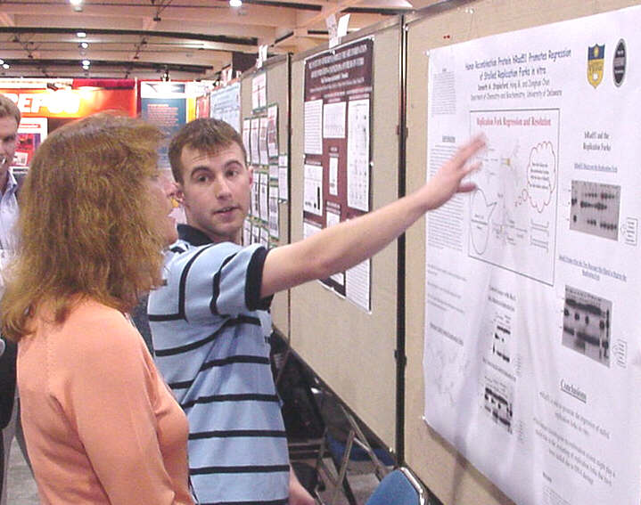
Promotes Regression of Stalled Replication Forks in vitro Kenny
Stapleford, Hong Bi, and Junghuei
Chen.
|
Homologous recombination is one of the major pathways for repairing DNA double strand breaks as well as DNA damages occurred during replication that would stall the replication fork. In prokaryotes, it has been shown clearly that recombination proteins; RecA, RecG, and RuvABC, can promote the regression of the stalled replication fork which leads to restart of the replication process. However, it has not been demonstrated that similar mechanism exists in the eukaryotic system. Using two synthesized three-stranded DNA replication forks, one with complementary sequences at the fork region and another that contains two mismatched bases at the junction of the fork to block spontaneous branch migration and in turn stall the regression of the replication fork, we find that with ATP hydrolysis, human Rad51 protein, human homolog of RecA in E. coli, can promote regression of the synthetic replication fork. Furthermore, we show that even in the presence of mismatched base pairs, human Rad51 is able to use the energy derived from ATP hydrolysis to overcome the mismatched bases and allow the fork regression to continue. Therefore, the results suggest that similar to the prokaryotic homologous recombination system, the human homologous recombination system might play a major role in restarting of the replication fork that has been stalled due to DNA damage. Funding for this project was received through the Howard Hughes Medical Institute. |
|
Arthur
T. Suckow,
Ulhas
P. Naik, Bharesh K. Chauhan, Ales Cvekl, and Melinda
K. Duncan
|
Junctional adhesion molecule (JAM-1) is a member of the immunoglobin superfamily involved in the organization of tight junctions and the regulation of leukocyte transmigration. Recently, a cDNA microarray analysis of transgenic mice overexpressing PAX-6 in lens fiber cells revealed that JAM-1 mRNA expression was 2.5 fold elevated over normal. This data suggested that JAM-1 gene expression is regulated by PAX-6, a transcription factor essential for normal eye development. The over-expression of JAM-1 in the PAX-6 transgenic lenses of adult mice was confirmed by RT-PCR. Further, JAM-1 mRNA was detected in the cornea, an epithelial tissue dependent on PAX-6 for normal morphogenesis. Immunohistochemistry revealed that the JAM-1 protein is first detected in the presumptive corneal epithelium of mice at 14.5 days post conception (dpc), coincident with the onset of PAX-6 expression and formation of the cornea from migrating neural crest cells. By 16.5 dpc, JAM-1 is also expressed in the pseudo-stratified epithelium of the salivary glands, the epidermis of the skin, the conjuctiva and the fusion line between the upper and lower eyelids. High levels of JAM-1 protein continue to be found in the corneal epithelium and conjuctiva of adult mice. Since JAM-1 binds to ZO-1 and recruits PAR-3 to tight junctions, it may be essential for normal morphogenesis and maintenance of the corneal epithelium. |
|
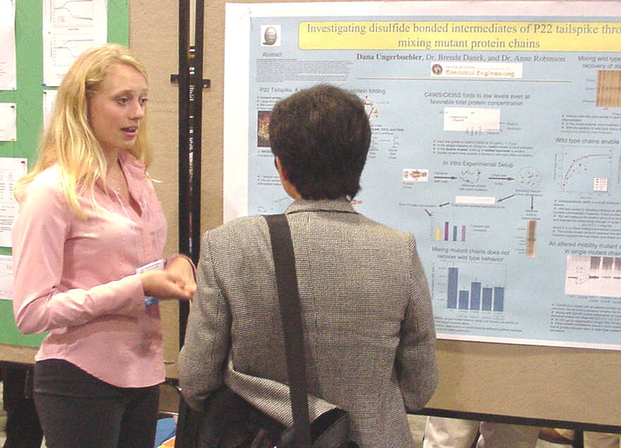
Interactions of P22 Tailspike Dana
Ungerbuehler and Anne
Skaja Robinson
|
Misfolded and aggregated proteins are implicated in several disorders such as Alzheimers, Cystic fibrosis, and prion diseases. The lack of productive folding is thought to play a major role in the progression of these diseases, and some treatments are being sought to minimize misfolding or maximize the yield of functional protein. The kinetics of protein folding are of great interest in biochemical engineering and pharmaceutical industries, and are extremely complex to model in vivo. P22 tailspike is a protein-folding model due to its complex molecular interactions and oligomeric structure. While the native trimeric structure is not disulfide bonded, evidence exists for a folding intermediate with oxidized sulfhydryl groups. A C-terminal truncation revealed that the cysteines at 496, 613, and 635 were the most likely sites of sulfhydryl activity. Cysteine to serine point mutations at the 496, 613, and 635 residues were purified in trimer form. Initial analysis of the single serine mutants revealed that they folded 2-3 times slower than wild type and with only 65-80% of wild type yields. In vitro refolding studies were performed by mixing different single mutants to investigate if wild type yield and assembly kinetics could be recovered. Combining single mutant chains indicated some qualitative improvement in the yields, however the kinetics of folding appeared to remain unchanged. Mixing wild type with the single mutant and double mutant chains revealed a recovery of folding intermediates trapped in the mutants. This indicates that molecular interactions between chains are able to rescue stalled intermediates. Thus, allowing mixed chain interactions do not produce more rapid folding, yet may produce more efficient folding. Two double cysteine mutants and one triple cysteine mutant were also created. Only one of the double mutants, C496S/C635S, folded into the native trimer. The yields of this tailspike mutant were significantly reduced, however it formed trimer at the rates of the single mutants. Monomer was overpopulated in the double mutant, whereas dimer is the dominant intermediate in the single mutants. This suggests that the limiting cysteine residue interactions occur in chain assembly of the folding intermediates. |
Miscellaneous Photos. Click on image for enlargement.
Alaina Brown with her research supervisor, Prof. Zondlo. |

Checking in upon arrival at Quality Inn. |

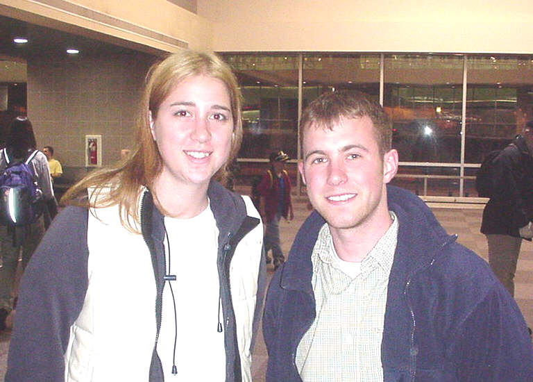
University of Delaware group at San Diego Airport. |

Headed home at the San Diego Airport |

Kenny Stapleford with Phillip Ortiz, principal organizer of the ASBMB Undergraduate Poster Competition. |
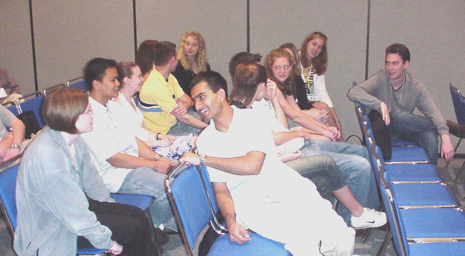
University of Delaware group awaiting the awards ceremony. |

Stephanie Miller presenting her talk in the scientific sessions. |
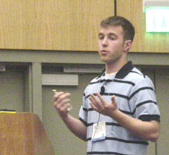
Kenny Stapleford presenting his talk in the scientific sessions. |

Award winners and competition organizers. |
University of Delaware group in sunny southern California. |
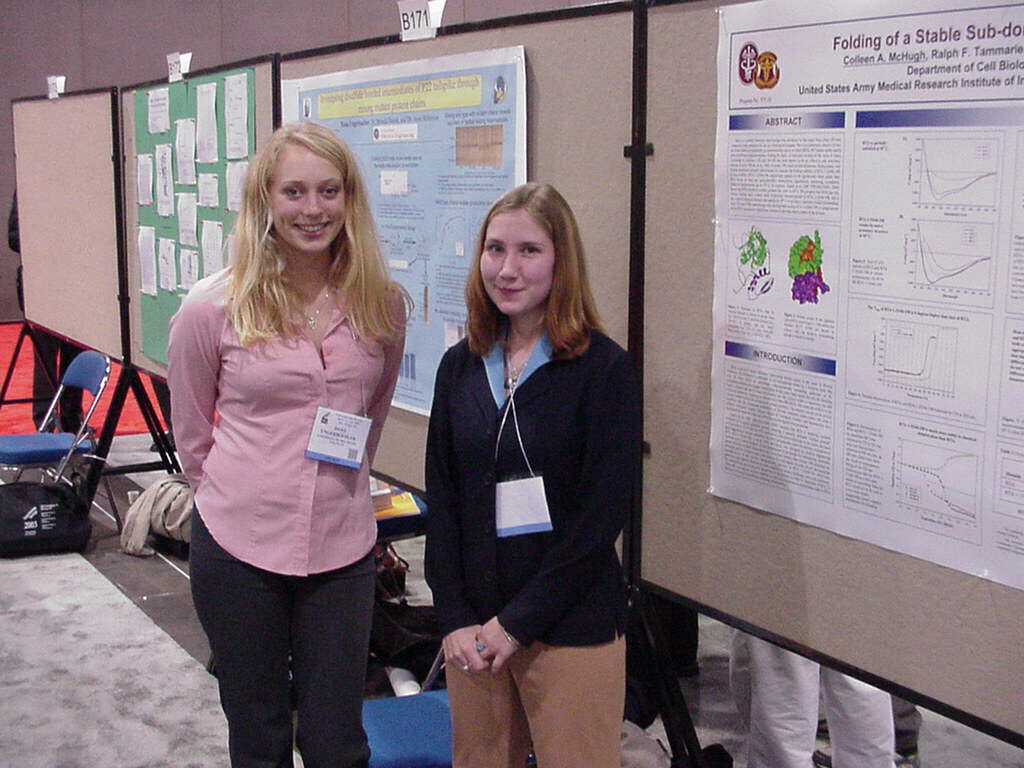
Dana Ungerbuehler with a long lost friend from grade school. |
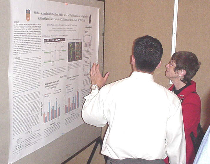
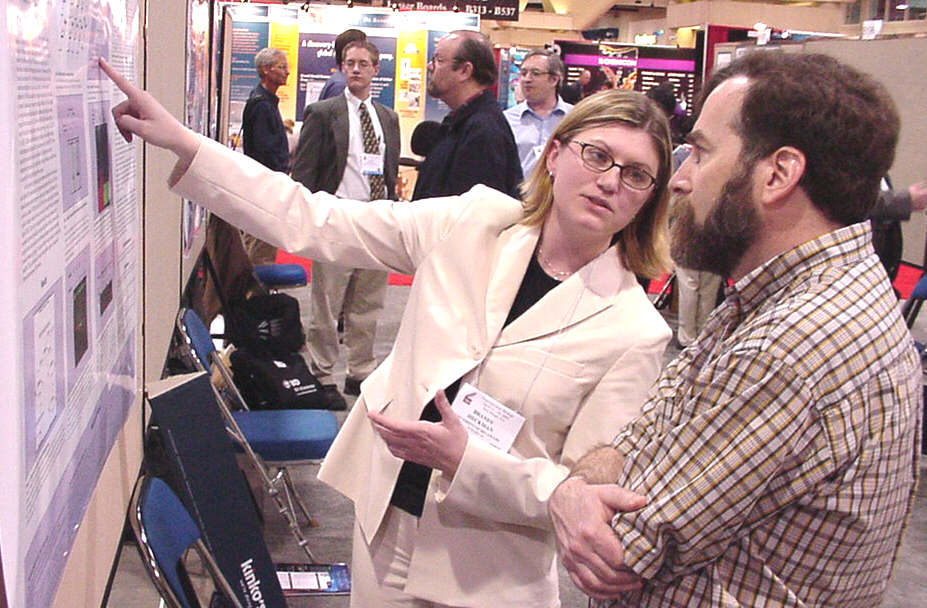
Erwin Puente describing his work to judge Judith Voet. |
The trip to the Experimental Biology Meetings
in San Diego was organized by the University of Delaware HHMI Undergraduate
Science Education Program with additional support from travel grants from
the American
Society for Biochemistry and Molecular Biology, the Beckman Scholars
Program, and the Women Scholars Program. The HHMI
Program, the Beckman
Scholars Program, Charles Peter White Fellowships, the NIH BRIN Program,
and the Undergraduate Research Program
supported research by the students.
 .
.