CELL PHYSIOLOGY

    Welcome to the Cell Physiology (BISC 305) Webpage. This site will have information about the course syllabus, news about the class, upcoming seminars of interest, and group problems that may be assigned in class. In addition, there will be a brief outline of material covered before each exam, and a sample exam to get acquainted with the style of questions used.


FLUORESCENCE MICROSCOPY OF MICROTUBULES

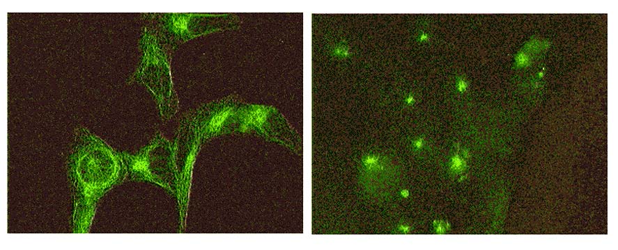
      Microtubules are one of the major components of the cytoskeleton of cells. Microtubules function both to provide stability to cells, and, more importantly, as "railroad tracks" for the directed movement of cellular organelles, vesicles, etc to specific locations within the cell. The figures below are images taken from one of our lab sections last year, using a monoclonal antibody to beta-tubulin and an FITC-conjugated seconday antibody. The cells are LLC-pK epithelial cells, a kidney cell line, grown on glass coverslips. Cells were permeabilized with detergent/stabilization buffer, fixed with formalin and extracted with cold acetone before antibody staining. On the left are control, untreated cells, showing an extensive microtubule network throughout the cells. On the right, cells were preincubated for one hour at 4 degrees C to de-polymerize the microtubules. The initial recovery/regrowth of the MTs demonstrates the localization of the centrosome or MTOC in these cells.


SPRING 2002 SYLLABUS

BISC 305, CELL PHYSIOLOGY

SPRING, 2002 COURSE DESCRIPTION AND GUIDELINES

GENERAL GOALS:  This course will concentrate on dynamic processes within cells, between cells  and between cells and their environment. The major areas that will be covered are: cell membranes, cell junctions and adhesion to the extracellular matrix, cytoskeleton and cell motility, signal transduction processes, protein trafficking, vesicular transport in cells and cell cycle. For each topic we will try to incorporate the most recent information from the primary research literature. An understanding of the dynamic nature of these processes will be a major goal. There will also be an emphasis on experimental approaches and findings. Finally, students will be asked to demonstrate critical analysis of research findings.

INSTRUCTOR:  Gary Laverty, Associate Professor, 205/247 Wolf Hall, 831-8180
                                      (laverty@udel.edu)

GTA: Kimberly Robinson, biokim@udel.edu

OFFICE HOURS: M,W, 9:30-11:00;  F, 14:00-16:00 (or whenever I am in the office)

 LECTURES: T,R 11:00-12:15, 205 Kirkbride

 COURSE WEBSITE: http://www.udel.edu/Biology/laverty/cells.html

 TEXT: Karp, "Cell and Molecular Biology. Concepts and Experiments" 3rd Ed, Wiley.

 GRADES:  Final grades will be based on a 300 point scale, as follows:

                                             Exam I (in class)                               100 points
                                             Exam II (finals week)                         100 points
                                             In-class quizzes (3 @ 20)                    60 points*
                                             Research Assignment                         40 points#
                                              ___________________________________
                                                           TOTAL                               300 points

          * In-class quizzes: 10-15 minute quizzes will be given on the following dates:
Feb 21; March 12 and April 23

        # Research Assignment: details to be announced


CLASS SCHEDULE

     DATES             TOPICS                                                              CHAPTER/PAGES

    Feb 5                 Introduction                                                                 1 (1-30)
                                      (also review Chapters 2 and 3 as needed)

    Feb 7, 12           Biomembranes, composition, structure, fluidity           4 (122-150)

    Feb 14, 19         Membrane transporters and channels                          4 (150-165)
                                                                                                                   (173-177 EP)

    Feb 21*, 26,      Cytoskeleton I: IFs and MTs                                          9 (333-366)
     28                                                                                                     14 (590-607)

    March 5, 7         Cytoskeleton II: actin and cell motility                           9 (366-392)

    March 12*,        Cell signaling                                                               15 (628-665)
     14, 19, 21, 26

    March 28         MID-TERM EXAM (in class)

    April 2, 4           Spring Break!

    April 9, 11         Cell surface, extracellular matrix, adhesion                  7 (243-272)
      16, 18             integrins, cell junctions                                               15 (654-658)

    April 23*,         Cell compartments, protein trafficking                          8 (279-329)
      25, 30,             endoplamsic reticulum, Golgi, lysosomes
      May 2, 7            endocytosis

    May 9, 14         Cell cycle                                                                     14 (580-590)


REVIEW FOR MIDTERM EXAM
(Biomembranes, Transport and Cytoskeleton)

I. Membranes
II. Transport Systems
III. Cytoskeleton: general functions, 3 types of structural elements
A. Intermediate Filaments
        1. extended, rope-like filaments with conserved helical regions
        2. families: keratins, Vimentin-like, Neurofilaments, Lamins
        3. Role of nuclear lamins in mitotic disassembly/reassembly of nuclear envelope
B. Microtubules
        1. polymers built from dimeric subunits (alpha/beta tubulin), 13 protofilaments
        2. Role of GTP binding/hydrolysis
        3. polarity of MTs
        4. In vitro behaviors (treadmilling, gliding, organelle movement, shearing, etc)
        5. dynamic instability (slow growth/ catastrophic disassembly): role of GTP cap
        6. Interphase MTs
                a. MTOC/centrosome: role as nucleation site (minus ends); gamma tubulin
                b. structure of centrioles/basal bodies
                c. MAPs help to stabilize MTs and crosslink
                d. MT motors -role in intracellular trafficking of vesicles, organelles
                        1. kinesins (plus end): structure and function (eg, fast axonal transport)
                                ATPase, MT binding domains, cargo binding
                        2. cytoplasmic dyneins - minus end directed
                        3. "walking" behavior; ATP cycle (dissociation, hydrolysis, movement, etc)
        7. MTs in cilia and flagellae (axonemes): "9 + 2" organization of MTs
                a. role of stabilization/crosslinking proteins (eg nexin, etc)
                b. ATP cycle: changes in MT affinity, movement of head groups, etc
                c. axonemic dynein creates shearing forces translated into sliding
        8. MTs in the mitotic spindle apparatus
                a. mitotic stages: prophase, prometaphase, metaphase, anaphase, telophase
                b. 3 types or arrangements of MTs: polar, kinetochore, aster
                c. role of kinesin related proteins (KRPs) and dyneins
                        1. separation of centrosomes in early prophase
                        2. anchoring of MTs to spindle "poles" within centrosome
                        3. side capture of chromosomes (walking to + ends
                        4. "congress" of chromosomes during early metaphase (alignment) and anchoring
                                of + ends to kinetochore (attachments stable only under tension)
                        5. Exp showing MTs depoymerize from + end; eploymerization may provide
                            poleward driving force alone
                        6. separation of poles during anaphase
                c. structure and function of kinetochores
                        1. metaphase/anaphase checkpoint: Mad-2; exp showing even one unattached
                            chromatid generates "wait" signal; tension on both sides of chromatid pair
                            releases Mad2
                        2. kinetochore MTs shorten at + end (anaphase A)
                        3. polees move apart (anaphase B) due to forces and elongation at polar MTs
C. Actin Microfilaments
        1. polymer structure, polarity of filaments treadmilling behavior, etc
        2. Examples of actin organization in cells: microvilli, contractile ring (cytokinesis)
                adhesion belts, cytoplasmic streaming, cell cortex and stress fibers
        3. Drugs: cytochalasins, phalloidin
        4. Actin binding proteins
            a. nucleating proteins: Arp2/3
            b. monomer binding proteins: thymosin
            c. capping proteins : Cap-z
            d. polymerizing-promoting: profilin (acts to accelerate ADP/ATP exchange and orients
                subunits to polymer
            e. depolymerizing and severing: gelsolin
            f. cross-linking: filamen (orthagonal arrays); villin (tight bundles)
        5 . Actin motors (myosins)
            a. myosin II (conventional): forms dimers and bipolar filaments
            b. myosin I: monomer binds membranes, cargo
            c. myosin V: binds cargo, walks in "big" steps without twisting
        6. Cell crawling: lamellipod extension, adhesion, traction (force generation), tail retraction
              a. Actin polymerization drives lamellipod extension
               b. recruitment of Arp2/3 to leading edge and to branch points along filaments
               c. stabilize cross links with filamen
               d. break down filaments behind leading edge (gelsolin); profilin recharges and carries
                    subunits to leading edge (actin filaments "treadmill")
               e. myosins I and V carry membrane vesicles, Arp2/3 and profilin-actin complexes to
                    leading edge
               f. myosin II creates tension around adhesion points (stress fibers)
               g. spatial coordination (tail retractions, etc); membrane recycling, integrin insertion
                    in front
IV. Cell Signaling
        A. Cell-specific responses: enzyme activation, gene transcription, channels, cytoskeleton
        B. Receptors: channels, G-protein coupled, receptor tyrosin kinases
        C. G-protein coupled receptors: (eg epinephrine, acetylcholine, ADH, PTH etc)
                1. 7  transmembrane spans
                2. cytoplasmic domains interact with trimeric G proteins---GDP release
        D. G proteins: monomeric vs trimeric
                1. active with GTP bound; inactive with GDP bound
                        a. GDP to GTP exchange activates
                        b. GTP hydrolysis inactivates
                2. trimeric: beta/gamma subunits inhibit alpha subunit activity
                        a. activated GPCRs cause GDP/GTP exchange
                            release of alpha subunit (active)
                        b. intrinsic GTPase activity slowly inactivates (reassociation of subunits)
                3. Monomeric (Ras family)
                        a. require GEFs (guanine nucleotide exchange factors) to help release GDP
                            (act as activators)
                        b. require GAPs (GTPase activating proteins) to help hydrolyze GTP
                            (act as inactivators)
        E. Kinases vs Phosphatases (phosphorylation and dephosphorylation of proteins acts as
            switching mechanism: allosteric hanges in proteins
        F. Enzyme cascades
                1. allows for amplification of downstream reactions
                2. allows for branch points (divergence of cellular responses to one signal)