

Shedding new light on infertility
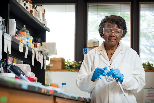
OUR FACULTY | We’ve all heard about the birds and the bees. But it’s what UD’s Patricia A. Martin-DeLeon is learning from mice that might help us understand the intricacies of infertility on another level.
DeLeon has studied reproductive biology for decades and has witnessed the critical point of conception—where the sperm finally enters the egg—many times in her studies of fertility in mice, the closest genetic model to humans (and with a much faster reproductive cycle).
It’s what happens right before this pivotal interaction that DeLeon and her team have discovered: The sperm gets some assistance.
This previously unknown exchange with microscopic particles from the fallopian tube “helps prepare the sperm for its big push into the egg,” says DeLeon, the Trustees Distinguished Professor of Biological Sciences at UD.
Sperm, meet egg
But to understand what makes this interaction so important, we must first begin with the basics.
The fertilization process starts with the egg: Once it is released from an ovary and enters the fallopian tube, the hair-like cilia that line this tiny tube sweep it toward the uterus. While in the fallopian tube, the egg will either meet the sperm and be fertilized (within a 12- to 24-hour window), or they won’t meet, and the egg will dissolve.
DeLeon and her UD team have identified previously unknown particles in the secretions from the fallopian tube that help the sperm get ready for its all-important drive into the end zone. They termed these bodies “oviductosomes,” combining “oviduct” (another name for the fallopian tube) and “exosomes” (small sacs found in all bodily fluids to help cells survive).
Hormones trigger the release of these oviductosomes, which are only 100 nanometers in diameter. The tiny cargo-filled sacs then attach to the sperm like decorations on a Christmas tree, before the sperm fuses with the egg.
Once these sacs are in place, they transfer special proteins to the sperm for use when the time is right. One of those proteins includes a “calcium clearance pump,” which the sperm uses to eliminate elevated levels of calcium to prevent a buildup of nitric oxide, which can cause increased amounts of free radicals and DNA damage. While removing potentially elevated calcium levels, the pump also takes in essential hydrogen ions from the extracellular space, providing an environment that is optimal for its final, critical push into the egg to start the zygote’s life.
IVF potential
Discovering these oviductosomes provided the researchers a window into the cargo being delivered by the female to the sperm.
Sperm doesn’t land into the egg unassisted, they found; the oviductosomes serve as members of the fertilization flight crew, lightening the load and ensuring a safe delivery.
This finding could help improve IVF, or in vitro fertilization, which currently has a 32 percent success rate.
If oviductosomes aren’t currently present in the IVF process, the researchers believe, perhaps they can be harvested to assist in the sperm-egg introduction.
“We’ve shown that these oviductosomes are carrying critical molecules that include not only proteins, but also nucleic acids such as RNA and lipids,” DeLeon says. “That gives us hope they can be used as vehicles for improving fertility and the chances of producing healthy embryos and offspring.”
Currently, DeLeon and her team are analyzing the protein-rich contents of this cargo to find out exactly what gives the sperm what it needs for its last push to penetrate the egg—always head first, tail out—to fertilize it.
“Our work may lead to the discovery of genes and gene products that cause infertility,” DeLeon says of the team’s continuing research. “We may identify proteins required to improve the efficiency of IVF, and improve the outcome and health of the offspring. It’s really another step in the direction of personalized medicine.”
The power of technology
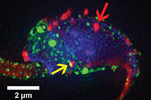
DeLeon and her team made their novel discovery using a high-powered, three-dimensional super-resolution microscope that UD acquired in 2012. The technology, whose inventors won the Nobel Prize in 2014, can illuminate what’s happening in a cell, right down to a single molecule.
At 10 millionths of an inch wide, the oviductosomes from a female mouse were pre-labeled with a fluorescent dye and incubated together with the sperm.
Within an hour, the oviductosomes were fused to the sperm’s surface. After two to three hours, the oviductosomes continued to accumulate, primarily on the sperm’s head and the midpiece of its tail.
Integrins, which are membrane receptors on both the sperm and the oviductosomes, helped to facilitate their bonding, along with fusion stalks on the sperm’s surface.
DeLeon is seeking funding for the next phase of her research, which aims to examine the microRNA and RNA molecules as well as the protein population present in the cargo of oviductosomes.
A leader in the field
DeLeon has been making major contributions to reproductive science since she published her first scientific article—“Cannabis and Chromosomes,” examining the impact of marijuana on embryonic cells—in The Lancet in 1969, as a master’s student at the University of the West Indies in her native Jamaica.
The response to that paper was so dramatic, with requests for copies from scientists around the world, that DeLeon set her sights on a career in research and teaching and never looked back, continuing on for her doctorate at the University of Western Ontario. Her post-doctoral studies were done at McGill University. She joined the UD faculty in 1976.
DeLeon’s co-authors on the study include Amal A. Al-Dossary, AS14PhD, now a professor at the University of Dammam in Saudi Arabia; Pradeepthi Bathala, AS18M; and Jeffrey Caplan, director of the BioImaging Center at the Delaware Biotechnology Institute.
Article by Tracey Bryant