Javascript is required to view this video

ADVERTISEMENT
- Rozovsky wins prestigious NSF Early Career Award
- UD students meet alumni, experience 'closing bell' at NYSE
- Newark Police seek assistance in identifying suspects in robbery
- Rivlin says bipartisan budget action, stronger budget rules key to reversing debt
- Stink bugs shouldn't pose problem until late summer
- Gao to honor Placido Domingo in Washington performance
- Adopt-A-Highway project keeps Lewes road clean
- WVUD's Radiothon fundraiser runs April 1-10
- W.D. Snodgrass Symposium to honor Pulitzer winner
- New guide helps cancer patients manage symptoms
- UD in the News, March 25, 2011
- For the Record, March 25, 2011
- Public opinion expert discusses world views of U.S. in Global Agenda series
- Congressional delegation, dean laud Center for Community Research and Service program
- Center for Political Communication sets symposium on politics, entertainment
- Students work to raise funds, awareness of domestic violence
- Equestrian team wins regional championship in Western riding
- Markell, Harker stress importance of agriculture to Delaware's economy
- Carol A. Ammon MBA Case Competition winners announced
- Prof presents blood-clotting studies at Gordon Research Conference
- Sexual Assault Awareness Month events, programs announced
- Stay connected with Sea Grant, CEOE e-newsletter
- A message to UD regarding the tragedy in Japan
- More News >>
- March 31-May 14: REP stages Neil Simon's 'The Good Doctor'
- April 2: Newark plans annual 'wine and dine'
- April 5: Expert perspective on U.S. health care
- April 5: Comedian Ace Guillen to visit Scrounge
- April 6, May 4: School of Nursing sponsors research lecture series
- April 6-May 4: Confucius Institute presents Chinese Film Series on Wednesdays
- April 6: IPCC's Pachauri to discuss sustainable development in DENIN Dialogue Series
- April 7: 'WVUDstock' radiothon concert announced
- April 8: English Language Institute presents 'Arts in Translation'
- April 9: Green and Healthy Living Expo planned at The Bob
- April 9: Center for Political Communication to host Onion editor
- April 10: Alumni Easter Egg-stravaganza planned
- April 11: CDS session to focus on visual assistive technologies
- April 12: T.J. Stiles to speak at UDLA annual dinner
- April 15, 16: Annual UD push lawnmower tune-up scheduled
- April 15, 16: Master Players series presents iMusic 4, China Magpie
- April 15, 16: Delaware Symphony, UD chorus to perform Mahler work
- April 18: Former NFL Coach Bill Cowher featured in UD Speaks
- April 21-24: Sesame Street Live brings Elmo and friends to The Bob
- April 30: Save the date for Ag Day 2011 at UD
- April 30: Symposium to consider 'Frontiers at the Chemistry-Biology Interface'
- April 30-May 1: Relay for Life set at Delaware Field House
- May 4: Delaware Membrane Protein Symposium announced
- May 5: Northwestern University's Leon Keer to deliver Kerr lecture
- May 7: Women's volleyball team to host second annual Spring Fling
- Through May 3: SPPA announces speakers for 10th annual lecture series
- Through May 4: Global Agenda sees U.S. through others' eyes; World Bank president to speak
- Through May 4: 'Research on Race, Ethnicity, Culture' topic of series
- Through May 9: Black American Studies announces lecture series
- Through May 11: 'Challenges in Jewish Culture' lecture series announced
- Through May 11: Area Studies research featured in speaker series
- Through June 5: 'Andy Warhol: Behind the Camera' on view in Old College Gallery
- Through July 15: 'Bodyscapes' on view at Mechanical Hall Gallery
- More What's Happening >>
- UD calendar >>
- Middle States evaluation team on campus April 5
- Phipps named HR Liaison of the Quarter
- Senior wins iPad for participating in assessment study
- April 19: Procurement Services schedules information sessions
- UD Bookstore announces spring break hours
- HealthyU Wellness Program encourages employees to 'Step into Spring'
- April 8-29: Faculty roundtable series considers student engagement
- GRE is changing; learn more at April 15 info session
- April 30: UD Evening with Blue Rocks set for employees
- Morris Library to be open 24/7 during final exams
- More Campus FYI >>
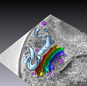
2:04 p.m., May 12, 2010----Cells can take minutes, or even hours, to die -- when preserved using standard chemical fixation and for decades this was the best available method to researchers. However, modern cryogenic methods of preservation nearly instantaneously stop all biological processes -- within milliseconds -- while maintaining the most life-like view of the cell.
The beginning of April marked the first University-wide cryo-preservation workshop for students, University of Delaware professors, and investigators from DuPont Pioneer and Johns Hopkins University.
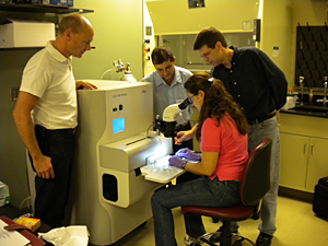
Hosted by the Delaware Biotechnology Institute's (DBI) Bio-Imaging Center, led by Kirk Czymmek, participants were able to test over $375,000 worth of microscopy-related instruments on loan from by Leica Microsystems, which also sponsored the workshop and coordinated outside speakers for the event.
“To conduct proper and efficient research, we need to avoid 'artifacts' which can lead to false or misleading representations of what is happening inside a cell,” said Czymmek, associate professor in UD's Department of Biological Sciences. “Cryotechniques provide the most life-like view of cells and are considered the gold-standard for rapidly immobilizing cells for microscopy. A workshop like this provides our students with the knowledge and hands-on experience to work with the best instrumentation available.”
A few of the technologies on hand during the three-day Cryo-Sample Preparation and Correlative LM-EM Workshop from April 6-8 were the HPM100 High Pressure Freezer, EMPACT High Pressure Freezer, the Grid Plunger and an automated Immuno-Gold Labeling System.
The freezers are able to rapidly freeze samples with liquid nitrogen, without changing the arrangement of molecules within the sample. Once samples are frozen -- tissues, cultured cells, plants, yeast, fungi and hydrogels -- they can be cut into very thin slices by a diamond and then viewed under a microscope. And because they are in their closest-to-natural state, scientists can gain a much better understanding of how cells and organisms work and respond to their environment.
“We routinely work with key institutions and labs because it benefits us all in terms of developing products and advancing research,” said Ann Korsen, director of sales and marketing for nanotechnology at Leica Microsystems.
Students were able to bring personal samples to the workshop to process, including plants, yeast and other fungi, bacteria, fruit fly embryos, and mammalian tissue.
“DBI's Bio-Imaging Center and team have been instrumental in the progression of my research interests by incorporating state-of-the-art technology and instrumentation, novel microscopic techniques, and unparalleled assistance from multiple institutions and collaborators,” said Carissa Young, a doctoral candidate in the Department of Chemical Engineering at UD.
As the University continues with its strategic plan to position UD as one of the best public institutions in America, the DBI Bio-Imaging Center is following along with vigor.
“It is an exciting time to be at the University of Delaware as it moves forward with its Path to ProminenceTM. To support the University's strategic plan, our researchers will need access to the same sophisticated technologies other leading institutions have in order to be competitive,” said Czymmek. “Technologies for examining cells at high-resolution are changing rapidly, and we hope to augment existing equipment at UD based on what we've learned during the workshop.”
Article by Laura Crozier


