 |
18th World Congress of Soil Science
July 9-15, 2006 - Philadelphia, Pennsylvania, USA |
 |
Metal contamination of surface and subsurface environments is a problem worldwide. Unique metallophyte plants (hyperaccumulators) accumulate high concentrations of trace metals in their harvestable biomass, and thereby offer a sustainable method for treatment of metal-contaminated sites (phytoremediation) and an opportunity to mine metal-rich soils (phytomining). Several species of
Alyssum (
Brassicacea family) have the ability to simultaneously hyperaccumulate Ni and Co in a mixed-contaminant system. Information on the localization, speciation, and associations of accumulated metals with other elements in hyperaccumulator plants can provide insight into the physiological and biochemical mechanisms of metal tolerance and accumulation. Synchrotron-based techniques such as microfocused X-ray absorption fine structure (XAFS) and X-ray fluorescence (XRF) spectroscopy and computed microtomography (CMT) enable the investigation of metal reactions and processes in natural systems at the micron scale. SXRF and CMT were utilized to observe
in situ metal localization and compartmentalization in the Ni/Co hyperaccumulator
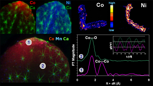
A. murale. SXRF images revealed preferential localization of Co at leaf tips and margins and a relatively uniform Ni distribution in
A. murale leaves. CMT cross-sectional images indicated aqueous Co was primarily limited to the plant vascular system, while Ni was enriched in vascular and epidermal tissue. Cobalt speciation in plant tissue was investigated
in situ with bulk and µ-XAFS. Primary Co species in the roots and leaves were aqueous metal complexes with organic and amino acids (e.g. malate and histidine). Preliminary µ-XAFS investigations indicated the Co deposited at leaf tips and margins formed a Co(OH)
2•
n H
2O precipitate with some degree of short range order. Results suggest different metal detoxification and sequestration mechanisms are utilized by
A. murale to tolerate elevated concentrations of Co and Ni in plant tissue.
Image: (upper row) Co and Ni µ-SXRF images of a leaf from metal hyperaccumulator A. murale (Note the stellate leaf trichomes depicted in the Ca channel) plus Co and Ni CMT (tomograms) cross-sectional images; (bottom row) Tri-color µ-SXRF image (Co, Mn, and Ca) of A. murale leaf plus Co-XAFS k3-weighted chi (inset) and the corresponding Fourier transforms (FT) for leaf tip and mid-leaf regions (spots 1 and 2 on the tricolor µ-SXRF map).


