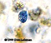
| The cyst of Chilomastix mesnili, shown on the right, is pear-shaped and measures 4 to 6 µm wide and 6 to 10 µm long. There is a single nucleus and a curved cytostomal fibril called the shepherd's crook. The image at right is a trichrome stain (1000x). | 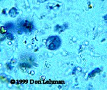 |
 |
The trophozoites of C. mesnili are also pear-shaped and measure from 6 to 24 µm in length and 4 to 8 µm wide. The single nucleus usually has a prominent karyosome. The anterior flagella are difficult to see. The oral groove (cytostome) is sometimes seen near the nucleus. The image on the left is an iron hematoxylin stain (1000x). |
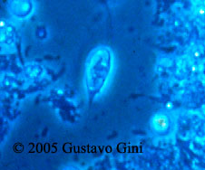
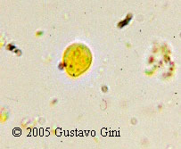
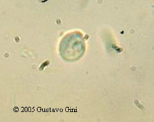
| Wet Mounts The image at right is a wet mount of a trophozoite viewed by phase-contrast microscopy. The image below left is an iodine stained wet mount of a cyst. The image right is unstained wet mount of a cyst, the plane of focus is on the nucleus. Images courtesy of Gustavo Gini. |
 |
 |
 |
Copyright 2008 Don Lehman