 |
 EB2016 EXPERIMENTAL BIOLOGY MEETINGS San Diego, CA APRIL 1-6, 2016  |
 |
|
|
EB2016 EXPERIMENTAL BIOLOGY MEETINGS San Diego, CA APRIL 1-6, 2016 |
|
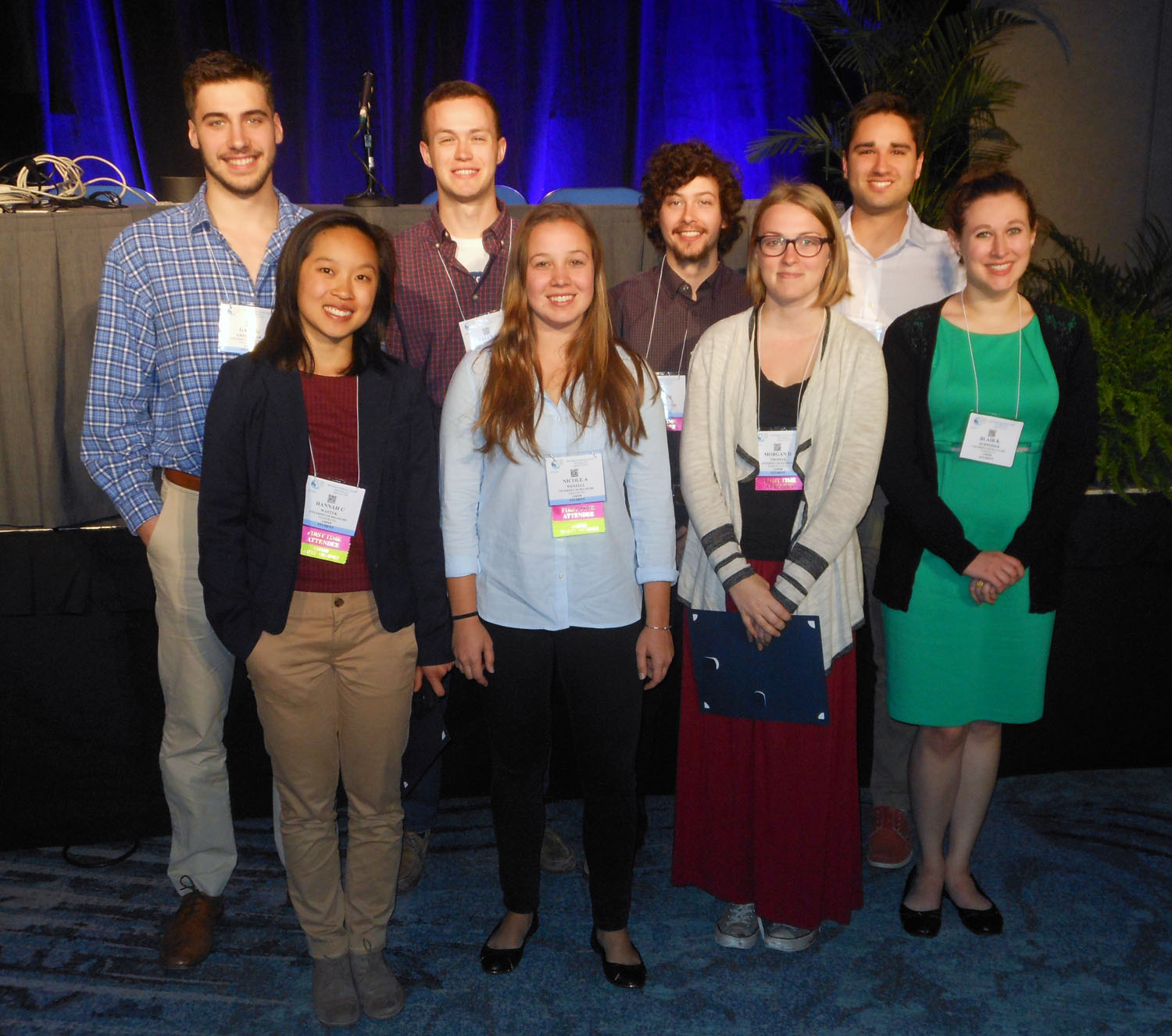
The University of Delaware group included three faculty and 8 undergraduates.
 |
||
| Prof.
Hal White, Chem & Biochem Prof. Seung Hong, Biol Sci Prof. Gary Laverty, Biol Sci |
Josh Barton, Biology Gabe Gregorzak, Chemistry Tyler Heiss, Chem and Biochem Blair Schneider, Biology |
Jay Subramoney, Chem and Biochem Morgan Thomas, Biology Hannah Wastyk, Chem and Biochem Nicole Wenzell, Chem and Biochem |
|
|
Molecular
Characterization of Two Human Lens Epithelial Cell Lines and Their
Suitability to Study Function of Cataract Genes
Joshua Barton, Archana D. Siddam, Deepti Anand, Salil A. Lachke. Biological Sciences, University of Delaware, Newark, DE The ocular lens is a transparent tissue that focuses light on the retina, allowing high-resolution vision. The loss of lens transparency causes an eye defect termed cataract, which is the leading cause of blindness worldwide. Half of the U.S. population above age 80 is affected by cataracts, for which surgical treatment is the principal therapeutic intervention. In 2014, over 3.6 million cataract surgeries were performed in the United States, and over 20 million surgeries were performed globally. Age-related cataracts are caused by a variety of stress conditions including elevated oxidative stress, increased sugar (diabetes), smoking or environmental insults such as exposure to ultraviolet (UV) radiation. Thus, characterization of biological and environmental factors that affect lens transparency is the first critical step toward the development of new therapies for cataracts. Recent discoveries have revealed a surprising link between proteins that are components of cytoplasmic RNA granules (RGs) and mammalian cataract. RGs like Processing Bodies (PBs) and Stress Granules (SGs) represent specialized cytoplasmic sites for regulation of RNA in the control of gene expression. PBs, the constitutive class of RGs, are RNA-protein complexes that silence or channel mRNA to decay, and therefore function to regulate the cellular proteome. SGs form as an early reaction to cellular stress, specifically allowing the translation of proteins that function in homeostasis. To investigate RG function in lens cells, we molecularly characterized two human lens epithelial cell lines (LECs), SRA01/04 and HLE-B3. These previously uncharacterized cell lines can serve as valuable in vitro tools for investigating the intricate molecular basis for human cataractogenesis. It is important in cell culture studies to validate the origin and identity of the cell line. Therefore, our first step was to confirm that the cell lines were of human origin. Analyzing genome-specific Short Tandem Repeats by PCR, we authenticated that the LEC lines are indeed human-derived. We next analyzed gene expression in these LECs by high throughput Illumina microarray analysis to investigate the extent of their retention of lens-like character. The microarray data demonstrates that both LECs retain expression of genes that are expressed and/or enriched in normal lens epithelial cells. Using a bioinformatics tool “iSyTE” developed to predict genes implicated in lens biology and cataract, we further validated the expression of cataract associated genes in these LECs using RT-PCR. Furthermore, we find that both LECs support formation of RGs, and exhibit formation of SGs under various stress conditions such as chemical and UV-stress. Thus, these studies present the first cellular models for investigating the molecular biology of UV-radiation exposure in lens epithelial cells, and therefore represent a new resource for study of factors that are linked to age-associated cataract in humans. Support or Funding Information Funding was provided by the 2015 Delaware Governor's Bioscience Fellowship to Joshua Barton, and University of Delaware Startup funds as well as a grant from The Pew Charitable Trusts awarded to Salil Lachke. Abstract No. 1058.1 Poster No. C66 Tuesday, April 5 |
|
|
Synthesis of
Artificial Bacterial Cell Wall Fragments as Tools to Study the Innate
Immune System
Gabriel Gregorzak, James Melnyk, Kristen DeMeester, Catherine Leimkuhler Grimes. Department of Chemistry & Biochemistry, University of Delaware, Newark, DE During
a bacterial infection, the body’s first line of defense is provided by
the innate immune system. This system is triggered by
pathogen-associated molecular patterns (PAMPs) that are recognized by
different members of the body’s innate immune system called pattern
recognition receptors (PRRs). One such PRR of interest is
nucleotide-binding oligomerization domain-containing protein 2 (Nod2).
Mammalian Nod2 is a cytosolic protein that binds to and signals in
response to carbohydrate containing bacterial cell wall fragments; the
smallest of which is muramyl dipeptide (MDP). Mutations in Nod2
increase the likelihood of the development of Crohn’s Disease and other
chronic inflammatory diseases. Previously, our lab has shown that Nod2
binds directly to MDP, but currently the molecular mechanisms of
activation and binding are still unknown1. Currently, we are addressing
this issue using synthetic chemistry. This project focuses on the
synthesis of an assortment of MDP dimers, which vary in the length and
functionality of their connecting linkers. To avoid troublesome
late-stage de-protection issues, the synthesis diverges in its early
stages to result in two differing MDP-like fragments. These two
fragments can then be coupled together and de-protected, resulting in
the MDP dimer. Once synthesized, these dimers will be used in in
vitro SPR binding assays, as well as in cell-based assays to elucidate
the binding and activation mechanisms used by Nod2. Ultimately, these
synthetic tools will aid in the advancement of our understanding of how
are body’s sense and respond to bacteria.
GG Supported Howard
Hughes Medical Institute Undergraduate Summer Scholars award
Abstract No. 612.4 Poster No. C202 Sunday, April 3 |
|
|
Chemical
and Chemoenzymatic Synthesis of UDP-N-Acetyl
Glucosamine Probes
Tyler Heiss, Kristen E. DeMeester, Hai Liang, Borja Barbero, and Catherine Leimkuhler Grimes Department
of Chemistry and Biochemistry, University of Delaware, 163 The Green,
Newark, DE,
19716
Uridine-diphosphate-N-acetylglucosamine
(UDP-GlcNAc) is a
ubiquitously used nucleotide sugar involved in bacterial cell wall
biosynthesis
and the posttranslational modification GlcNAcylation. If functionalized
versions of UDP-GlcNAc were more easily accessible, these chemical
probes could
be used to rapidly investigate biological systems. However, current
chemical
strategies for synthesizing UDP-GlcNAc derivatives are difficult and
lengthy.
Herein we describe the chemoenzymatic synthesis of strategically
functionalized
UDP-GlcNAc chemical probes from Glucosamine-1-Phosphate (GlcN-1-P) and
synthetic acetyl donors known as N-acetylcysteamine
thioesters (SNAcs) using the bacterial cell wall biosynthesis enzyme
GlmU. Hypothesis:
If GlmU is shown to use GlcN-1-P and derivatized SNAcs to produce
modified
UDP-GlcNAc’s, then these altered building blocks will be used to
investigate
bacterial cell wall biosynthesis and O-GlcNAc transferase (OGT)
activity.
Acknowledgments and Funding: We would like to thank the Koh Lab and the rest of the Grimes Group for their continuing help and support. We also thank the Howard Hughes Medical Institue (HHMI), the University of Delaware’s Summer Scholars program, and the Pew Foundation for funding my work. Recipient of an ASBMB Undergraduate Travel Award Recipient of an Honorable Mention Award in the ASBMB Undergraduate Poster Competition. Abstract No. 612.3 Poster No. C201 Sunday, April 3 |
|
TRANSCRIPTOME
ANALYSIS OF ILLINOIS ABDOMINAL AND CARDIAC ADIPOSE
Blair Schneider1,
Colin Kern2, Allen Hubbard 3, Wayne Treible 4,
John W. Finger Jr. 5 , Tracey Tuberville 6 ,
The domestic chicken is very
important in its use as a model organism for understanding
regulation of
numerous biological processes in avians. To better understand energy
storage
and mobilization, this work focuses on defining the transcriptome of
chicken abdominal
and cardiac adipose tissue. Transcriptome
libraries were prepared from Illinois abdominal and cardiac adipose
samples
with the following procedure: 1. Isolation of mRNA using mirVana™
miRNA Kit, 2. Verification of mRNA quality and integrity by Fragment
Analysis, 3.
Transcriptome libraries prepared using Illumina Stranded RNA Prep Kit
and sequenced
at the Delaware Biotechnology Institute Core Sequencing Facility, 4.
Transcription levels determined using the fRNAkenseq
software, and 5. Identification of
differentially expressed
genes, data analysis, functional clustering and pathway analysis. To begin our analysis we identified genes
that were differentially expressed between the abdominal and cardiac
fat
tissue. This identified differentially
expressed transcripts encoding adipokines, transcription factors and
enzymes
involved in processing fat. The same
procedure was performed on alligators, and the gene expression was
analyzed in
their fat bodies. Current effort focuses
on comparing alligator gene expression with that obtained from chicken
adipose
tissue to better understand the evolution of this tissue in Archosaurs.
Acknowledgements:
This material is based upon work supported by the National Science
Foundation
under Grant No. 1147029 and by the Agriculture and Food Research
Initiative
Competitive Grant No. by NIFA 2010-04233 from the USDA National
Institute of
Food and Agriculture.
|
|
Chemical Ligation
Synthesis
of Isotope-Labeling Compatible Selenoprotein S Jay Subramoney,
Zhengqi Zhang, Rujin Cheng, Sharon
Rozovsky Selenoproteins
contain the 21st amino acid
selenocysteine (Sec) and play vital
roles in human health, such as antioxidant defense. Although
selenoproteins are
expressed naturally in the human body, procuring sufficient quantities
for
biochemical and biophysical characterization by heterologous expression
is
challenging. Low yield in genetic
incorporation is due to the Sec codon being identical to the UGA stop
codon in
mRNA. An alternative approach for their preparation is chemical
ligation, a
semi-synthetic method that relies upon an amide bond formation to
generate
peptides and proteins from their complementary fragments. Here, we
describe a
new method for chemical ligation of selenoprotein S (SelS) which is
compatible
with simple and low cost isotopic labeling and does not rely on
complete chemical
synthesis of a selenoprotein. SelS is a member of the endoplasmic
reticulum associated protein degradation (ERAD) pathway, a
protein machinery devoted to relocating misfolded proteins from the ER
to a
cytoplasmic protein degradation complex. The active site lies close to
the C-terminal where a sole Sec is paired with a Cysteine 13 residues
away.
Previously we have characterized the activity of SelS prepared by
heterologous
expression, however, structural characterization with the Sec in place
was
prohibited by the low yield. In our approach a Sec-containing fragment
is
expressed in E. coli and used as a source of the selenopeptide during
the
chemical ligation. This has the advantage of a high yield, with simple
and
efficient ligation while being compatible with isotopic-labeling of
selenoproteins. Useful applications of this method include
incorporation
of Selenium-77 and Carbon-13 for structural and biochemical
characterization by
NMR spectroscopy. Abstract No.
595.5 Poster
C83 Sunday, April 3. |
|
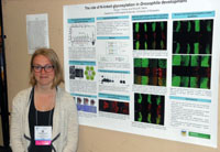
The Role of N-linked
Glycosylation in Drosophila Development
Morgan Thomas and Erica M. Selva Department of Biological Sciences, University of Delaware, Newark, DE Asparagine-linked or
N-linked glycosylation is an important post-translational modification
pathway that adds 14-sugar oligosaccharide chains to the Asparagine of
specific Asn-Xaa-Ser/Thr sequences in target proteins on the luminal
side of the endoplasmic reticulum. These glycan “tags” are necessary
for multiple cell functions including cell-cell recognition,
extracellular signal regulation, and proper protein folding. The focus
of this study is to examine the effect of loss of function mutations in
the N-linked pathway during development. A group of human diseases,
congenital disorders of glycosylation (CDG), arise from mutations in
the genes involved in various steps of this pathway. CDGs display
severe and pleotropic phenotypes, which reflect the universal
importance of this protein modification, but most cases display
phenotypes that arise from impaired neuronal function. The two specific
genes under study in this project are alg9
and alg10. Each gene encodes
a glycosyltranferase that adds a sugar residue to the growing
oligosaccharide chain before it is transferred en masse to a target
protein. The Drosophila eye is used as a model organ to study the
effects of loss-of-function mutations in these genes as it serves as a
surrogate for neuronal development. In adult flies, these mutations
yield a small rough eye phenotype, which is more severe in alg9, as it acts five steps before alg10 in the pathway. In order to
determine the molecular basis of this phenotype, larval eye discs that
are homozygous mutate for alg9
and alg10 were dissected,
stained for different glycoprotein and neuronal markers, and then
imaged using confocal microscopy. Using ELAV, a neuron specific marker,
we observed normally differentiated neurons in the eye disc.
However, these mutations interrupt proper glycoprotein trafficking, as
Chaoptin, a photoreceptor specific cell surface adhesion molecule,
accumulates in the cell bodies of alg9
and alg10 mutant
photoreceptors. Presence of cleaved Caspase-3 later in eye development
suggests that the accumulation of glycoprotein in the cell body
eventually leads to photoreceptor apoptosis. Photoreceptor death likely
continues through pupal development resulting in the reduced number of
photoreceptors seen in alg10
adult eyes and an almost complete absence in alg9 adult eyes. These results
indicate that CDG patients may have normal neuronal specification and
differentiation, but experience neuronal deficits due to intracellular
accumulation of glycoproteins leading to cell death. These data also
suggest induction of the unfolded protein response (UPR) resulting in
apoptosis may play a role in this process. Markers of endoplasmic
reticulum stress and the unfolded protein response such as XBP1 and BiP
will be studied in the future.
Support or Funding Information Howard Hughes Medical Institute Undergraduate Summer Fellowship, University of Delaware Supply and Expense Grants Abstract No. 619.1 Poster No. C236 Sunday , April 3 Recipient of an Honorable Mention Award in the ASBMB Undergraduate Poster Competition. |
|
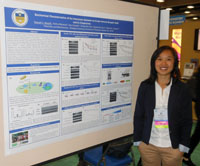
Biochemical Characterization of the
Interaction between Innate Immune Receptor Nod2 and its Chaperones
The human
body houses over 10 trillion bacterial cells, of which our innate
immune system must delineate between pathogenic and commensal. Disease
can arise during misregulation of this bacteria, and one example is the
debilitating inflammatory bowel disorder, Crohn’s disease (CD). The
molecular pathology of CD is known to be a mutation in Nod2, an innate
immune receptor responsible for binding directly to bacterial cell wall
fragment, muramyl dipeptide (MDP). MDP has recently been shown to
stabilize Nod2 in a cell based assay, and after binding to MDP, Nod2
signaling proteins activate the NF-κB pathway, a protective
inflammatory response. In Crohn’s variants, however, Nod2 is rapidly
degraded, thereby the downstream signaling is disrupted. Our initial
work identified Hsp70 as a chaperone molecule that binds to and
stabilizes Nod2. The purpose of this study is to characterize this
interaction and stabilization effect, such that the natural
stabilization of Nod2 within the cell can be extrapolated to a possible
treatment option that mimics Hsp70’s activity on unstable Nod2 Crohn’s
variants. Hsp70 is a highly ubiquitous protein that is able to confer
selective stability by tuning its specific activity via a number of
different cochaperones. Our data shows that Hsp40 contributes to the
chaperone complex, and through limited proteolysis, we show that both
Hsp70 and Hsp40 mediate Nod2 stability in vitro, and that this
stability is further enhanced with the addition of Nod2’s ligand, MDP.
Furthermore, we have biochemically characterized the importance of
Hsp70’s ATPase activity for Nod2’s stability. Significance of this
study extends beyond CD, as understanding Hsp70’s action and
determining a method of control for Nod2 could be extrapolated to
stabilize a large number of disease-relevant mutated proteins.Hannah C. Wastyk1, Vishnu Mohanan2, Amy Schaefer1, Ching-Wen Hou1, Mackenzie Lauro1, Catherine Leimkuhler Grimes1,2 1Chemistry and Biochemistry, 2Biological Sciences, University of Delaware, Newark, DE, 19716 Acknowledgements: The authors would like to thank the following sources of funding: the Chemistry-Biology Interface program of NIH, the Howard Hughes Medical Institute, and the Hofmann Fund through NUCLEUS. Recipient of an ASBMB Undergraduate Travel Award Recipient of a First Place Award in the ASBMB Undergraduate Poster Competition. Abstract No. 811.2 Poster No. C84 Monday, April 4 |
|
UNDERSTANDING
A FUNDAMENTAL FORCE IN PROTEIN FOLDING: TUNING THE n π* INTERACTION
VIA DESIGNED PEPTIDES
Nicole A. Wenzell, Himal K. Ganguly, Anil K. Pandey, Glenn P.A. Yap and Neal J. Zondlo* Department of Chemistry and Biochemistry, University of Delaware, 339 Brown Laboratory, Newark DE 19716 An n→π* interaction is a local interaction that orders two consecutive residues by delocalizing the lone pair of electrons from oxygen of the donor carbonyl to the antibonding orbital of the acceptor carbonyl carbon. This interaction is a recent discovery that gives insight into the inherent biases that proteins have to drive them to their folded state. By changing the electronics of the donor carbonyl on model proline-based peptides, we can tune the strength of this n→π* interaction. Proline can readily populate both the trans and cis conformations in a peptide bond. The n→π* interaction is shown to stabilize the trans conformation. Thus, in our model system the equilibrium constant of trans-cis isomerization (Ktrans/cis) acts as a readout for the strength of the n→π* interaction. Greater electron donation on the donor carbonyl causes a stronger interaction, resulting in higher Ktrans/cis values. The electronics of the donor carbonyl in the model peptides were modulated by synthesizing a series of derivatives with varying electron-donating capabilities on the donor carbonyl. The series of derivatives synthesized were selected in order to compare their electron donating capabilities to their n→π* interaction strength. The derivatives synthesized for analysis were, Boc, pivaloyl, formyl, acetyl, monochloroacetyl, dichloroacetyl, trichloroacetyl and trifluoroacetyl. These successfully synthesized derivatives provide a range of electronic character to be analyzed for the strength of the n→π* interaction in each compound. The determination of the strength of the n→π* interaction was accomplished through combined analysis via 1H and 13C NMR and X-ray crystallography. NMR analysis provides the Ktrans/cis values for each derivative and thus the strength of the n→π* interaction. Electron donation involved in the n→π* interaction is seen via the chemical shifts of the 13C NMR. The chemical shift of the acceptor carbonyl moves downfield with stronger electron-donating substituents, while carbonyls uninvolved in the interaction remain constant. X-ray crystallography of the model peptides reveals insights into the n→π* interaction as it indicates the distance and angles of the interaction. Specifically, the more electron donating character of the acetyl compared to the formyl group results in a 0.3Å shorter distance (3.3-3.0Å) and an angle (115˚vs 90˚) that more closely resembles an ideal nucleophilic attack (109.5˚). The comparison of the Ktrans/cis values, the carbonyl chemical shifts, and the angles and distances of the donor-acceptor carbonyl pair serve as direct readouts for the strength of the n→π* interaction, which varies depending on the electron donating character of the donor carbonyl. Recipient of an ASBMB Undergraduate Travel Award Abstract No. 598.8 Poster No. C107 Sunday, April 3 |
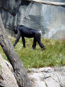 Bonobo at the San Diego Zoo |
 San Diego from our plane as we departed. |
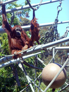 Moma and baby orangutan at San Diego Zoo |
|
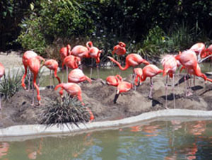 Flamingos at San Diego Zoo |
 Aerial Tram at San Diego Zoo |
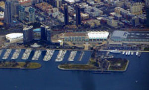 The Marriott and the San Diego Convention Center from the air |
 The Marriott Hotel in the morning fog |
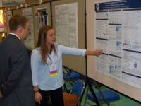
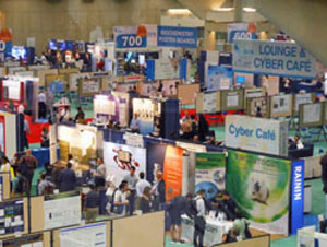 Can you find your poster? |
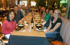 Dinner at Sovereign |
 Dinner at Sovereign |
 Snowy Mountains in New Mexico |
 Hannah receiving her award |
|
 Dinner at Chianti |
|
The trip to the Experimental Biology
2016 Meetings
in San Diego was organized by a legacy from the University of Delaware
HHMI
Undergraduate
Science Education Program with additional support from travel grants
from
the American
Society for Biochemistry and Molecular Biology.