|
in San Diego, April 2 - 6, 2005 |

|
|
|
in San Diego, April 2 - 6, 2005 |
|
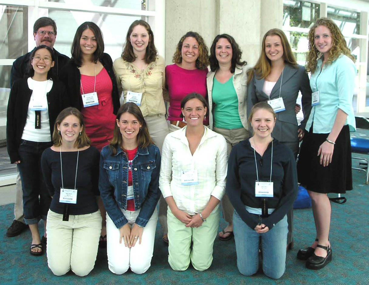
The University of Delaware group included four faculty and twelve undergraduates.
| Prof. Hal White Chem &
Biochem Prof. David Usher Biol Sci Prof. Gary Laverty, Biol. Sci. Seung Hong, Biol Sci |
Agata Bielska,
Chem & Biochem Meghan Bills, Biol Sci. Meghan Cashman, Chem & Biochem Glenn Christman, Chem & Biochem |
Madaline
Gregorits, Biological Sci. Liang-I Kang, Biol Sci Erin Kenaley, Biol Sci Elisabeth Mari, Biol Sci |
Kimberly Miller, Biol Sci Amanda Peters, Biol Sci Kristen Reese, Biol. Sci Mariclair Yandon, Del Biotech Inst. |
|
| Undergraduate
students from the University of Delaware who participated in the American
Society for Biochemistry and Molecular Biology's 9th annual Undergraduate
Research Poster Competition, San Diego, CA. 4 April 2005. From Back left
to front right: Glen Christman, Liang Kang, Maddy Gregorits, Aggie
Bielska, Meghan Cashman, Lis Mari, Kim Miller, Kristen Reese, (front row)
Meghan Bills, Erin Kenaley, Marci Yandon, and Mandy Peters. |
|

|
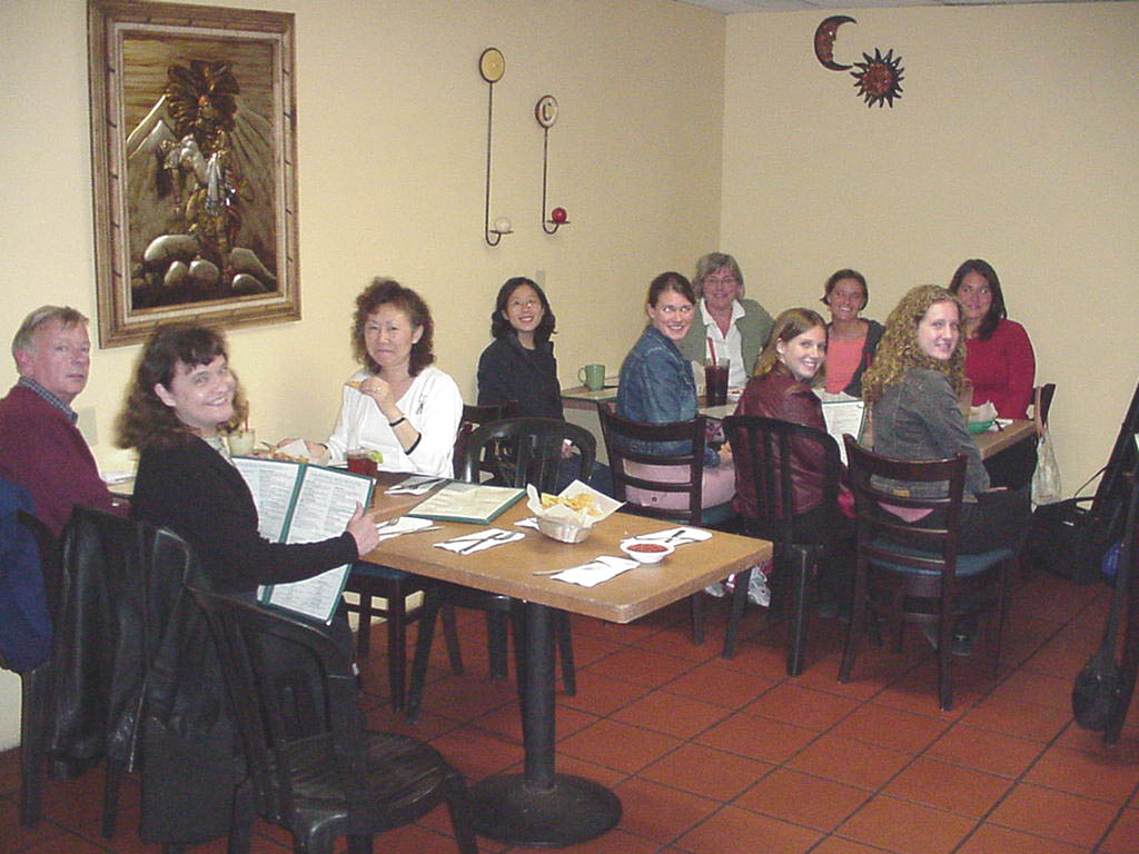
| Alumni Dinner at Cafe Coyote in Old
Town San Diego, 2 April 2005. Left to right: Mr. Schmieg, Florence Schmieg,
Seung Hong, Liang Kang, Erin Kenaley, Marci's mom, Meghan Bills, Marci
Yandon, Kristen Reese, and Maddy Gregorits. |
From left to right: Gary Laverty,
Mandy Peters, Dan
Dries '00, Glenn Christman, Aggie Bielska, Hal White's empty chair,
Artie Suchow
'04, Allison
Olszewski '02, Jen Risser
'04, Meghan Cashman, Lis Mari, Justin DiAngelo
'02. (Note-Justin apears twice-look in mirror) |
|
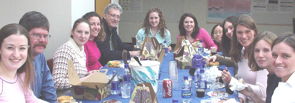
| Lunch gathering under the McKinly Lab
skylight after practice presentations of posters on Wednesday, 9 March.
From left to right: Kim Miller, Glenn Christman, Aggie Bielska, Meghan Cashman, Prof. Hal White, Kristen Reese, Elisabeth Mari, Liang Kang, Maddy Gregorits, Erin Kenaley, Meghan Bills, and Marci Yandon. Mandy Peters not shown. |
|
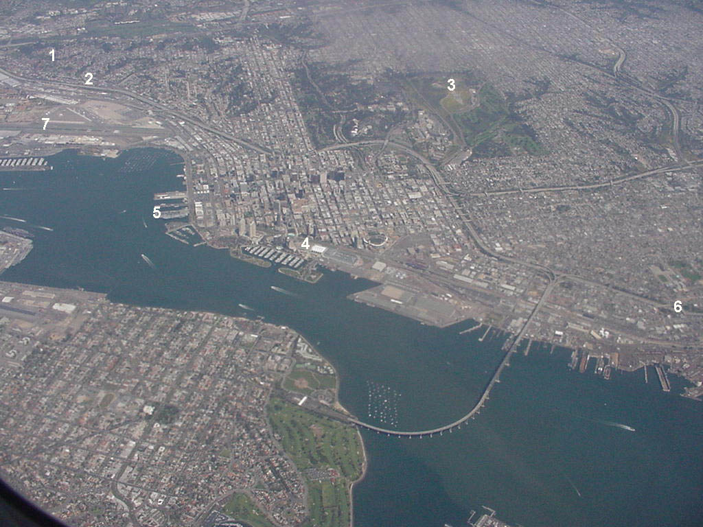
| Our
last view of San Diego. Things looks close but it took about an hour by
trolley with waiting and transfers to get from the Convention Center (4)
three miles to the Comfort Inn (2) right next to Interstate 5 near
Old Town (1) and the airport (7). Interstate 5 (6) goes south to Tijuana.
Those who walked from the transfer station may have seen the aircraft carrier
midway in dock (5). The San Diego Zoo (3) is situated in Balboa Park.
|
|
Conformational
Effects of Protein Phosphorylation 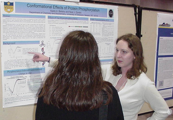
|
Phosphorylation-induced conformational
change is an important tool used by cells to regulate metabolic pathways,
signal transduction pathways, and transcription. The objective
of this research is to characterize the specific conformational effects
of protein phosphorylation using the proteins tau and cJun as a model. Hyperphosphorylation
of tau has been shown to cause its dissociation from microtubules and
subsequent precipitation into neurofibrillary tangles (NFTs) in Alzheimer’s
disease. In order to study mechanisms by which phosphorylation could
lead to NFT formation, various tau-derived peptides were synthesized.
Phosphorylated and non-phosphorylated versions of the peptides were then
analyzed using circular dichroism (CD) and NMR spectroscopy. Significant
conformational changes were found in regions which, when hyperphosphorylated,
are associated with NFT formation. These conformational changes
were consistent with an increase in type two polyproline helix (PPII) character. The oncoprotein, cJun, is a transcription factor regulated by phosphorylation. Peptides derived from cJun were synthesized and analyzed in non-phosphorylated, mono-phosphorylated, and di-phosphorylated forms. Upon phosphorylation, significant conformational changes were observed favoring PPII formation. The magnitude of these changes increased as the number of phosphorylation sites increased. |
|
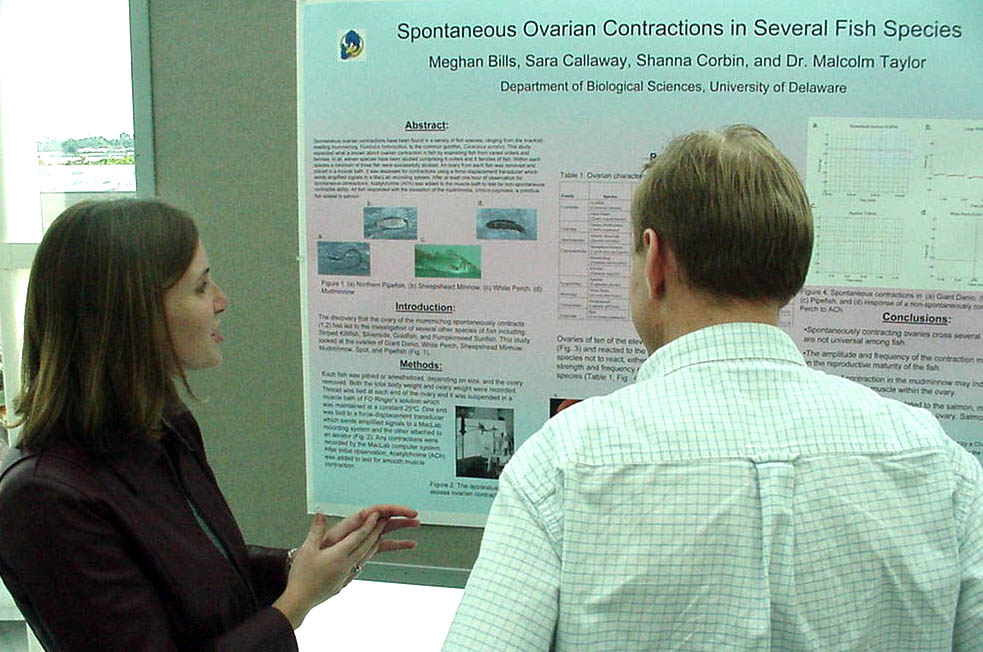
Spontaneous Ovarian Contractions in Several Fish Species Meghan
Bills and Malcolm Taylor
|
Spontaneous ovarian contractions
have been found in a variety of fish species, ranging from the brackish
dwelling mummichog, Fundulus heterclitus, to the common goldfish, Carassius
auratus. This study sought to expand what is known about ovarian contraction
in fish by examining fish from varied orders and families. In all, eleven
species have been studied comprising 6 orders and 8 families of fish.
Within each species a minimum of three fish were studied. An ovary from
each fish was removed and placed in a muscle bath. It was assessed for
contractions using a force-displacement transducer which sends amplified
signals to a MacLab recording system. After at least one hour of observation
for spontaneous contractions, Acetylcholine (ACh) was added to the muscle
bath to test for non-spontaneous contractile ability. All fish responded
with the exception of the mudminnow, Umbra pygmaea, a primitive fish
related to salmon. Ovarian contraction in fish may be a useful model
for study of mammalian smooth muscle function. This research was supported
by a Charles Peter White undergraduate research fellowship to MLB. |
|
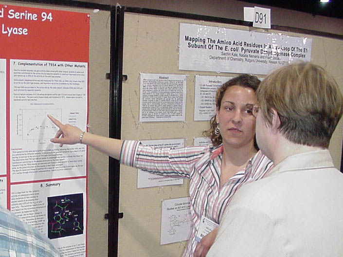
Evaluation of
the Role of T93 and S94 in the Function of Adenylosuccinate Lyase |
Adenylosuccinate lyase
(ASL) of Bacillus subtilis is a homotetramer that catalyzes the conversion
of adenylosuccinate to AMP and fumarate. The enzyme has 4 active
sites/molecule each of which has amino acids contributed from 3 subunits.
T93 and S94 are conserved in all ASLs and are close to other amino acids
previously identified by mutagenesis as being in the active site.
To test their involvement in the enzyme’s function, T93 and S94 were replaced
by alanine. T93A and S94A exhibit circular dichroism spectra superimposable
on that of wild-type enzyme, indicating there is no change in secondary
structure. T93A exhibits 0.5% of the Vmax of wild-type ASL with a 10-fold
increase in Km for adenylosuccinate. Intersubunit complementation yields
increased activity for T93A when mixed with other inactive ASL mutants with
replacements for residues contributed to the active site by different subunits.
The largest increase in activity accompanies the pairing of T93A and K268Q
mutants. H68Q and T93A mutants do not show complementation since both
are provided to the active site by the same subunit. S94A has 65%
of the Vmax of wild-type ASL with little change in Km. pH-Vmax profile
measurements reveal a pK2 value for S94A of 7.83 in contrast to 8.32 for
the wild-type enzyme. T93 may orient adenylosuccinate optimally for
catalysis; while S94 stabilizes protonated H68/H89, the determinants of pK2.
(Supp. By HHMI & NIH DK60504.) |
|
Characterization
of the ThiI-persulfide Intermediate Formed During 4-Thiouridine Biosynthesis
|
Two enzymes, ThiI and IscS, are
involved in the modification of uridine to 4-thiouridine in some prokaryotic
tRNAs. IscS removes a sulfur atom from free cysteine and transfers
it to ThiI, and indirect evidence indicates that a persulfide group is
formed at Cys-456. A fluorescent derivative of iodoacetamide, I-AEDANS,
has been used to trap the putative persulfide group. Both thiol and persulfide
groups react with I-AEDANS to displace iodide and form a carbon-sulfur
bond. Reduction is expected to release the labeled persulfide group
(which has a disulfide linkage) but not a thiol group (which has a thioester
linkage). In a non-reducing environment, both ThiI and IscS are fluorescently
labeled by I-AEDANS. Treatment with reducing agents (either DTT, 2mercaptoethanol,
or TCEP) greatly diminished the fluorescence of ThiI, with residual labeling
from a small amount of ThiI in the thiol form before the addition of
IAEDANS. The tryptic fragment of ThiI containing Cys-456 connected to
the fluorescent label was detected using MALDI mass spectroscopy.
The observed mass included one "extra" sulfur, indicating that a persulfide,
not a thiol or trisulfide group, was modified by I-AEDANS. As expected,
this fragment was labile to reduction. This research was supported
by the HHMI Undergraduate Science Education Program and the National Institutes
of Health (GM59636). |
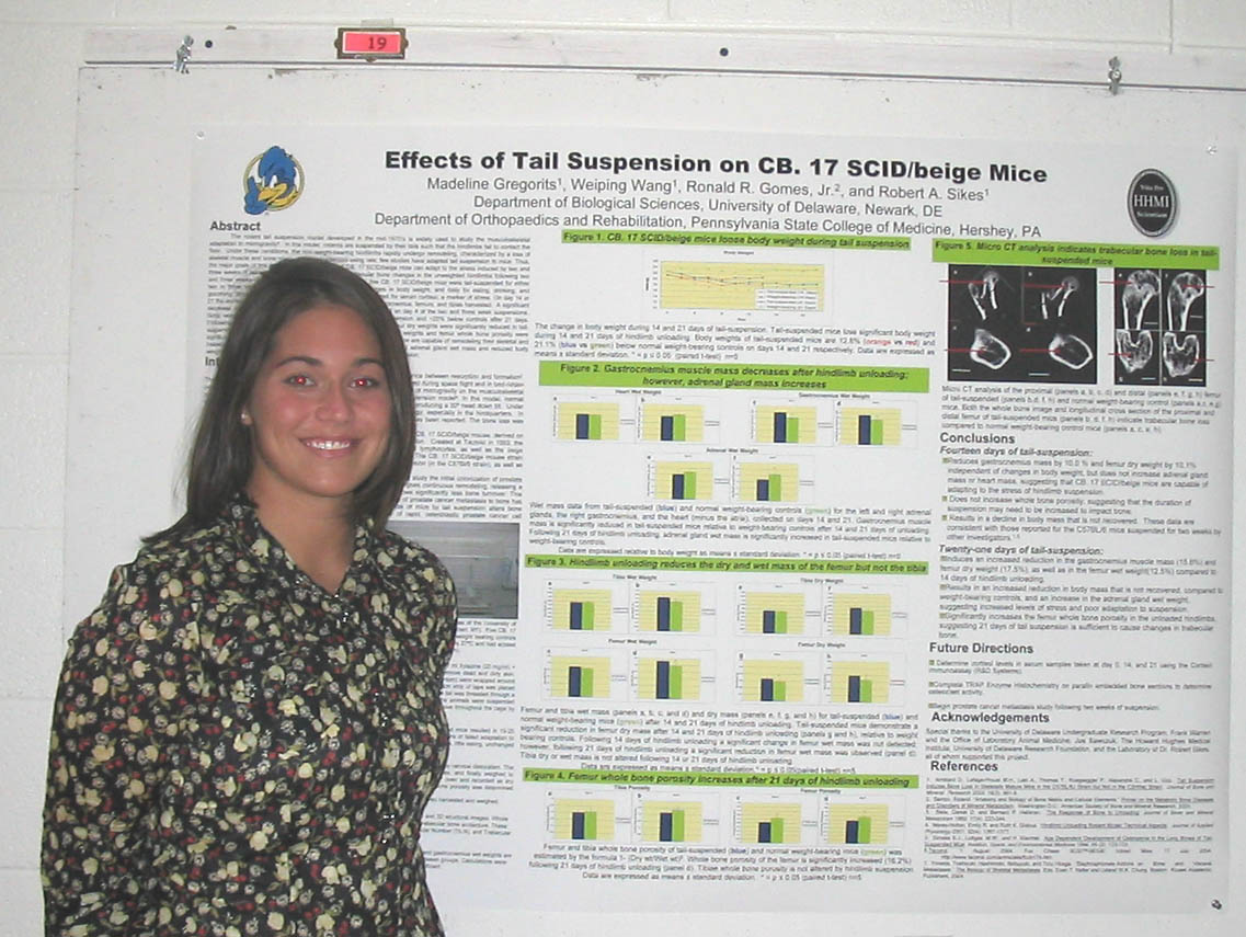
Effects of Hindlimb Suspension on CB. 17 SCIB/beige Mice Madeline
Gregorits, Ronald R. Gomes, Jr. and Robert Sikes |
The rodent tail suspension model
developed in the mid-1970’s is widely used to study the musculoskeletal
adaptation to microgravity. In this model, rodents are suspended
by their tails such that the hindlimbs fail to contact the floor.
Under these conditions, the non-weight-bearing hindlimbs rapidly undergo
remodeling, characterized by a loss of skeletal muscle and bone mass.
Best characterized using rats; few studies have adapted tail suspension
to mice. Thus, the major goals of this study were: 1) Determine if
CB. 17 SCID/beige mice can adapt to the stress induced by two and three
weeks of tail suspension; 2) Characterize the trabecular bone changes in
the unweighted hindlimbs following two and three weeks of tail suspension.
To test our hypothesis, five CB. 17 SCID/beige mice were tail-suspended
for either two or three weeks. Mice were monitored regularly for changes in
body weight, and daily for eating, drinking, and grooming. Blood was collected
on day 0, 14, and 21 to be analyzed for serum cortisol, a marker of stress.
On day 14 or 21 the animals were sacrificed and the heart, adrenal glands,
gastrocnemius, femurs, and tibias harvested. A significant decrease
in body weights of the tail-suspended mice were observed on day 4 of
the two and three week suspensions. Body weights remained ~10% below
controls after 14 days of suspension and ~20% below controls after 21
days. Following 14 days of suspension, gastrocnemius muscle mass and
femur dry weights were significantly reduced in tail-suspended mice. Following
21 days of suspension, adrenal gland wet weights and femur whole bone porosity
were significantly increased. These findings suggest that CB. 17 SCID/beige
mice are capable of remodeling their skeletal and trabecular bone with changes
in load-bearing activity; however, increased adrenal gland wet mass and
reduced body weights may suggest increased stress levels and poor adaptation
to suspension. |
| The Role of Junctional Adhesion
Molecule A (JAM-A) in the Corneal Epithelium Liang-I Kang, Vesselina Cooke, Ulhas P. Naik, and Melinda K. Duncan Department of Biological Sciences 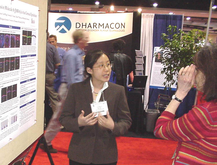
|
Junctional Adhesion Molecule A (JAM-A)
is a ~38 kDa protein implicated in platelet activation and adhesion, angiogenesis,
and the structural integrity of endothelial and epithelial cells. Recently,
we showed that JAM-A expression is upregulated in the ocular lens in response
to Pax6, a transcription factor important for eye development. The presence
of JAM-A, JAM-B, and JAM-C mRNA in the wildtype lens and JAM-A mRNA in
the wildtype cornea was confirmed through RT-PCR. Using immunohistochemistry,
JAM-A protein was found in the blood vessels of the developing mouse eye
as early as 12.5 dpc and in the corneal epithelium by 13.5 dpc. High levels
of JAM-A remain in the corneal epithelium throughout adulthood. Mice with
an insertion of a promoter-less GEO genetrap construct in the JAM-A gene
lack JAM-A staining in the cornea, validating the JAM-A antibody specificity.
Both heterozygous and homozygous JAM-A knockout mice express both gene
products of the genetrap cassette, placental alkaline phosphatase and
-galactosidase, in the corneal epithelium, further confirming this expression
pattern. While the eye appears to develop normally in JAM-A knockout mice,
older animals have subtle abnormalities in the corneal epithelium. We hypothesize
that JAM-A is important to maintain the cornea and future studies will be
directed toward determining the function of JAM-A. Funding: NIH and the Arnold and Mabel Beckman Foundation |
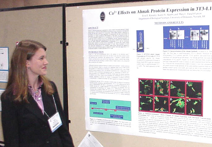
| Calcium Effects
on Ahnak Protein Expression |
Ahnak is a 700 kDa protein that
is expressed in a variety of cells. The precise function of Ahnak remains
unknown. Ahnak has been proposed to be involved in actin cytoskeleton
rearrangement in some cell types and in calcium homeostasis. Ahnak cellular
location is influenced by intracellular calcium levels. In this
study, we assessed the effect of calcium on the expression of Ahnak in
3T3-L1 preadipocytes. RT-PCR and Western blot analyses revealed that
Ahnak is present in the 3T3-L1 preadipocyte cell line. Immunostaining of
3T3-L1 cells with Ahnak specific antibodies showed that Ahnak is localized
in the cytoplasm and along the plasma membrane. Immunostaining of proliferating
3T3-L1 cells (in exponential growth phase) cultured in varying levels of
extracellular calcium showed that Ahnak protein expression may increase
with increasing extracellular calcium levels. When cytoplasmic calcium
levels were raised with thapsigargin or calcium ionophore, Ahnak protein expression
increased in the treated cells as compared to control. These data suggest
that calcium is an important regulator of Ahnak expression in 3T3-L1 cells. |
| Effects of Hip/Rpl29
Gene Disruption in Murine Embryonic Stem Cells Elisabeth Mari, Daniel Carson, and Catherine Kirn-Safran Department of Biological Sciences 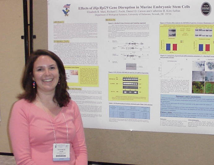
|
HIP/RPL29 is a multifunctional
ribosomal protein with heparan sulfate binding properties that is highly
expressed in developing tissues including the inner cell mass of early
preimplantation mouse embryos. Recently, we found that HIP/RPL29 transcript
and protein are among the most abundant in embryonic stem (ES) cells and
are strongly detected in limited amounts of total lysates. Because of
HIP/RPL29 abundance in ES cells, we hypothesize that HIP/RPL29 expression
is required for basic cell survival and growth. To evaluate the
effect of HIP/RPL29 monoallelic expression, we performed RT-PCR on Hip/Rpl29
+/- ES cell lines using specific primers for HIP/RPL29. Our results
showed that there was no significant change in the steady-state level of
both HIP/RPL29 mRNA and protein in +/- versus wild type ES cell lines.
This observation indicates that gene compensation mechanisms are taking
place to restore normal levels of expression. Preliminary transcriptional
profiling studies of +/- ES cell lines by RT-PCR suggested that Oct-3/4,
a transcription factor known to sustain stem cell renewal and pluripotency,
is differentially regulated in +/- ES cell lines when compared to control
cell lines. Future efforts will consist in determining whether concomitant
changes in Oct-3/4 protein expression levels are seen in +/- versus +/+
ES cells. In order to investigate the importance of HIP/RPL29 for ES cell
growth and survival we are currently performing experiments that will
induce the loss of the remaining wild type allele by culturing +/- ES
cells carrying the neomycin phosphotransferase gene in the presence of
high concentrations of G418. The outcome of this work will determine whether
HIP/RPL29 null cell lines are viable. All the in vitro data generated
with Hip/Rpl29 +/- ES cells constitute valuable tools for the disruption
of Hip/Rpl29 gene in animals and will help understand HIP/RPL29 function
in ES cell metabolism and embryo survival. (This work was supported by Charles
Peter White Fellowship [to E. R. M.] and NIH grant HD25235 [to D.D.C.]). |
|
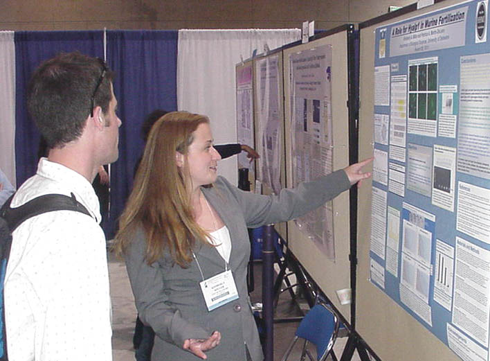
A
Role for Hyalp1 in Murine Fertilization
|
The cumulus cells surrounding the
unfertilized egg have an extracellular matrix rich in hyaluronic acid
and form a formidable barrier to sperm penetration. The digestion
of hyaluronic acid and subsequent penetration of the cumulus cells, required
for fertilization, is aided by enzymes dubbed ‘reproductive hyaluronidases,’
of which Spam1 remains the most extensively characterized. Located
on mouse chromosome 6, the gene family also contains Hyalp1, whose encoded
protein remains almost fully uncharacterized, in addition to Hyal4, Spam1
(PH-20), and Hyal5. Recently, it was discovered that sperm from
Spam1 knockout mice were able to penetrate the cumulus and successfully
fertilize oocytes, indicating that other proteins involved in this process
can functionally compensate for a lack of Spam1 (Baba, et al. 2002).
The aim of this study is therefore to investigate the role of Hyalp1 in
murine fertilization. Testicular developmental RT-PCR and immunohistochemistry,
and immunocytochemistry on epididymal sperm show colocalization of Hyalp1
on the acrosome cap of mature sperm. Hyaluronic Acid Substrate Gel
Electrophoresis and In vitro fertilization vestment penetration assays demonstrate
hyaluronidase activity of Hyalp1 testicular protein and the role of Hyalp1
in the breakdown of hyaluronic acid and the penetration of the cumulus cells
surrounding the unfertilized oocyte by mature caput sperm. Vestment
penetration was retarded by the inhibition of Hyalp1 with antiserum.
This implicates a significant role for the murine reproductive hyaluronidase
in fertilization. This work is supported by NIH grant# R01 HD38273 and the Howard Hughs Medical Institute Summer Research Scholar Grant Program. |
|
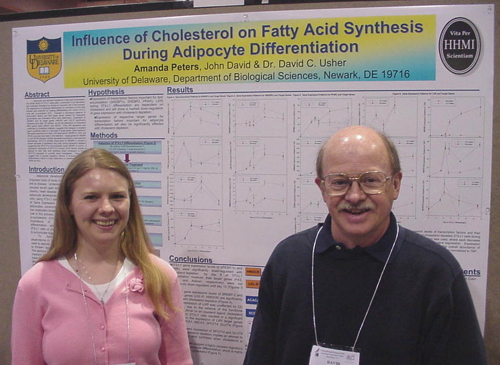
Influence of
Cholesterol on Fatty Acid Synthesis During Adipocyte Differentiation
|
Adipocytes are important regulators
of fatty acid homeostasis. The SAGE library of 3T3-L1 adipocytes,
constructed in our laboratory, has implicated cholesterol as having
an important role in this process. The dependence of adipocyte
differentiation on cholesterol to form lipid droplets was examined by
treating differentiating and control 3T3-L1 cells with -cyclodextrin
(CD). Changes in expression patterns of transcription factors
and their target genes needed for triglyceride accumulation and cholesterol
efflux were determined. Expression of SREBP2 and its target genes
HMGCR and LDL-R significantly increased soon after CD addition, as did
the expression of cholesterol efflux genes ApoE, ABCA1, and ABCG1.
That LXR expression was unaffected by cholesterol depletion, suggests
that changes in oxysterol ligand availability leads to a decrease in
these genes under treatment. Decreased expression for other LXR
target genes SREBP-1c, FAS, and PPAR occurred much later in the treatment.
Target genes for SREBP-1c and PPAR showed a similar decreased expression
in the treatment. Interestingly, SPOT14, another target gene of
SREBP-1c, showed a severe decrease in expression very early during cholesterol
depletion, suggesting that SPOT14 is not exclusively under SREBP-1c control.
A decrease in expression of SPOT14, a known G-6-P sensor, implies an attempt
to slow fatty acid synthesis when cholesterol is limited. SPOT14
regulation is currently being investigated. These results suggest
that a complex regulatory network is involved in adipocyte differentiation,
which is highly dependent on cholesterol. Funded by HHMI and SDI. |
| The Characterization of Hyal3 in
Murine Testicular Cells Kristen L. Reese and Patricia A. Martin-DeLeon, Department of Biological Sciences 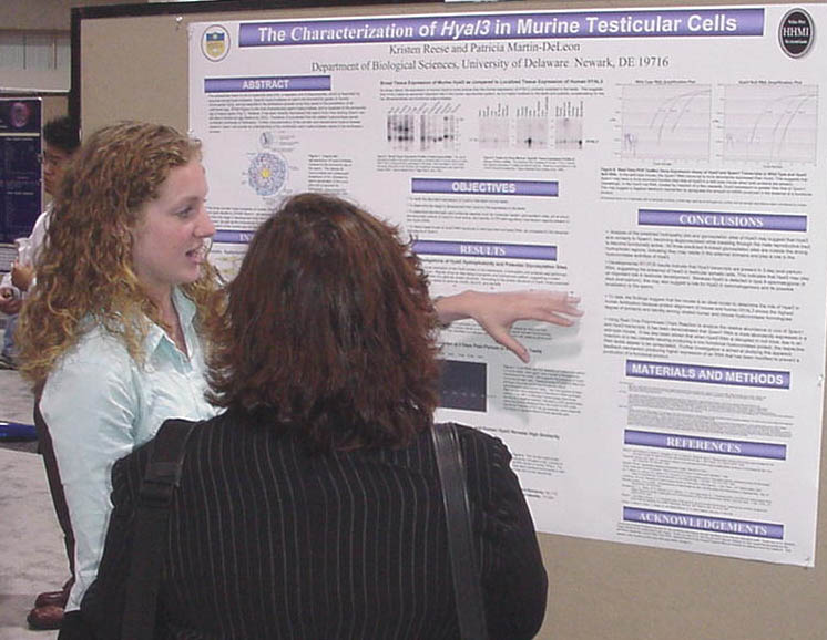
|
The digestion of hyaluronic acid is
required for penetration of the cumulus cells surrounding the egg during
fertilization and is implemented by a family of enzymes termed hyaluronidases.
Hyal3, one of the three murine somatic hyaluronidases, has high similarity
to the ‘reproductive’ hyaluronidases and is most highly expressed in the
testes, but its contribution to the fertilization process and intracellular
characteristics are not yet known. It has recently been determined
that sperm from mice lacking a primary reproductive hyaluronidase, Spam1,
are able to fertilize oocytes due to a predicted contribution of related
hyaluronidases. However, humans lack a functional reproductive hyaluronidase
other than Spam1. Hyal3 is particularly important to characterize
because it has the highest amino acid identity to its human homologue, with
similar testicular expression patterns and identity to Spam1. Therefore,
the knowledge obtained in the mouse model can then be applied to humans.
It is the goal of this study to investigate developmental expression of
Hyal3 in the testes, its location in sperm, and its role in the dissolution
of the extracellular matrix of the cumulus. Briefly, the methods used
are as follows: RT-PCR and Real-Time PCR for quantitative, qualitative, and
developmental analyses and Hyaluronic Acid Substrate Gel Electrophoresis
to determine hyaluronidase activity. Thus far, our results demonstrate
that Hyal3 is made in germ cells, suggesting its presence in sperm and that
the protein demonstrates hyaluronidase activity. We are currently determining
its role in cumulus penetration using antibodies for Hyalp1 and Hyal5 and
sperm from Spam1 knockout mice. This research is funded by the Howard
Hughes Medical Institute and NIH grant #R01 HD38273. This research is funded by the Howard Hughes Medical Institute (KLR) and NIH grant #R01 HD38273 (PAM-D). |
|
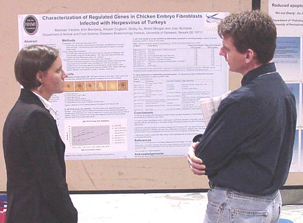
Characterization Of Genes Regulated By Infection Of Chick Embryo Fibroblasts (CEFs) With Herpesvirus Of Turkeys (HVT).
Mariclaire Yandon, Erin Bernberg, Amarin Cogburn,
|
HVT
is used as a vaccine against Marek’s disease virus (MDV), an alphaherpesvirus
that grows in chickens, chick embryos, and chick embryo fibroblast
(CEF) cell culture. Infection of CEFs with either MDV or HVT causes
the formation of characteristic plaques and relatively few cells in
a culture become infected. The low level of infectivity impairs global
gene expression studies of host responses to infection, since the infected
cells are likely to have a very different gene expression profile than
surrounding or uninfected cells. We coupled laser capture microdissection
(LCM) with microarray analysis of HVT-infected CEFs to identify regulated
genes. To confirm differential expression, a quantitative reverse transcription-polymerase
chain reaction was used to compare gene expression in plaques, uninfected
cells, as well as cells adjacent to plaques. A large number of regulated
genes have no match to any entry in GenBank. Additional sequence information
was obtained on several clones, and expression profiles evaluated by dbEST
data. This approach adds functional information to previously uncharacterized
genes and will improve our understanding of the mechanism of viral disease.
Funded by NIH BRIN and USDA NRI Programs. |
The trip to the Experimental Biology Meetings
in San Diego is being organized by the University of Delaware HHMI Undergraduate
Science Education Program with additional support from travel grants
from the American
Society for Biochemistry and Molecular Biology, the Beckman Scholars
Program, and the University of Delaware's Women Scholars Program.
The HHMI Program,
the Beckman
Scholars Program, Charles Peter White Fellowships, the Biomedical Research Infrastructure
Network (BRIN) Program, and the Undergraduate
Research Program supported research by the students.
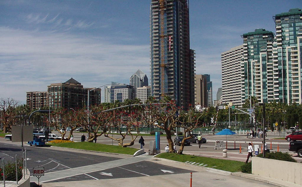
Downtown San Diego from Convention Center |
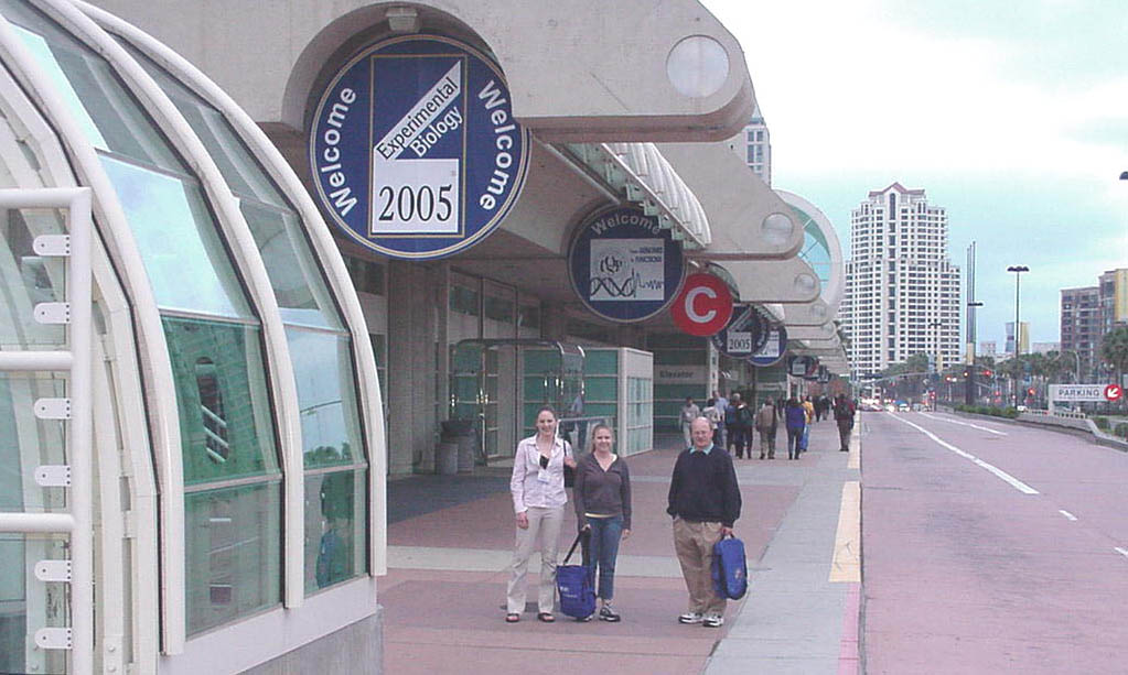
Aggie, Mandy, & Dr. Usher at Convention Center |
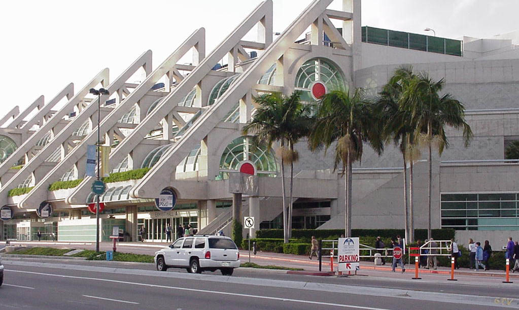
Convention Center from across the street. |
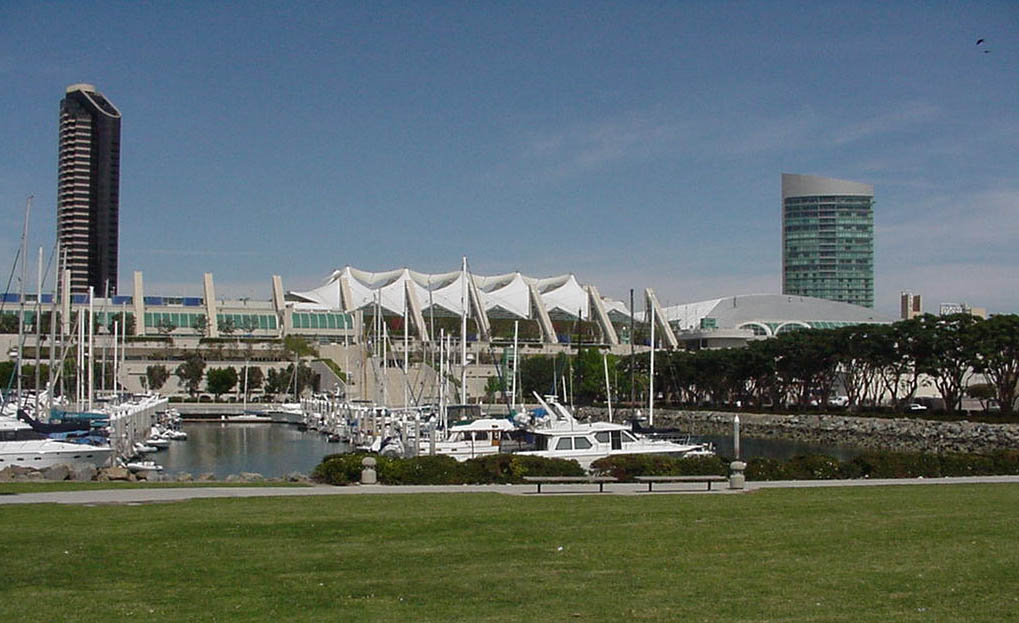
Convention Center from the marina. |
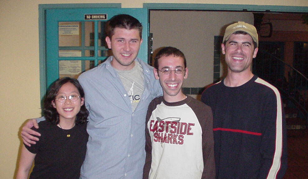
Reunion of successive students from Melinda Duncan's lab. Liang Kang '06, Artie Suckow '04, Justin DiAngelo '02, Dan Dries '00. |
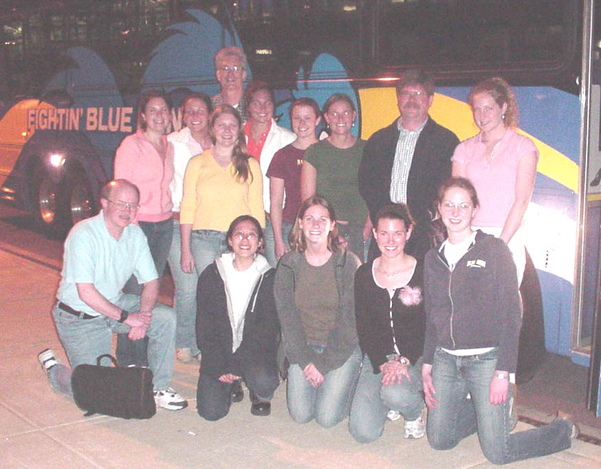
Blue Hen bus pickup at PHL. |
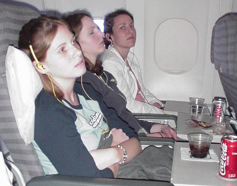
Meghan B., Aggie, and Meghan C. on their way to San Diego. |
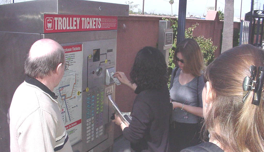
Liang discovers that the San Diego Trolley Machine doesn't like one dollar bills. |
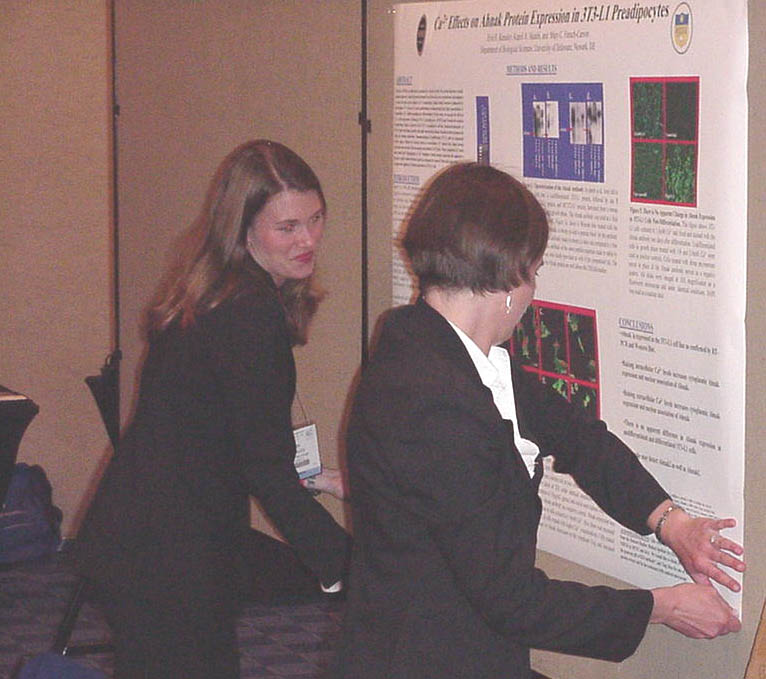
Erin and Marci putting up a poster. |
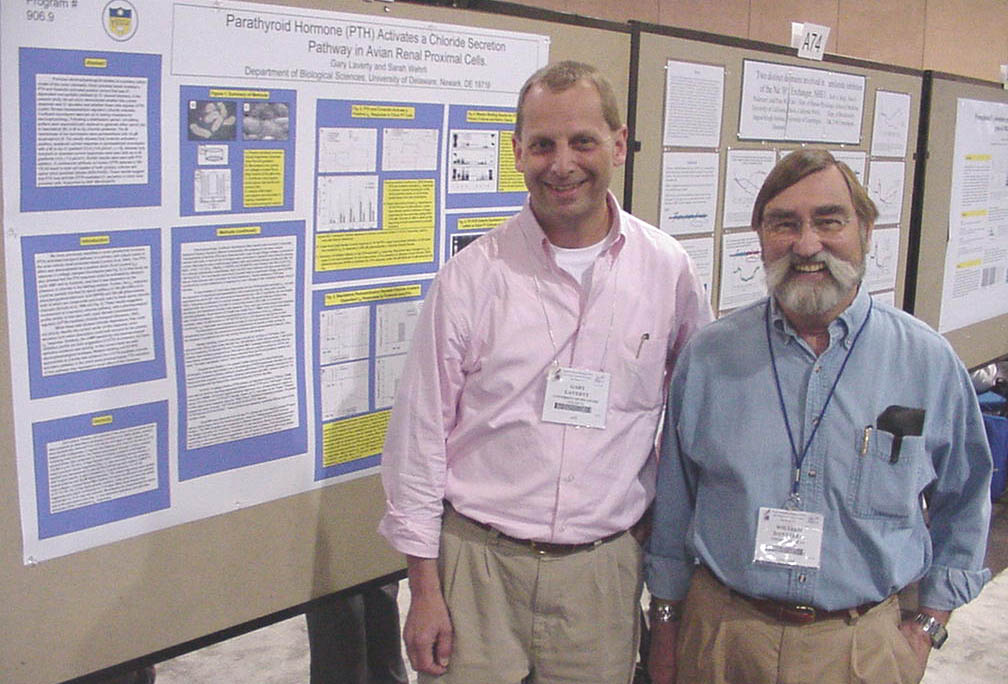
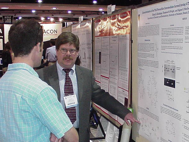
Gary Laverty with his poster and his postdoc advisor. |
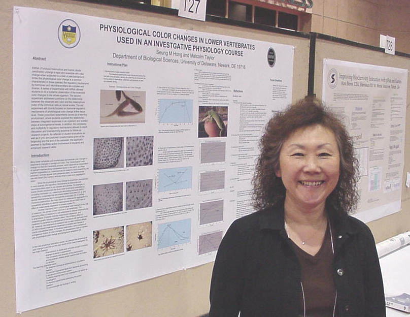
Seung Hong with her poster. |

Don't let that poster get away! |
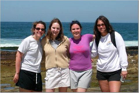
Yes gang, that is the Pacific Ocean behind us. |
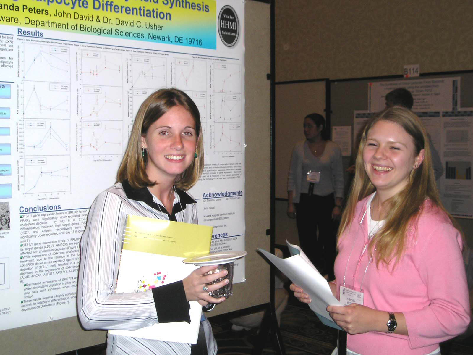
Meghan Bills and Mandy Peters at the ASBMB Undergraduate Poster Competition |
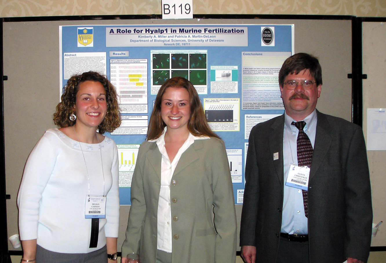
Meghan Cashman, Kim Miller, and Glenn Christman at the ASBMB Undergraduate Poster Competition. |

The University of Delaware Delegation at the ASBMB Awards Ceremony. |