Abstracts from the Department of Chemistry and Biochemistry
Undergraduate Summer Research Symposium August 14, 2003

Ordered alphabetically by student's last name

De Novo Design of Lanthanide Fingers

Chris am Ende, Mao Ye, Neal Zondlo, and Douglass Taber
Department of Chemistry and Biochemistry

A series of peptides has been synthesized using the 25 amino acid zinc finger motif as a model.  The sequence has been altered to allow for lanthanide, instead of zinc, binding.  The binding has been analyzed using a several methods.  Circular dichroism (CD) was used to monitor the extent of protein folding.  The CD data showed characteristic metal bound zinc finger CD spectra upon titration with various lanthanides.  Metal binding was also analyzed using fluorescence of tryptophan.  In addition, fluorophores have been conjugated to selected amino acids to monitor peptide folding using fluorescence resonance energy transfer (FRET).  FRET data gave further evidence of the conformational changes associated with metal binding.  Funding for this project was provided by the The Arnold and Mabel Beckman Foundation and start up funds from the University of Delaware. 


Formation of Supported Bilayer for the Separation of Membrane Proteins

Vincent Asiago, Stuti Christie, and Mary Wirth
Department of Chemistry and Biochemistry

We are investigating whether supported phospholipids bilayers can be used for the separation of membrane proteins. To this end, we are studying the use of thin (10nm) films of poly acryl amide to support thin bilayers. The question is whether or not these lipids are mobile. Supported phospholipids were formed on thin films of poly acryl amide film surface using 1, 2 dioleyol-sn-glycero-3-phosphocholine (DOPC) and 1 mol% 1, 2-dioleoyl -sn-phosphoethanolamine-N-carboxyflourescein (DOPE). The poly acryl amide film was grown by atom transfer radical polymerization on fused silica. The fluorescence images of the supported phospholipids were acquired using an upright microscope and an ICCD camera. Uniform bilayers become non-uniform when they were exposed to air, which can be reconstituted by applying water on the surface. Funding provided by the C. Eugene Bennett Professorship.


Role of Metal Ions in Oxidative Protein Folding: 
Do Flavoprotein Sulfhydryl Oxidases Need Metals? 

Stephen G. Brohawn, Irena Rudik Miksa, and Colin Thorpe
Department of Chemistry and Biochemistry

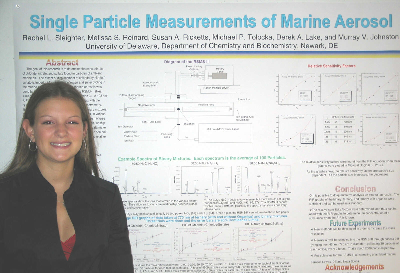
Each disulfide bond generated during the oxidative folding of secreted proteins requires removal of 2 electrons.  In higher eukaryotes, sulfhydryl oxidases have been found to catalyze this process with reduction of oxygen to hydrogen peroxide.  Both metal-dependent and flavin-dependent classes of these oxidases have been described.  The first protein sequence of a metal-dependent enzyme, from a copper-containing skin sulfhydryl oxidase, has been recently published and is 51% identical to the sequence for a flavin-dependent sulfhydryl oxidase already studied extensively in our laboratory.  Thus we were concerned that our work on the emerging flavin-dependent sulfhydryl oxidases had missed an important additional copper cofactor.  The present study establishes that the best-studied flavin-dependent oxidase contains no significant copper or other metals, and that copper and zinc are inhibitors of disulfide bond formation.  Studies with zinc as a model divalent metal show that it binds to CXXC centers and drastically perturbs the flow of reducing equivalents in this multi-domain enzyme.  Similarly, copper binds within the active center close to the flavin prosthetic group.  Our metal analyses show that the flavin-dependent oxidase is prone to bind metals and this may have misled earlier workers.  Thus the evidence that the skin sulfhydryl oxidase requires copper (rather than flavin) for enzyme activity should be reexamined. (Supported by Beckman Scholars Program and NIH  GM26643) 


A Thermodynamic Propensity Scale 
for Type II Polyproline Helices

Alaina M. Brown, and Neal J. Zondlo
Department of Chemistry and Biochemistry


Although type II polyproline (PPII) helices and proline-rich peptides are common structures in globular proteins, little is known about their stability, dynamics, and fundamental energetics. The ability to predict regions of high propensity for PPII helices is crucial to the analysis of PPII-mediated intermolecular and intramolecular interactions.  Sequences of three or more consecutive prolines induce PPII helix formation.  A model peptide (Ac-GPPXPPGY-NH2) was designed with two proline residues on each side of a randomized position, X.  Peptides with each of the twenty amino acids in the randomized position were tested using circular dichroism (CD) spectroscopy to determine the relative stabilization effect on propagation of a PPII helix.  The CD signal of each peptide was compared to the CD signal of the reference peptide (X=P) at 228 nm to determine the relative PPII helix stability.  Beta-branched amino acids (deltadeltaG~ 0.6 kcal/mol), cysteine, serine, and asparagine (deltadeltaG~ 0.3 kcal/mol), were all found to have a significant destabilizing effect on the PPII helix, while all other amino acids were moderately worse than proline (deltadeltaG~ -0.2 kcal/mol).  Helicity was temperature-dependant and pH-dependant for charged residues, but independent of salt and peptide concentration.  This research was funded by a Howard Hughes Medical Institute grant, the American Heart Association, and start-up funds from the University of Delaware for Dr. Zondlo.

 
Controlling Neurite Outgrowth: 
AFM Analysis of Comb-Polymer-Patterned Surfaces

Heather Egolf-Fox, Zhanping Zhang, 
and Thomas P. Beebe, Jr
Department of Chemistry and Biochemistry

Some neurons, particularly those of the central nervous system (CNS), do not regenerate completely when severed. Patients with injuries to the CNS would benefit from the discovery of a method for promoting regeneration after injury.  To study neuron growth, a surface was patterned with biomaterials. In order to understand the outgrowth of neurons, surface patterns must first be prepared and characterized. The method of microcontact printing (µCP), widely used in other areas of self-assembled monolayers, was used here to produce micron-sized protein and polymer features.  Despite its widespread use, very few of the important parameters for µCP have been studied systematically.  An elastomeric stamp made of polydimethylsiloxane was coated with a comb polymer known to inhibit neuron outgrowth, and then the pattern was transferred using µCP to a polystyrene surface.  Atomic force microscopy (AFM) was used to analyze the patterned surfaces.  Increasing the concentration of the comb polymer, as well as the pressure used to transfer the comb polymer pattern onto the surface resulted in greater feature thickness.  Furthermore, apparent feature thickness increased when AFM analysis was done in fluid rather than in air.  The Howard Hughes Medical Institute and the National Institutes of Health (EB000463, NIBIB Institute) provided funding.

 
Cysteine Mutagenesis to Study the Role of Disulfide Bonds in GPCRs Folding

Jordyn Gamiel*, Damien Thévenin*, and Clifford R. Robinson *†‡
*Department of Chemistry and Biochemistry, †The Delaware Biotechnology Institute, and ‡Department of Chemical Engineering

G-Protein Coupled Receptors (GPCRs) are a family of seven-helix transmembrane proteins that mediate signaling and control a wide range of cellular behavior in animals. GPCRs are implicated in many human diseases, and are attractive and proven drug targets. However, little is known about their folding and their structure. We hypothesize that cysteines in the transmembrane helices interact forming disulfide bonds to direct the receptors onto the productive folding pathway. The main goal of this study is to determine if cysteine-cysteine disulfide bonds contribute to the folding and structure of the adenosine A2a receptor (A2aR). The six cysteines present in A2aR transmembrane domains are mutated to alanine: single and double mutations are generated by PCR. The mutated A2a genes are then transfected into Human Embryonic Kidney cells in order to perform radio labeled ligand-binding assays. Pair-wise analysis of the effects of cysteine mutations on ligand binding is performed, to determine whether interactions between the cysteines, possible via disulfide bonds, affect the GPCRs folding and structure. Elucidating the principles of stability, folding, and assembly for GPCRs can have a major impact on our understanding of GPCR structure and function, and our ability to produce and characterize these receptors. This project was funded by NIH BRIN (JG), and by NIH grant 1-P20-RR015588


Molecular Cloning and Expression of Pig Phopholipase A2 

Sookhee Hwang, Ryan Cox, and Junghuei Chen
Department of Chemistry and Biochemistry

To investigate the structural-function relationship of Phopholipase A2(PLA2), the wild type of  PLA2 and mutant PLA2 were constructed and purified. A recombinant DNA which had a wild type of PLA2 was made by using two different plasmids. Each of the Plasmid A(pAB3) which contains PLA2 and Plasmid B(pET-25b(+)) which was used as an expression vector were digested with the two restriction enzymes BamH1 and HindIII which formed sticky ends. We   inserted 450 bp genes that contained PLA2 into a vector Plasmid B which had 5.5 kb. The recombinant DNA plasmid was transformed with host E. coli cells with ampicillin selectivity. To  make sure if we have a recombinant plasmid pET-25b(+)-pLA2 that we expected, the same restriction enzymes were used to digest again with the recombinant DNA plasmid and confirmed   that we had the both 450 bp and 5.5kb. After sequencing, we constructed the mutant PLA2 with K10M by using site-directed mutagenesis method. To express the protein, we transformed the plasimid into E. coli strain BL21(DE3)pLYSS, and then purified. This research is funded by HHMI Undergraduate Science Education Program. 


Kholiswa Laird and Mary Wirth
Department of Chemistry and Biochemistry

Research has shown that the tailing of peptides in HPLC has a significant dependence on the presence of the amino acid, lysine, in the sequence. To further investigate this phenomenon, two synthetic peptides were studied, Ala-Lys-Gly-Tyr-Pro-Ser and Ala-Lys-Gly-Tyr-Pro-Lys, which differ in sequence by the substitution of a terminating lysine. The chromatograms were fit to Gaussian functions to assess the amount of tailing.  Results indicated that the peptide having the additional terminating lysine in the sequence had more tailing. However, increasing the flow rate revealed that the tailing is mainly due to the presence of two conformations, presumably due to the proline present in both peptides.  To study tailing without contribution from multiple conformations, a third peptide was synthesized, Ala-Lys-Gly-Tyr-Ala-Lys.  In this peptide, the proline is substituted with alanine.  No multiple conformations were evident. Some tailing persists, and the effect of having fewer lysines is presently being studied. Research funding was provided by Howard Hughes Medical Institute.


Cloning and Expression of the Third Transmembrane Helix of the Adenosine A2a Receptor 

Oluwapelumi Magbagbeola, Yu-Sung Wu, and Clifford R. Robinson
Department of Chemistry and Biochemistry

Adenosine is said to be one of the most important neuromodulators in the central and peripheral nervous system due to its function: regulating the speed at which a nerve cell fires. Adenosine receptors, which have been categorized into four subtypes (A1, A2a, A2b, and A3), are members of a super family of guanine nucleotide-binding protein coupled receptors - GPCRs.  The Robinson laboratory is interested in understanding the folding, assembly, and stability of GPCRs - in particular the A2a receptor.  The goal of this study is to generate each of the seven transmembrane helices by recombinant methods in E. coli. This accomplishment will provide the foundation for studying the stability of individual helices, assembly and association of various helix pairs, expression of two-helix units, and larger GPCR fragments.  Our approach is to produce TM3 as a fusion to soluble proteins, including GST and KSI. We present here the progress of subcloning the third transmembrane helix (TM3) of A2a receptor into expression vectors pGEX and pKSI, and methods of subsequent expression and purification of the corresponding fusion proteins. Funding for OM provided by HHMI Undergraduate Biological Sciences Education Program. 


In vitro Examination of the Molecular Bases of Resistance to Thyroid Hormone 
Caused by Mutations in the Thyroid Hormone Receptor

Jonathan Marshall and John Koh
Department of Chemistry and Biochemistry

Resistance to thyroid hormone (RTH) is a genetic disease caused by mutants of the thyroid hormone receptor (TR).  TR is a nuclear hormone that controls the expression of genes that regulate growth and metabolism.  The Koh research group works on developing hormone analogs that restore activity to mutant forms of TR associated with RTH.  RTH mutations can affect coactivator association and/or corepressor release from TR.  The contributions of these interactions can not be accurately assessed in cell based assays, there for I have established an invitro "GST Pull-down" assay for directly measuring coactivator association.  This project involved transformation and expression of the coactivator protein GRIP1 in E. coli, poly His-tag purification, and loading on to a GST Resin.  Ligand dependant association of radio labeled TR made by invitro transcription/translation could be determined by this invitro assay. 


Purification and Characterization of Paraoxonase (PON1)

Chris McAndrew, Ahkil Khanal, and Brian Bahnson
Department of Chemistry and Biochemistry

Human plasma paraoxonase (PON1) has been shown to have arylesterase and paraoxonase activity. This high-density lipoprotein (HDL) associated enzyme exhibits antiatherogenic properties and acts as a detoxifying agent for several chemical warfare agents and insecticides. We show that the reported purification process (Gan et al.) contains a ~68kDa co-purifying contaminant. We have developed a modified procedure using size exclusion chromatography to obtain pure PON1 from human serum. In order to support the current homology model of PON1 developed using the crystal structure of DFPase as a model, a CD spectrum of pure, monodisperse PON1 was measured. Previous attempts were inconclusive due to a high background caused by detergent micelle light scattering. The detergent free form of PON1 has been characterized to exist as monomer, dimer, and higher order soluble aggregates. Thus, detergents are necessary to retain native, monodisperse enzyme. In conjunction with our modified purification procedure, Triton X-100 was exchanged for n-Octyl-beta-D-glucopyranoside (bOG), a detergent with a critical micelle concentration (CMC) of 20mM. Also, bOG was used because of its characteristically low absorbance from 190-250 where CD measurements are made. Buffer concentrations were manipulated to obtain the lowest absorbance in this region without losing enzymatic activity to obtain CD spectra. Although the activity and oligomeric states of PON1 have been characterized in a number of detergents, no data was previously available for its presence in bOG. PON1 was found to have a slightly lower rate of phenylacetate hydrolysis in the presence of this detergent, indicating it may cause slight conformational modifications to the structure of the enzyme. With this information and conclusive CD spectra data, the structure of PON1 and the current homology model can be further characterized. Supported in part by the HHMI Undergraduate Science Education Program.


Epoxide Opening at the More Substituted Carbon using Grignard Reagent

Jung Ha Park and Douglass F. Taber
Department of Chemistry and Biochemistry

Department of Chemistry and BiochemistryOpening the epoxide at the more substituted carbon using Grignard reagent, which usually attacks the less substituted carbon, has been studied. The addition of butyl lithium to a ketone of p-methoxyacetophenone led to a tertiary alcohol, which was further reacted with sulfuric acid and acetic acid to produce an alkene structure. The alkene structure was purified by column chromatography. The epoxidation was carried out by reacting the purified alkene structure to m-chloroperoxybenzoic acid (mCPBA), the most commonly used reagent in literature for epoxidation of alkenes. The Grignard reagent was used to open the epoxide at the more substituted carbon. This research was funded by the Ronald McNair Scholars Program.


Bromination of  p-hydroxybenzaldehyde Using Br2 and NBS to Obtain 
3-Bromo-4-Hydroxybenzaldehyde to Synthesize Vanillin

Shweta Patel and Douglass F. Taber
Department of Chemistry and Biochemistry

Elemental bromine is very toxic, yet it is the most widely used reagent to brominate aromatic compounds.  Crystallized N-bromosuccinimide is relatively safe and is an excellent replacement for Br2 and can efficiently brominate p-hydroxybenzaldehyde in acetonitrile.  The reaction is extremely fast and must be done at low temperature to avoid forming the side product 3,5-bromo-4-hydroxybenzaldehyde.  It was also necessary to quench the reaction immediately because the desired product, 3-bromo-4-hydroxybenzaldehyde, is very unstable and the equilibrium shifts towards the formation of p-hydroxybenzaldehyde.  The monobromo could not be separated from p-hydroxybenzaldehyde by chromatography or crystallization, so the mixture had to be used in the next step of making vanillin, a naturally occuring aromatic compound in vanilla beans.  The mixture of monobromo and starting material was reacted with NaOMe and copper catalyst and heated to reflux to synthesize 3-methoxy-4-hydroxybenzaldehyde. SP supported by HHMI Undergraduate Science Education Program.


New Water Soluble Phosphines for Biochemistry

Sarah E. Redding, Daniel J. Cline, and Colin Thorpe
Department of Chemistry and Biochemistry

Abstract withheld by request of the authors.
SER supported by the HHMI Undergraduate Science Education Program


Helix-Helix Interactions Between Transmembrane Peptides of Adenosine A2a Receptor

Matthew F. Roberts1, Tzvetana Lazarova1,3, and Clifford R. Robinson1,2,3
1Departments of Chemistry and Biochemistry and 2Chemical Engineering, 
and 3Delaware Biotechnology Institute

The adenosine A2a receptor (A2a) is a G Protein Coupled Receptor (GPCR) characterized by seven transmembrane helices. GPCRs are especially important to human health and play key roles in cellular function.  However, little is known about their molecular mechanism of activation and function, in part due to the proteins’ low expression levels.  Our goal is to improve understanding of folding and assembly of A2a, to increase knowledge of GPCR structure and stability, and to improve our ability to refold membrane proteins to active conformations. In the present study, peptides corresponding to A2a transmembrane (TM) domains were used to study helix-helix association and interactions in SDS micelles.  By comparing experimental circular dichroism (CD) spectra of mixed peptides with calculated averages of lone peptide spectra, we showed that specific association occurs for some TM helix pairs. The most significant is the pair of TM 2 and 3; TM 4 and 5, and TM 6 and 7 also appear to interact.  Detailed analysis of TM5 by CD spectroscopy and gel electrophoresis illustrates an ability to form oligomers that are very resistant to unfolding by urea and temperature.  These findings suggest TM5 may self-associate to form coil-coiled structures, which may participate in GPCR dimerization. MFR supported by HHMI Undergraduate Science Education Program.


Synthesis and Characterization of Tpt-Bu,Me Ligand and Fe(I) Dinitrogen Complex 

Julie Schwarzwalder and Klaus Theopold
Department of Chemistry and Biochemistry

Iron is essential to the human body and is important in several biological oxygenases. Synthesizing an Fe(I) dinitrogen complex is of interest because of the extraordinary reactivity of iron in the natural heme species.  This research centers around creation of the bulky organic tridentate ligand, t-butyl methyl tris(pyrazolyl)borate (Tp'). The ligand can be used to form Tp'FeI upon reaction with iron iodide under nitrogen gas.  Characterization of previously prepared compounds was done using H¹NMR spectroscopy.  Tp'FeI can be reacted with magnesium metal and nitrogen gas to reduce the iron metal center forming an Fe(I) dinitrogen complex. This highly air sensitive compound was characterized using H¹NMR and IR spectroscopy.  Future research interests include characterization of Tp'FeN2 by X-ray crystallography as well as a study of its reactivity.  Funding for this project was provided by the HHMI Undergraduate Science Education Program. 


Single Particle Measurements of Marine Aerosol 

Rachel L. Sleighter, Melissa S. Reinard, Susan A. Ricketts
Michael P. Tolocka, Derek A. Lake, and Murray V. Johnston
Department of Chemistry and Biochemistry

The goal of this research is to determine the concentration of chloride, nitrate, and sulfate found in particles of ambient marine air.  The extent of displacement of chloride into nitrate / sulfate is important for studies of nitrogen and sulfur cycling in the marine environment. This study of marine aerosols was done by laser mass spectrometry, using the RSMS-III (Real Time Single Particle Mass Spectrometer, version 3).  A 193 nm ArF Excimer Laser is used to ablate the particles, with the resulting ions analyzed by time of flight mass spectrometry. NaCl, NaNO3, and Na2SO4 were used to make binary mixtures, ternary mixtures, and ternary plus organic mixtures, in various mole ratios.  Laboratory generated aerosols of these mixtures were then sampled into the RSMS-III to study the relationship between signal intensity and concentration.  These data make it possible to estimate the concentration of an anion in sea-salt aerosols, by using the relative ion response (RIR) and relative sensitivity factors.  Support for this research was provided by the Science and Engineering Scholars program and the National Science Foundation, grant number CHE 0098831.


Expression of the Adenosine A3 Receptor in E.coli Through the use of Fusion Partners

Mary E. Stant-Boggs1, Bryan Berger3, and Clifford R. Robinson2,3,4
1Wesley College, 2Dept of Chemistry and Biochemistry, 3Dept of Chemical Engineering, and 4Delaware Biotechnology Institute

The adenosine A3 receptor (A3) is a member of the G-protein coupled receptor superfamily of seven-helix transmembrane receptors.  This receptor has been reported to help protect ventricular heart cells against exposure to ischemia; it also may have a role in reproduction.  Like most human membrane proteins, A3 is poorly expressed in E.coli, probably because of its extreme hydrophobicity. This obstacle limits use of A3 in biophysical studies such as circular dichroism or fluorescence spectroscopy.  Our goal is to enhance expression of A3 by fusion to a variety of partners with different sizes, expression levels, and solubility. These partners include the OmpF, KSI, and OmpX proteins. Expression of these fusions would make it possible to isolate A3 in larger quantities. Construction of A3 fusions is achieved by splicing the gene for the fusion partner to the 5’ end of the A3 gene to produce a fusion protein-A3 construct that can be more easily expressed in E.coli.  Expression is monitored through SDS-PAGE and Western blotting.  Currently, fusion proteins have been produced and transformed into E. coli; analysis of expression in E. coli is underway.  Protease activity may cause low expression, so fusing the partner to A3‘s C-terminus is being considered to enhance expression. This research has been funded by NIH-BRIN and NASA.


Links: Summer 2003 Undergraduate Research Symposium, Symposium Abstracts from other Colleges and Departments,
Undergraduate Research Summer Enrichment ProgramUnversity of Delaware Undergraduate Research Program, Howard Hughes Undergraduate Program.
Created 8 August 2003. Last up dated 25 August 2003 by Hal White
Copyright 2003, University of Delaware