 |
Thursday, 9 August 2001 McKinly Laboratory University of Delaware |
 |
|
Thursday, 9 August 2001 McKinly Laboratory University of Delaware |
|
Links to Student Abstracts by Department or College
| Agriculture | Chemical Engineering | Medical Technology |
| Biological Sciences | Chemistry and Biochemistry |
Links to Student Abstracts by Alphabetical Order
Poster
Abstracts for students working in the Department of Medical Technology
Blue Poster Nos. 1 6
Is DRIP130 a Metastasis Suppressor
Gene in Human Melanoma?
Sarah Baisch, Carrie Paquette-Straub, and Mary Beth Miele, Department
of Medical Technology
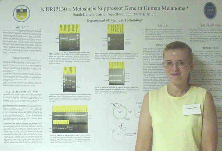
Vitamin D receptor (VDR) polymorphisms have been linked with breast and prostate cancers. DRIP130 is one of the 13 components of the Vitamin D Interacting Protein (DRIP) complex, which regulates transcription in cells via interaction with Vitamin D-VDR complexes. The gene encoding DRIP130 maps to ~6q22-q24, which is in a region frequently deleted in malignant melanoma. Colonies of bacteria containing plasmids with a 500 bp portion of DRIP130 and full length DRIP130 have been isolated. A Phage-derived artificial chromosome (PAC), RPCI-5-914N13, containing the DRIP130 gene sequence has also been isolated. This PAC cannot be transfected into mammalian cells because it does not contain a mammalian selection marker. Therefore, experiments to retrofit the PAC insert into the pPAC4 vector are in progress. This procedure will allow selection by blasticidin for future transfection studies to determine its effect on human melanoma metastasis.
Establishing a Panel of Antibodies
to Integrin Subunits
to Study Eristostatins Interaction with Melanoma
Cells.
Melissa Kuchar, Carrie Paquette-Straub, Mary Ann McLane, Department
of Medical Technology
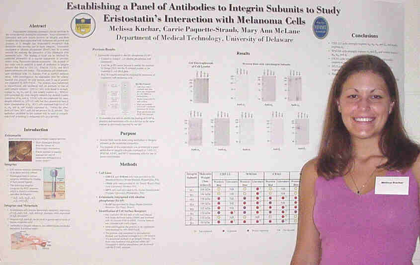
Experimental melanoma metastasis can be inhibited by the venom-derived disintegrin eristostatin. Since eristostatins interaction with cells usually involves an integrin, and these receptors have been associated with melanoma cell growth and invasion, it is thought that eristostatins interaction with melanoma cells involves one or more integrins. Eristostatin conjugated to alkaline phosphatase (ErAP) may be a useful material for studying the interaction of this disintegrin with melanoma cells. The binding of ErAP can be inhibited by unlabeled eristostatin in a manner comparable to previous studies using fluorescent-labeled eristostatin. The purpose of this study was to establish a panel of antibodies to integrin subunits that bind to 1205 LU, WM164, C8161, and MV3 human melanoma cell lysates. The melanoma cell membranes were solublized with 1% Nonidet P-40 in HEPES buffered saline. After centrifugation, the supernatant from the cellular material was assayed for total protein, and 25 m g of protein was separated by SDS-PAGE. The proteins were transferred to nitrocellulose and incubated with an antibody to one of eight integrin subunits. 1205 LU cells were found to strongly express a3, a4, a5, and b3, and weakly express av. WM164 cells possessed the same integrin subunits but showed weaker expression of a3 and a5. C8161 cells also expressed the same integrin subunits as 1205 LU cells, but they appeared to have a lower concentration of a4. MV3 cells expressed high levels of a3, a4, and a5 and weakly expressed av. Unlike the other three cell lines, MV3 cells did not possess the b3 subunit. The antibodies identified by this method will be used to compete with ErAP in binding to melanoma cells on a dot blot.
Connexin 43 Expression in Human
Melanoma Cell Lines
Lauren Palmer, Carrie Paquette-Straub, and Mary Beth Miele,
Department of Medical Technology
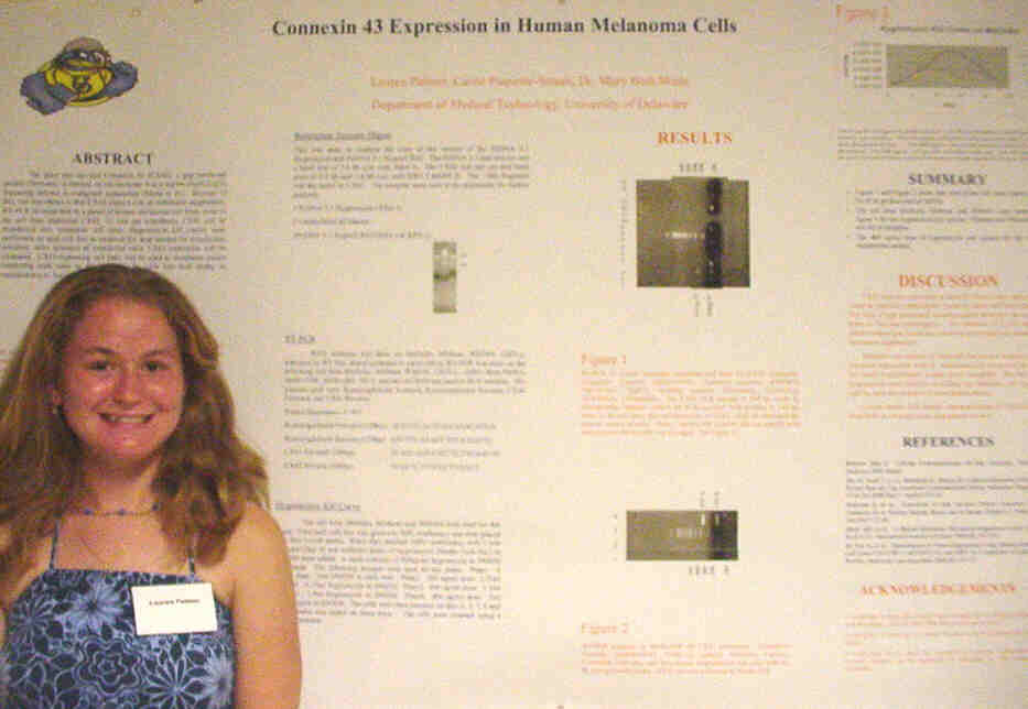
Connexin 43 (CX43), a gap junctional protein, is located on chromosome 6 in a region (6q21-q23) frequently deleted in malignant melanomas. Because of this, our hypothesis is that CX43 plays a role in metastasis suppression. RT-PCR revealed that in a panel of human melanoma cell lines, none of the cell lines expressed CX43. To test our hypothesis, CX43 will be transfected into metastatic cell lines. Hygromycin kill curves were performed on each cell line to establish the dose needed for transfection selection. After selection of transfected cells, CX43 expression will be measured. CX43-expressing cell lines will be used in metastasis assays employing nude mice to determine if the cells lose their ability to metastasize (i.e., metastasis suppressed).
Gene Expression on Chromosome 6
Courtney Smith, Carrie Paquette-Straub, Mary Beth Miele, Dept.
of Medical Technology
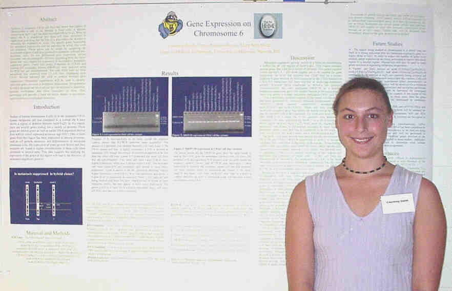
Analysis of metastatic C8161 cell lines has shown that regions of chromosome 6 (chr 6) are missing in these cells. A deletion occurring at 6q16.3-q23 has been described. When an intact chr 6 is transferred into these cell lines, metastasis is suppressed indicating that the loss of a gene within the deletion is necessary for metastasis to occur. The specific genes responsible for metastasis suppression and the pathways by which they work are unknown. These genes can be found by comparing the expression of gene(s) and gene products in metastatic cells and non-metastatic cells. To test differential gene expression, various metastatic and non-metastatic cell lines originating from the C8161 parent line were studied for expression of two putative metastasis-suppressor genes. These two genes, Connexin 43 (Cx43) and Vitamin D3 interacting protein (DRIP130), were analyzed using RT-PCR and gel electrophoresis. The total RNA used for these procedures was collected from 15 cell lines originating from C8161. Results indicated the need to analyze multiple gene interactions involved in regulation of metastasis suppression. Microarray experiments will be used to identify additional genes involved in melanoma progression. The pathways in which the genes are involved can then be analyzed to determine possible mechanisms that allow metastases to form. This knowledge will provide a basis for future studies in prevention and/or treatment of malignant melanoma.
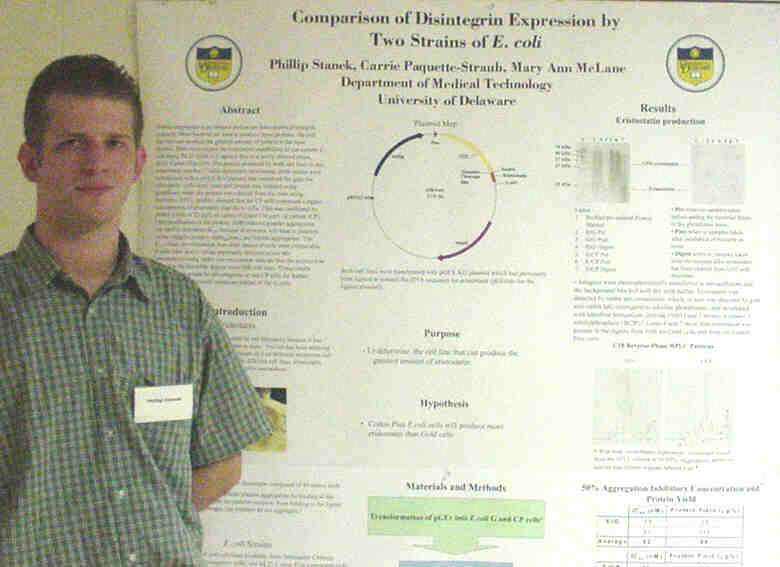
Protein expression is an integral part of our laboratory's disintegrin research. Since bacteria are used to produce these proteins, the cell line that can produce the greatest amount of protein is the most desired. Here we compare the expression capabilities of our current E. coli strain, BL21 Gold (G), against that of a newly derived strain, BL21 Codon Plus (CP). The protein produced by both cell lines in this experiment was the 7.5 kDa disintegrin eristostatin. Both strains were transformed with a pGEX-KG plasmid that contained the gene for eristostatin. Cells were lysed and protein was isolated using glutathione resin; the protein was cleaved from the resin using thrombin. HPLC profiles showed that the CP cells expressed a higher concentration of eristostatin than the G cells. This was confirmed by protein yields of 23 µg/L of culture (G) and 116 µg/L of culture (CP). Upon purification of the protein, ADP-induced platelet aggregation was used to determine IC50, because eristostatin will bind to platelets via the integrin receptor "IIb$ 3, and inhibit aggregation. The IC50 values for eristostatin from both strains of cells were comparable to each other and to values previously obtained in our lab. Immunoblots using rabbit anti-eristostatin indicate that the protein was present in the thrombin digests from both cell lines. These results suggest that it would be advantageous to use CP cells for further expression of eristostatin mutations instead of the G cells.
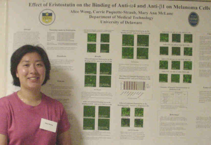
The disintegrin eristostatin, a protein isolated from the venom of
the viper Eristocophis macmahoni, has been shown to inhibit experimental
melanoma metastasis in a mouse model. The receptor for eristostatin is
unknown, but eristostatin may inhibit melanoma cell metastasis by
binding to the integrin alpha4beta1. Expression of alpha4beta1 increases
in metastatic melanoma cells, and antibodies to alpha4 have been shown
to inhibit melanoma cell metastasis. In this study, we examined the effect
of disintegrins on the binding of alpha4 to melanoma cells. We expressed
bacterial recombinant eristostatin, echistatin, and seven mutations targeting
amino acids in the RGD loop. Human MV3, 1205 LU, and WM164 melanoma cells
were incubated with fluorescent-labeled anti-alpha4 and an unlabeled disintegrin
and were observed by confocal microscopy. Eristostatin was able to inhibit
binding of alpha4beta1 to MV3 and 1205 LU. Truncated eristostatin inhibited
binding of anti-alpha4 to WM164 cells when asparagine at position 31 was
replaced with a methionine. Echistatin was unable to inhibit binding of
anti-alpha4 to the three melanoma cell types. These findings suggest that
the RGD motif of eristostatin is required to prevent anti-alpha4 from binding
to MV3 cells, and that the asparagine at position 31 is necessary for eristostatin
to prevent the binding of anti-a4 to 1205 LU cells. The RGD sequence and
the tryptophan at position 30 are critical for eristostatin to prevent
the binding of anti-alpha4 to WM164 cells. In a similar study, the effect
of disintegrins on the binding of anti-beta1 to MV3, 1205 LU, WM164, and
C8161 melanoma cells was examined. Finally, characterization of a number
of integrins present on the surface of the melanoma cells was determined
using antibodies to alphavbeta3, alpha2, and alpha6.
Poster
Abstracts for students working
in
the Department of Chemistry and Biochemistry
Blue Poster Nos. 7 12
Structural and Mechanistic Enzymology
of N-Terminal ThiI
Kevin Crotty, Matthew Kuhl, and Eugene Mueller, Department of
Chemistry and Biochemistry
Relatively little is known about RNA modification reactions and the
metabolism of sulfur-containing biomolecules, and s4U generation
provides a paradigm for both of these critical, ubiquitous biochemical
processes. The s4U generation in tRNA imparts near-UV sensitivity
to tRNA through a photo-induced crosslink with cytidine-13. The formation
of s4U is catalyzed by two enzymes, IscS and ThiI. ThiI from
E. coli appears to have a large N-terminal and a smaller C-terminal
domain. N-Terminal domain of ThiI contains a "P loop" motif that is found
in a family of enzymes called the PPi synthatase family. The
"P loop" motif catalyzes the adenylation of carbonyl groups for oxygen
substitution. This poster presents experiments to determine the roles of
the N-terminal and C-terminal domains of ThiI in the activation of O4
uridine in the sulfur transfer.
The Characterization of Animal
Glues
by Thermal Desorption Gas Chromatography Mass Spectroscopy
Michele Jablonski, Michael Lassman, and Burnaby Munson, Dept
of Chemistry and Biochemistry

Forensic scientists and art conservators occasionally need to identify
glues or other binding media. A reliable identification procedure could
be useful to detect fraudulent or highly restored pieces. Animal glues
are high molecular weight proteins. Many modern glues are high molecular
weight polymers. Information on the composition of glues is not readily
available. Samples of different types of glues were decomposed (pyrolyzed)
in a Thermal Desoprtion unit (SIS). The volatile products were trapped
at the front of a gas chromatographic column, separated and detected by
temperature programmed gas chromatography/mass spectroscopy (HP). Thermal
desorption (decomposition) conditions (temperature and time) were varied
for the glues. Chromatographic conditions (initial temperatures and temperature
program) for the separation were also varied. Chromatograms of products
from samples of rabbit skin glue, aged hide glue and white glue that were
decomposed under similar conditions were reproducibly different.
Phosphines as Substrates of Sulfhydryl
Oxidase: Studies with Model Disulfides
James Psathas and Colin Thorpe, Department of Chemistry and
Biochemistry
Water soluble phosphines, such as tris-(2-carboxyethyl)-phosphine
(TCEP) and tris-(2-cyanoethyl)-phosphine (TCNP), will reduce disulfide
bonds. TCEP is a good substrate of sulfhydryl oxidase; however, the closely
related TCNP is not a significant substrate. Kinetic studies using a rapid-reaction
stopped-flow spectrophotometer show that TCEP reacts 30-100 times faster
than TCNP with a range of model disulfides. These reactions are first-order
in both phosphine and disulfide. The reactivity of phosphines presumably
depends on the rate-limiting nucleophilic attack of a phosphine on a disulfide.
Therefore, the nucleophilicity can be quantified by the pKa value of the
phosphorus atom. The pKa of TCEP was determined to be 7.8 by P31
NMR a value much higher than that quoted for TCNP (pKa=1.3). This pKa
shift correlated with the increased nucleophilicity of TCEP as expected.
These model studies explain, in part, why TCEP is a much better substrate
of sulfhydryl oxidase.
Will introduction of an E.
coli protein lead to 4-thiouridine in yeast tRNA?
Jaimie Robinson and Eugene Mueller, Department of Chemistry
and Biochemistry
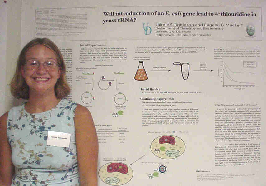
In E.coli, the proteins IscS and ThiI work together in the
transformation of the uridine at position 8 of tRNA into 4-thiouridine
(s4U). The s4U acts as a near-UV sensor forming a
crosslink with cytidine-13 on the same tRNA, thus leaving the tRNA a poor
substrate for amino acyl synthetases. Examined eukaryotes contain neither
s4U nor ThiI homologs, but many have IscS homologs, such as
Nfs1p in S. cerevisiae. This project examines the ability of Nfs1p
to substitute for IscS as a first step towards generating s4U
in yeast by expressing thiI. As Nfs1p has been characterized as
both mitochondrial and nuclear, the mitochondrial targeting sequence from
Nfs1p is being fused to thiI in a yeast expression vector. To conduct
in
vitro tests, Nfs1p is being expressed in E.coli cells.
Designing Molecules to Compensate
for Mutations in the Thyroid Hormone Receptor
Aditya Sharma, Marc C. Putnam, and John T. Koh, Department of
Chemistry and Biochemistry
The disease RTH occurs due to mutations in the thyroid hormone receptor.
Many of these mutations happen at the ligand-binding domain or near regions
involved in ligand-dependent gene activation. Proper receptor activity
can be recovered by rationally designing ligands that target the receptor
and compensate for the mutation.
Selective Reductions of Conjugated
Ketones with Control of
Absolute Configuration Using "TADDOL" Derivatives
Benjamin Thuma and Douglass Taber, Department of Chemistry and
Biochemistry
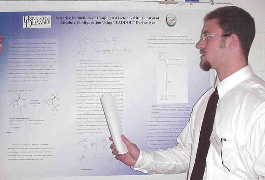
The development of highly enantioselective reducing processes for
conjugated carbonyls into their corresponding alcohols is of great importance
in biological related synthesis. The use of chiral ligands in conjunction
with reducing agents such as NaBH4 and LiAlH4 have shown to reduced conjugated
carbonyls with relatively high control of absolute configuration. The use
of a, a, a', a'-Tetraaryl-2, 2-dimethyl-1, 3-dioxolan-4, 5-dimethanol (TADDOL)
has been shown to be a highly effective moiety for production of the necessary
reducing ligand. In our research, we hope to show that "TADDOL" derivatives
5a-e complexed with LiAlH4 will selectively reduce 1, 1-dimethylethyl 7-oxo-phenylheptanoate
4 into 1, 1-dimethylethyl
(7S)-7-hydroxy-phenylheptanoate 3 in good yield with high enantioselectivity.
Investigating Isotopic Exchange
at C5 during 4-Thiouridine Biosynthesis
Todd M. Greco and Eugene G. Mueller, Department of Chemistry
and Biochemistry
Many bacteria modify uridine-8 of tRNA to 4-thiouridine (s4U),
which serves as a photosensor for near-UV light. In early s4U
generation assays using cell extracts and tRNA transcript containing [5-3H]uridine
([3H]tRNA), no [5-3H]s4U was detected
even though [35S]s4U was detected in parallel reactions
run with [35S]cysteine. What happened to the tritium? Some aminoacyl
tRNA synthetases catalyze the wash-out of tritium from [5-3H]uridine
at position 8 in tRNA, and their presence in cell extracts could explain
the lack of [5-3H]s4U. Alternatively, the tritium
wash-out might be inherent to s4U generation, which would provide
mechanistic information. To resolve the source of wash-out, s4U
generation with [3H]tRNA will be repeated using purified enzymes
rather than cell extracts. From preliminary experiments, the [3H]tRNA
appears to be radiochemically pure and competent as a substrate. Next,
s4U generation assays will be performed using the verified [3H]tRNA
substrate.
Poster
Abstracts for students working in the College of Agriculture
Blue Poster Nos. 19 21
The Study of Nitric Oxide in
the Hypersensitive Response in Arabidopsis
Chiara Ciotoli, Kyle Dorkoskie, Carrie Jacobus, Bevan Kirley,
Barbara Farnworth, and Allan Shapiro,
Department of Plant and Soil Sciences
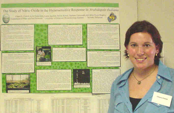
This research focuses on the plants natural defense reaction, the
hypersensitive response and the involvement that nitric oxide may have
in it. The possibility of increasing or accelerating the hypersensitive
response could be discovered through the genetic manipulation of the gene
or genes responsible for the biochemical pathway, which causes the hypersensitive
response. Arabidopsis plants were mutated two ways: through point
mutations, and through the insertion of activation tags. These two different
screens each required inoculating the plants with Pseudomonas bacteria
and an inhibitor of the biochemical pathway responsible for the hypersensitive
reaction. Positive plants from both screens are then, crossed with another
plant or self-pollinated, brought to seed, and inoculated again. The end
goal of both screens is to determine the location of the gene or genes
responsible for encoding the nitric oxide synthase enzyme.
Screening and Mapping of NOS
gene in Arapidopsis thaliana
Carrie A. Jacobus, Barbara Farnworth, Allan D. Shapiro, Department
of Plant and Soil Sciences
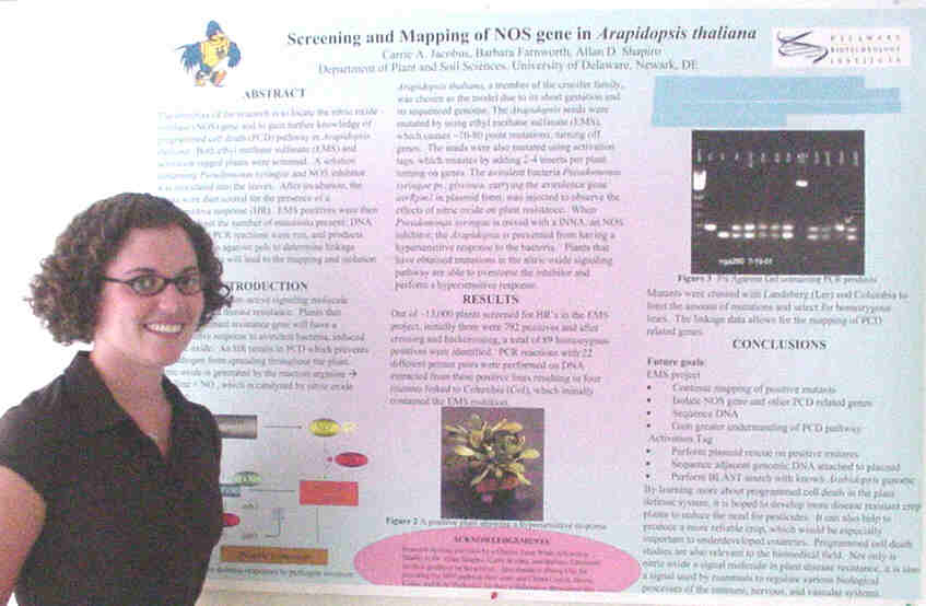
The objective of the research is to locate the nitric oxide synthase (NOS) gene in Arapidopsis thaliana. Both ethyl methane sulfanate (EMS) and activation tagged plants were screened. A solution containing Pseudomonas syringae and NOS inhibitor was inoculated into the leaves. After incubation, the plants were then scored for the presence of a hypersensitive response (HR). EMS positives were then crossed to limit the number of mutations present; DNA was extracted, PCR reactions were run, and products were then run on agarose gels to determine linkage groups. This data will lead to the mapping and isolation of the NOS gene.
Characterization of an Avian Antigen Presenting
Protein, chDEC205
Lacy Weisenberg and Calvin Keeler, Department of Animal and
Food Sciences
Macrophages are important modulators of the immune response. We have
constructed an expressed sequence tag (EST) library from an avian macrophage
cell line (HD11) stimulated with bacterial lipopolysaccharide (LPS). Over
1,200 clones from this library were sequenced and subjected to BLAST analysis.
Eleven percent of the clones represent genes with potential immunological
functions. Antigen recognition and processing is one essential step in
the immune response. One gene that we have identified from the library
and are studying in greater detail is the avian homolog to DEC205/gp200mr6.
This transmembrane protein is a member of the mannose receptor family.
It is also expressed by dendritic and thymic epithelial cells. This protein
is believed to be responsible for presenting antigens to T cells. The initial
EST clone contained only part of the DEC205 gene. We are using primers
designed from the amino acid sequences of the mammalian homologs to DEC205
in RT-PCR to clone the remainder of the avian gene. The avian DEC205 gene
is approximately 50% identical to its mammalian counterparts. In order
to study the chDEC205 protein, another aspect of this project entails the
cloning of a portion of the gene in a prokaryotic expression vector. We
will use the protein to elicit the formation of polyclonal antisera in
rabbits, which can then be used to study the synthesis and localization
of this protein in vivo.
Poster
Abstracts for students working in the Department of Chemical Engineering
Blue Poster Nos. 14 16
Relation of Protein Solubility
to Solution Thermodynamic Properties
John Bishop, Abraham M. Lenhoff, and Stanley I. Sandler, Department
of Chemical Engineering
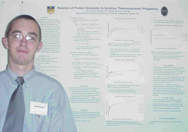
Although many scientists have proposed relationships between the
solubility of a protein and electrolyte concentration, the formulations
have all been empirical in some way. In order to find a more predictive
relationship, an expression for the partial molar Gibbs free energy of
a protein (![]() ) in an aqueous electrolyte
solution was derived. The derivation stems from the hypothesis that the
excess Gibbs free energy of the protein can be constructed from two terms:
one term accounting for the protein-solution interactions by way of the
osmotic second virial coefficient and the other term accounting for the
change in salt-solution interactions when adding protein. Satisfactory
results are seen at dilute protein concentration and low ionic strength
of electrolyte. However, calculated values of
) in an aqueous electrolyte
solution was derived. The derivation stems from the hypothesis that the
excess Gibbs free energy of the protein can be constructed from two terms:
one term accounting for the protein-solution interactions by way of the
osmotic second virial coefficient and the other term accounting for the
change in salt-solution interactions when adding protein. Satisfactory
results are seen at dilute protein concentration and low ionic strength
of electrolyte. However, calculated values of ![]() at
the solubility limit for various electrolyte concentrations were not identical
as expected. This result could be due to interactions between the solid
protein crystals and the electrolyte solution that were neglected in the
calculation. Also, the contribution from the protein-solution interaction
term was negligible indicating that the behavioral characteristics of the
protein in solution are unimportant, or that the osmotic second virial
coefficient relation is inappropriate for this model.
at
the solubility limit for various electrolyte concentrations were not identical
as expected. This result could be due to interactions between the solid
protein crystals and the electrolyte solution that were neglected in the
calculation. Also, the contribution from the protein-solution interaction
term was negligible indicating that the behavioral characteristics of the
protein in solution are unimportant, or that the osmotic second virial
coefficient relation is inappropriate for this model.
Chiral Separations Using Affinity
Ultrafiltration:
Effects of Chloride Ion and Ethanol on Selectivity
and Process Optimization
Richard Dombrowski, Jonathan Romero, and Andrew Zydney, Department
of Chemical Engineering
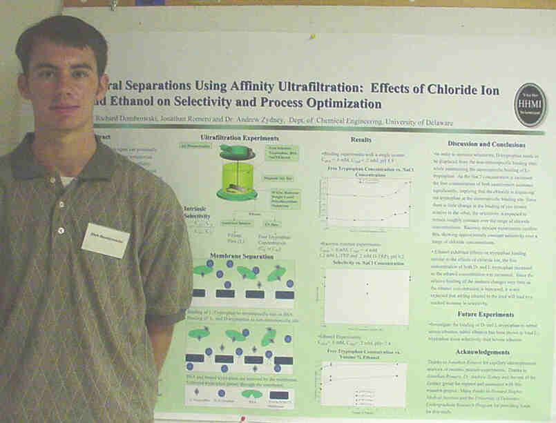
Ultrafiltration using a stereospecific binding agent can potentially
provide a high throughput, cost-effective means for commercial separation
of chiral molecules. The feasibility of using affinity ultrafiltration
for chiral separations as been demonstrated in recent studies with selectivities
as high as 11 reported for a model system of DL-tryptophan with bovine
serum albumin as the stereospecific binding agent. The goal of this study
was to investigate the effects of chloride ion and ethanol on the binding
of L- and D-tryptophan to bovine serum albumin. Ultrafiltration experiments
were performed to determine the concentration of unbound tryptophan at
various chloride or ethanol concentrations. The data showed an increase
in the free tryptophan concentration as the concentration of chloride or
ethanol was increased. Ultrafiltration experiments with a racemic mixture
at different chloride concentrations gave a maximum selectivity of 9 for
the separation of tryptophan stereoisomers.
The Creation of C496/635S
Double Mutant Tailspike
Dana Ungerbuehler and Anne Skaja Robinson, Department of Chemical
Engineering
The field of biochemical engineering is currently experiencing an
explosion of interest and importance in the world of science. In the past,
oligomeric protein folding has been understudied due to the lack of technology
with which to characterize the intermediates. As our knowledge of protein
folding increases, the complexity of the processes which drive these reactions
becomes apparent. Such is the case with P22 tailspike, a model protein
which has been studied for years. This protein is not disulfide bonded
in its final trimeric conformation, and for the first time evidence exists
for an intermediate with oxidized sulfhydryl groups not present in the
native state. In this project, the 496 and 635 cysteine residues suspected
to be involved in disulfide bonding were removed and replaced with serine
residues. This double mutant was expressed at 15, 20 and 30 degrees Celsius,
and it was found to make trimer. The highest yield occurred at the lowest
temperature, where productive folding is most favorable. In light of past
folding studies, it can be speculated that either the 613 disulfide bonds
to its parallel residue in another chain, or an alternate pathway exists
in which disulfide bonding is avoided. In conclusion, the creation of this
mutant form presents many interesting questions and more research must
be conducted to solve the mystery of the elusive disulfide- bonded intermediates.
Poster
Abstracts for students working in the Department of Biological Sciences
Red Poster Nos. 1 21
Viability of Helicobacter
pylori- Effect of Other Microorganisms
Lisa M. Attanasio and Diane S. Herson, Department of Biological
Sciences
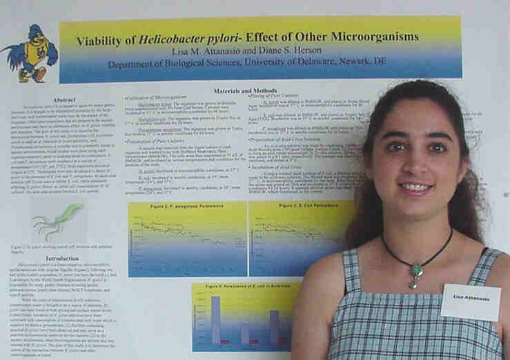
Helicobacter pylori is a causative agent for many gastric
illnesses. It is thought to be transmitted primarily by the fecal-oral
route, and contaminated water may be the source of the organism. Other
microorganisms that are present in the aquatic environment may have an
inhibitory effect on H. pylori viability and detection. The goal
of this study is to examine the interactions between H. pylori and
Escherichia
coli, a coliform which is used as an indicator of water potability,
and Pseudomonas aeruginosa, a microbe that is commonly found in
aquatic environments. Initial studies were done using each organism separately
prior to studying them in combination. E. coli and P. aeruginosa
were
incubated in a variety of temperatures (10°
, 23° , and 37°
C). Both organisms persisted longest at 37°
C. Techniques were also developed to detect H. pylori in the presence
of E. coli and P. aeruginosa. An acid-urea solution (pH 3)
was used to inhibit E. coli, while minimally affecting H. pylori.
Below an initial cell concentration of 107 cells/mL, the acid-urea
solution limited the cell growth of E. coli.
Genomic Walking of the Turtle (Pseudymes
scripta) Apolipoprotein C-III Gene
from the 3' End
Lauren Baker, Erin M. Hill, Jennifer A. Rutan, Robin Davis,
and David C. Usher,
Department of Biological Sciences
Apolipoprotein C-III (apoC-III), a protein that influences the catabolism
of triglyercide-rich lipoproteins, has previously been observed only in
mammals. A cDNA sequence was isolated from a turtle, Pseudymes scripta,
liver expression library, and was shown to be the equivalent of mammalian
apoC-III. Like mammalian apoC-III, turtle apoC-III is associated with both
high-density lipoproteins and triglyercide-rich lipoproteins and is synthesized
mainly by the liver. The human apoC-III gene is located in a linkage group
consisting of the apoA-I, apoC-III and apoA-IV genes. The apoC-III gene
is transcribed on the opposite strand of DNA as the apoA-I and apoA-IV
genes. The turtle apoC-III gene contains four exons and three
introns. The apoC-III gene has been sequenced from the 3' end to
intron 2. Therefore, leaving the promoter/enhancer region, exon 1 and intron
1 to be sequenced. My goal is to sequence that region and then eventually
"walk" toward where apoA-IV should be located. A 1.1 kb KpnI fragment,
0.9 and 1.2 kb KpnI fragments and a 1.5 kb SacI fragment were found that
should span from intron 2 to the promoter/enhancer region. The 1.1 and
1.5 kb fragments were purified, cloned, transformed and sequenced. However,
the sequences were not as expected due to the vector being rearranged.
In order to correct this, the 1.1 and 1.5 kb fragments were reamplified,
so cloning and transformation could be performed again. In the future,
primers will be designed from the promoter/enhancer sequence to allow for
"walking" toward the apoA-IV gene to occur.
Spontaneous Ovarian Contraction
in some Teleost Fish
Sara B. Callaway and Malcolm H. Taylor, Department of Biological
Sciences
Contraction of ovaries has been demonstrated in the mummichog, Fundulus
heteroclitus and several other teleosts. We have expanded this information
by recording spontaneous contractions and the response to acetylcholine
in ovaries of the striped killifish, Fundulus majalis; the Atlantic
silverside, Menidia menidia; the pumpkinseed, Lepomis gibbosus;
and the goldfish, Carassius auratus suspended in a smooth muscle
bath containing a modified Ringer's solution. Ovaries of all five species
produced similar contractions in response to 10-5 M acetylcholine. Spontaneous
contractions in F. majalis, and M. menidia occurred in a
series of single peaks with frequencies and amplitudes similar to the pattern
previously observed in F. heteroclitus. Contractions observed in
C.
auratus appeared in single peaks with an increased frequency and amplitude
compared to F. heteroclitus. Those seen in L. gibbosus were
more variable and had greater amplitude, typically with a series of superimposed
higher frequency contractions. These results suggest that ovarian contractions
may be a common phenomenon in the Atherinidae and Fundulidae, which share
many reproductive characteristics, including the occurrence of semilunar
reproductive cycles. The Centrarchidae are nest-building Percoids, which
are taxonomically distant from the other species analyzed.
Susceptibility of Ribozyme
Transfected ROS 17/2.8 Cells to Apoptosis
Laurelle Cheeseboro, Lincoln University and Cindy Farach-Carson,
Dept. of Biological Sciences
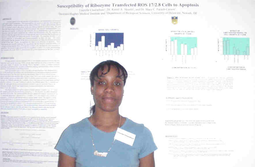
Calcium signals in the cell promote cell proliferation, cell differentiation
cell excitation and further signal transduction. The major pathway for
calcium to enter the cell is through the plasma membrane voltage sensitive
calcium channel (VSCCs). A major subunit of this channel that we studied
is the a1c that serves as the pore
forming subunit for calcium entry. We used three sub lines in these experiments:
ROS17/2.8, parental line, ribozyme transfected ROS17/2.8, and ribozyme
control. A ribozyme that eliminates the a1c
transcript
and a control scrambled ribozyme were previously transfected into cultured
rat osteosarcoma cells. We examined the effect of ribozyme against the
VSCCs on basal growth, and we concluded that the presence of ribozyme in
the ROS17/2.8 affects the growth of these cells. We examined the effect
of two steroid hormones (17-b -estradiol and
2-methoxyestradiol) on these cell lines. Our results demonstrated that
the presence of the active ribozyme does not affect the hormone-stimulated
growth of the cells. Lastly we examined the effect of the ribozyme on apoptosis.
These experiments are currently underway. We hypothesize that the ribozyme
transfected ROS17/2.8 will grow slower than the ROS 17/2.8 parental or
control cells, and might be more susceptible to apoptosis. Our results
to date indicate that this hypothesis is likely to be correct, demonstrating
the essential role of the VSCC in osteosarcoma cell growth.
The Role of L1-CAM in Glioma Cell
Invasion
Alexandra Cretu and Deni Galileo, Department of Biological Sciences
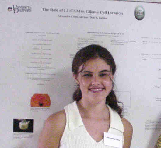
Cell adhesion molecules play an important role in cell-cell interactions
during many critical stages of embryogenesis as well as tissue repair in
the nervous system. One such molecule is L1, a transmembrane recognition
molecule that belongs to the immunoglobin supergene family and mediates
dynamic neurological processes such as neural cell migration, neurite extension
and adhesion. This cell adhesion molecule may facilitate in the invasiveness
of glioma brain tumors. By modifying the L1 expression directly in several
human and rat cell lines, the impact of this molecule on the invasiveness
of glioma brain tumors will be better understood. For rat 9L gliosarcoma
cell line, which does not express L1, the chick homologue NgCAM was stably
transfected into these cells. For rat C6 and human U-87 glioma cells, which
heterogeneously express L1, cells will be sorted to positive homogeneity
utilizing FACs. Then, L1 will be attenuated by direct gene conversion followed
by cell sorting. Invasiveness of the original tumor cells will be compared
to the cells that are modified for L1 expression. For in vivo experiments,
tumor cells are injected into the early chick embryo brain. After allowing
the injected embryos to develop for several days to two weeks, the distance
of migration is measured in tissue sections. For future experiments, in
vitro assays will also be performed by adding tumor cells to brain cell
aggregates and measuring the distance of tumor cell migration intothe
aggregate.
Cloning and Purification of DNA
polymerase a/primase and Replication Protein
A
Amanda K. Debes, Rupa Roy, Rebekah Parsons, and Daniel T. Simmons,
Dept. Biological Sciences
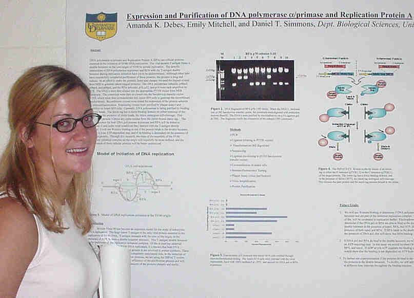
DNA polymerase a/primase and Replication
Protein A (RPA) are cellular proteins essential to the initiation of SV40
DNA replication. The viral protein T antigen forms a double hexamer on
the core origin of SV40 to initiate replication. The specific interactions
of DNA polymerase a/primase and RPA with the
T-antigen double hexamer during replication initiation have yet to be undetermined.
Although other labs have successfully completed purification of these proteins,
the process is long and tedious. In an effort to make the proteins faster
and cheaper we used the Impact system from NEB to generate intein-tagged
proteins. The DNA polymerase subunits; p48pol, p58pol, and p68pol, and
the RPA subunits; p70, p32, and p14 were each amplified by PCR. The DNAs
were then cloned into the appropriate PTYB vector from NEB constructs.
The constructs were then recloned into the baculovirus transfer vector
p1393, which were then co-transfected into insect SF9 cells to generate
the recombinant baculoviruses. Recombinant viruses were tested for expression
of the protein subunits by immunoflourescence. Expressing viruses were
purified by plaque assays and amplified on insect SF9 cells. Currently,
DNA polymerase is being purified by binding to chitin beads. The intein
tag has a chitin-binding domain on either terminus of the protein. In the
presence of chitin beads, the intein undergoes self-cleavage. This releases
the protein without any extra residue from the chitin-bound intein tag.
The purified protein for both DNA polymerase a/primase
and RPA will be tested to determine if and under what conditions they interact
with the T-antigen double hexamer. I will use Western blotting to test
if the protein binds to the double hexamer, whether it is an ATP-dependent
step, and if its binding is dependant on the presence of any other protein.
Through this research, the steps of the assembly of the SV40 replication
initiation complex on the origin will hopefully be more defined, and the
involvement of these cellular proteins will be better understood.
The Role of Bcl-2 in Brain Cell
Migration and Survival
Melanie Evans and Deni Galileo, Department of Biological Sciences
During brain development neurons migrate along radial glia to their
final destinations. Throughout migration, a signaling pathway, involving
interactions betweens substrates and integrins, facilitates migration and
suppresses apoptosis ( a normal period of widespread cell death.) In other
systems, integrin substrate interactions are known to cause expression
of the anti- apoptotic protein Bcl-2. Bcl-2 is expressed in early chick
optic tectum (midbrain), but its specific role in the signaling pathway
is uncertain. Immunostaining at various embryonic days (E) shows that Bcl-2
is present in the optic tectum, but the distribution in neurons along radial
glia is still unclear. Aside from identifying Bcl-2 in tissue sections,
cultured cells on cover slips were also stained and analyzed by confocal
and fluorescence microscopy. Further immunostaining along with analysis
by flow cytometry will be performed to further analyze Bcl-2, focusing
on E6, before migration takes place, and E9, after mass cell death occurs
(E7.5-E8). After the tissue has been analyzed for Bcl-2, its role in apoptosis
and migration will be tested in vivo by expressing the protein using
both a replication competent and replication incompetent retroviral vector.
Neuronal progenitors will be infected with the Bcl-2 retroviral vector
to determine its effect on the number of neurons that survive within a
clone throughout the period of early cell death. It is hypothesized that
the Bcl-2 expressing clones will contain statistically significant greater
numbers of neurons and they will be distributed differently. This will
show that Bcl-2 plays an important role in the migration of neurons up
radial glia during development.
The Involvement of Extracellular
Matrix Molecules in Neuronal Migration and Survival
Erin Henniger and Deni Galileo, Department of Biological Sciences
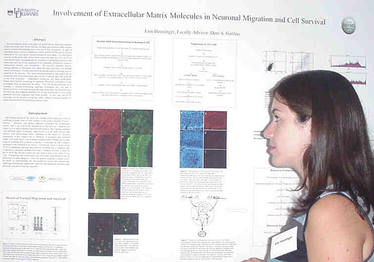
Neurons migrate along radial glial cell guides during brain development.
It has previously been shown that the cell adhesion molecule a
8b 1-integrin and an unidentified substrate
play a key role in this interaction. In order to determine exactly which
extracellular matrix (ECM) protein acts as the key substrate in this mechanism
several procedures were taken. Cryostat tissue sections of the chick optic
tectum from embryonic ages day 6-12 (E6-E12) were double-label immunostained
for vimentin (a cytoskeleton protein in the radial glia) and one of the
suspected ECM molecules: fibronectin, tenascin, osteopontin, laminin, vitronectin.
The sections indicated specific staining patterns of fibronectin (Fn) along
the radial glia and a less-defined pattern for tenascin. Although the other
ECM molecules were present in the sections, positive staining was not apparent.
The same immunolocalization was carried out in suspended cells of the optic
tectum from various ages and analyzed on the flow cytometer. Quantitative
results are still being determined. Future aims include attempting to attenuate
fibronectin expression in the developing brain to determine the effect.
This will be accomplished by injecting a fibronectin-blocking antibody
hybridoma cell line and a retroviral vector in ovo and allowing
the brain to develop. It is hypothesized that blocking the proposed substrate
(Fn) in the developing OT will cause abnormal neuronal migration and brain
growth. In this way, the ECM molecules can be identified as the key a
8b 1-integrin substrate needed for
proper interactions of the migrating neuronal cells.
Progress of the sequencing of the
Apolipoprotein A-II gene in Pseudemys scripta
Erin M. Hill, Lauren
E. Baker, Liz Manning, Stacey Karr, Robin Davis,
Gregory Stephens, Robert Hodson, and David Usher, Department of Biological
Sciences
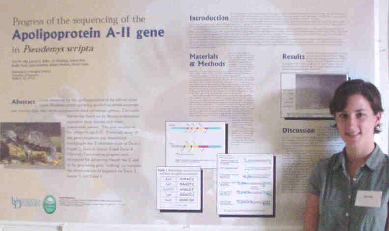
Gene sequences for the apolipoproteins of the red-ear slider turtle
Pseudemys
scripta are being studied to obtain structure and function data that
can be compared to other vertebrate species. The turtle was chosen based
on its distant evolutionary separation from human and other mammalian species.
The gene studied in this project is apoA-II. Previously some of this genes
sequence was determined, including in the 3 direction most of Exon 2,
Intron 2, Exon 3, Intron 3, and Exon 4. Currently I am making progress
with techniques for advancing toward the 5 end of the gene using gene
"walking" to complete the determination of sequence for Exon 2, Intron
1, and Exon 1.
A Possible Oncogenic Function
of the Novel Protein CIBB23.3
Melissa Krupski and Ulhas Naik, Department of Biological Sciences
To attain a better understanding of cancer, it is crucial for researchers
to continue identifying and characterizing oncogenes. Although originally
identified through platelet aggregation studies, the novel calcium and
integrin binding protein CIB became linked to oncogenesis when found to
be expressed in breast cancer cell lines. Inquiry into CIBs role in breast
cancer resulted in the isolation of the second novel protein CIBB23.3 (CIB
binder, clone 23.3) by a yeast-two-hybrid screening of the mammary epithelial
cell library. Preliminary focus formation studies showed the ability of
NIH3T3 cells, transfected with a mammalian expression vector (pcDNA 3.1)
containing 23.3 to form foci. The purpose of this investigation is to demonstrate
or disprove the possible oncogenic function of 23.3 however, the initial
experimentations were aimed at confirming the preliminary data. NIH3T3
cells were transfected with the vector containing 23.3, using Kras, a known
oncogene, as a positive control and the empty vector as a negative control.
The foci were allowed to develop for a duration of fourteen days, after
which the results were quantified. Although the data is inconclusive due
to difficulties with the controls, foci were present in abundance on plates
containing the 23.3 over-expressed cells. Though these results appear promising,
modification and repetition of this assay is necessary. Furthermore, to
determine if 23.3 is indeed an oncogene, additional assays commonly used
for this purpose (i.e. Soft agar colony formation assays, gene expression
assays) will be performed.
Characterization of Pre-Adipocyte
Cell Gene Expression using the Mouse 3T3 Cell Line
Devon Lump, John David, and David Usher, Department of Biological
Sciences
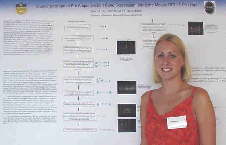
We are using the 3T3 L1 cell line from mice to characterize preadipocyte
gene expression. By constructing a SAGE (Serial Analysis of Gene Expression)
library from the 3T3 L1 cell line as well as four libraries using adipocyte
cells from two human samples we hope to gain further knowledge of adipocyte
gene expression and aid in the determination of what genes lead to the
onset of CAD. We began by isolating RNA, which we reverse transcribed to
cDNA. Subsequent modifications to the cDNA yielded a 13 bp tag, which can
be used to identify a unique transcript. These tags were ligated to form
ditags with adapters, which were later amplified. The adapters were then
cleaved and the ditags were purified and ligated to form concatemers, which
range in size from 200 bp 2 kb. The 500 800 bp region was found to
be the most effective for cloning. PCR was used to screen the transformants,
of which 40% were found to have inserts larger than 250 bp, equivalent
to approximately 10 ditags. We have accumulated more than 100 samples for
sequencing, resulting in 3,000 or more tags per library. Our goal is to
accumulate at least 30,000 tags per library, which is a sufficient number
of tags to identify unique gene transcripts. We hope to analyze both the
pre adipocyte and adipocyte 3T3 L1 cells for comparison purposes and to
use the information we gather to base our human adipocyte libraries from.
Eventually we will be looking at unique protein expression in both types
of human adipocyte cell gene sequences to learn more about how gene expression
correlates to the different hereditary and acquired coronary artery diseases.
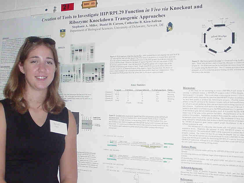
Heparin/Heparan Sulfate (HP/HS)-binding proteins (HIP) have been
linked to a variety of cell functions in vitro including growth regulation,
modulation of blood coagulation and cell adhesion. It is also identical
at the amino acid level to a previously described murine ribosomal protein.
To create a model in vivo, chimeric male mice carrying a line of cells
heterozygous (+/-) for HIP/RPL29 and agouti coat color were mated with
C57BL6 females (black coat color). Possible transmission of the mutation
can be identified in offspring by agouti coat color and a positive identification
through PCR and Southern Blot analysis. A single agouti mouse has
been produced thus far and data seems to show a negative result.
However, the ambiguity of the results makes a positive ID impossible at
this point. The other approach used, ribozyme technology, lowers
the amount of HIP/RPL29 mRNA leaving the nucleus by cleaving its specific
transcript. Transgenic mice carrying this ribozyme within their cells
will most likely be a more successful approach than the knockout mice.
Due to the widespread function of HIP/RPL29, it is possible that loosing
even one copy of the gene could be lethal. Therefore a knockout mouse
would not be able to survive embryogenesis. Production of the transgenic
mice should be a better investigative tool into the function of HIP/RPL29
in vivo.
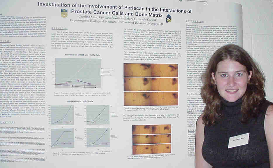
Because prostate cancer preferentially metastasizes to bone and exhibits
osteoblastic features, it is important to understand the bone microenvironment
to know how and why this occurs. Perlecan, an extracellular matrix HSPG
most likely has a role in interactions between prostate cancer and bone
matrix stromal cells. A ribozyme has been developed to reduce the production
of perlecan by cutting the transcript and is used in in vitro studies to
verify whether perlecan affects growth and proliferation of prostate cancer
cells in bone like conditions. The bone environment in vitro was
represented by human bone marrow stromal cells. Proliferation and motility
of prostate cancer cells, both normal and ribozyme transfected cells, were
analyzed in different conditions. No difference in growth was observed
between the parental line and the ribozyme-transfected clones. Wound healing
assays did not allow us to establish evidence of reduced motility of the
ribozyme-transfected prostate cancer cells. Despite some problematic results,
we obtained postive indications regarding the in vitro modeling
of the bone environment.
Characterization of P38 Protein
Found in Pseudemys scripta
Adrienne Nash, William Cain, Donna Maslak, Robert Hodson, and
David C.Usher
Department of Biological Sciences
Coronary artery disease is the leading cause of death in the United
States (1). Subsequently, an increased knowledge in this field is crucial
to developing treatments. The focus of this research is to identify and
characterize the lipoproteins of the red-eared slider, Pseudemys scripta,
with the expectation of gaining a better understanding of cholesterol and
triglyceride transport throughout the body. Apolipoproteins, the proteins
found on the surface of lipoproteins, direct the metabolism and the transport
of lipids in the body. The turtle was chosen because of its important evolutionary
links; many of the apolipoproteins important today evolved around the time
birds and reptiles branched off evolutionarily. A 38kD apolipoprotein (P38)
was found on a blot of an intermediate-density lipoprotein (IDL)/remnant
fraction in a turtle with a highly unusual lipoprotein profile. It is hypothesized
that P38 is apoliporptoeinA-IV (apoA-IV). The N-terminal portion of the
protein was sequenced and then synthesized (15 amino acids with a C-terminal
cysteine). The peptide was conjugated to human low-density lipoprotein
(LDL) as well as to ovalbumin. The conjugates were used to stimulate an
immune response in mice, and the antiserum was collected. In addition,
the spleen lymphocytes from an immunized mouse were fused with a B cell
myeloma line (P3) to produce hybridomas secreting monoclonal antibodies.
The titers of the immune response from the mouse after a tail IV injection
were promising (1:10,000). Although the fusion did not produce a very high
number of hybridoma primaries (64 wells with cell growth/ 372 wells plated),
the initial screen indicated that nine wells contained hybridomas secreting
anti-peptide antibodies. The future plans are to probe phage expression
libraries from the turtle intestine and liver to identify P38 in its native
form. The protein will then be sequenced and analyzed for apoA-IV homology.
Expression of RYR1 Ca2+
Release Channel in Mouse Osteoblastic Cells
Karyn G. Oberman, Jeremy M. Lyons, and Norman J. Karin, Department
of Biological Sciences
Lysophosphatidic acid (LPA) is a potent calcium signaling agent in
osteoblasts, but the endoplasmic reticulum (ER) channel that is activated
is unknown. We hypothesize that the channel responsible for Ca2+
release from the ER is the type 1 isoform of the ryanodine receptor (RYR1).
RT-PCR revealed that mouse osteoblastic cells (MC3T3-E1 cells) express
mRNA for RYR1, and the sequenced PCR product had a 99.3% similarity to
the previously published sequence of mouse RYR1. Western blot analysis
showed RYR1 protein expression in chicken breast, but not in MC3T3-E1 cells,
possibly because the expression level in the osteoblasts was too low to
be detected. RT-PCR also revealed that MC3T3-E1 cells express mRNA for
FKBP12, a binding protein closely associated with RYR1 in skeletal muscle.
Antisense experiments are underway to downregulate RYR1 expression and
test the function of the RYR1 protein in MC3T3-E1 cells.
Characterization of an N-Terminal
domain
inhibitory to DNA binding in SV-40 Large T-Antigen
Kevin M. OHayer, Rebecca Parsons, Rupa Roy, and Daniel T. Simmons
Department of Biological Sciences
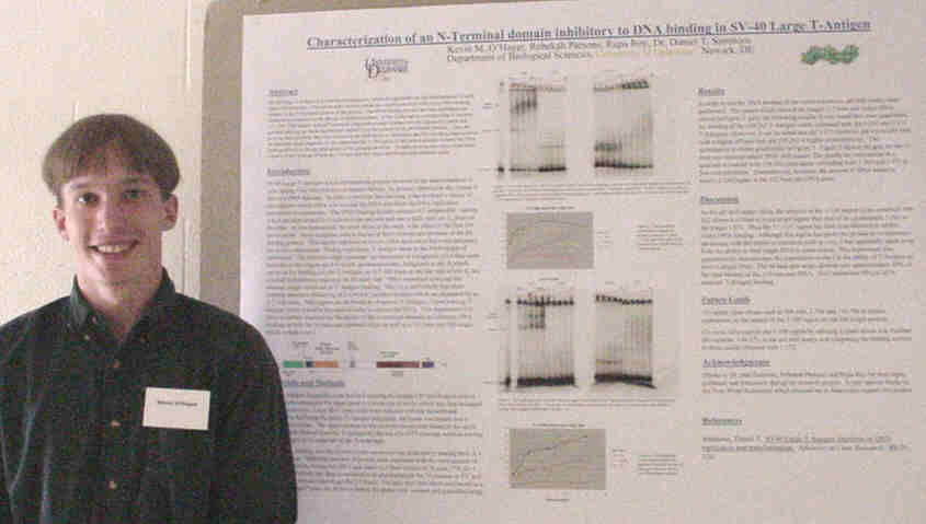
SV-40 Large T-Antigen is a multifunctional protein, which is responsible
for the transformation of cells upon viral infection. The region of the
protein, which this experiment deals with, is the DNA binding domain at
the N-Terminal portion of the protein. The DNA binding domain has been
elucidated and characterized extensively by the use of deletion mutants.
It lies in the region corresponding to residues 131-259. This inquiry utilized
N-terminal fragments created in a baculovirus expression system and purified
utilizing an intein tag that was cleaved from the protein in the purification
process. Once the proteins were purified, they were utilized in gel shift
assays to determine the DNA binding characteristics of each individual
fragment. It was found that the 1-109 region of the protein actually lowered
the DNA binding activity in the gel shift assays of the proteins involved.
Proteins missing this region were better suited to DNA binding of both
the 112 base pair full origin and 64 base pair minimal origin.
Preparation of a Standard Curve
as a Quantitative Tool
in Analyzing the Effects of Mechanical Loading on
Mouse Osteoblastic Cells
Erwin C. Puente, Joel J. Bergh, and M.C. Farach-Carson,
Department
of Biological Sciences
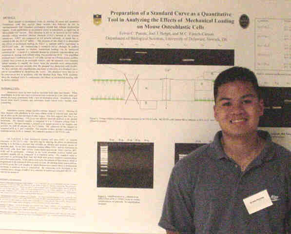
Bone adapts to mechanical stress by altering its mass and geometry.
Osteoblasts, cells that secrete bone matrix, are believed to act as mechanosensors
that transduce mechanical stimuli into cellular biochemical signals. Upon
application of mechanical stress to osteoblasts, a rapid rise in intracellular
Ca2+ occurs. This elevation is due to an increase in Ca2 influx through
voltage sensitive calcium channels (VSCC) located in the plasma membrane.
VSCC are composed of 4-5 proteins, of which the alpha1 subunit is the site
for Ca2+ influx. The purpose of this study is to determine the effects
of mechanical loading on VSCC a1 subunit mRNA
expression in MC3T3-E1 cells. Changes in mRNA expression in response to
chronic mechanical loading are done by comparing RT-PCR results to a standard
curve. A stock of alpha1 subunits standards of known concentrations was
prepared by cloning each subunit using Taq-polymerase PCR. The amplified
products were transformed into E. coli cells, spread on LB-amp plates,
positive colonies were grown in an overnight culture, and the plasmids
were isolated. Initial attempts to amplify the insert from the plasmid
were unsuccessful. Amplification was only possible after the plasmid was
linearized with EcoRV. We thus conclude that amplification and hence production
of a standard curve can be accomplished by linearizing the vector. The
standard curve has yet to be constructed due to problems with the BioRad
Real Time PCR machine. Once the standard curve is constructed, the effects
of mechanical loading will be further studied.
DNA-Protein Interactions at
Human Ori Sequences
Earl Stoddard and Daniel Simmons, Department of Biological Sciences
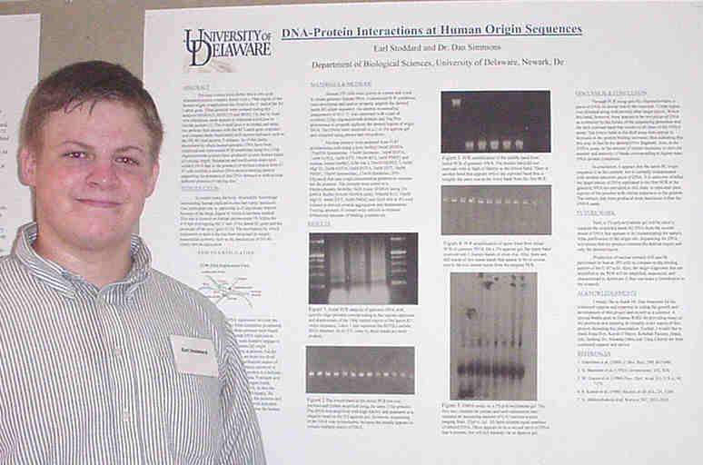
Previous studies have shown that a cell-cycle dependent protein complex
forms over a 74bp region of the human origin of replication site fixed
to the 3 end of the B2 Lamin gene. Three proteins were isolated during
this analysis (HOXA13, HOXC10 and HOXC13), but by their own admission,
none appears to represent a helicase or similar protein [Stanchina et al.(2000)
J.
Mol. Biol, 299, 667-680]. . The overall goal is to isolate and
study the proteins that interact with the B2 Lamin gene sequence and compare
them functionally with known helicases such as the SV-40 viral protein,
T-antigen. As of this point, procedures to obtain human genomic DNA have
been employed and customized PCR conditions using two 21bp oligonucleotide
primers have produced several distinct bands of varying length. Separation
and purification steps have yielded DNA that in the presence of nuclear
extracts from U-87 cells exhibits a distinct DNA-protein binding pattern
supporting the presence of and DNA interaction with several different proteins
of varying size.
The Expression Of Perlecan In
The Lens Capsule During Murine Embryonic Development
Rozie Townsend1, Patrick B. Kelley2, Melinda
K. Duncan2
1Department Biology, Delaware State University, 2Department
of Biological Sciences, Univ. of Delaware
The lens capsule is a thickened basement membrane surrounding the
lens. Since the adult capsule is known to contain the HSPG (heparan sulfate
proteoglycan) perlecan, which is proposed to be important for lens differentiation,
its developmental expression pattern was studied during murine lens development.
We hypothesized that a high expression of perlecan would be found in prenatal
murine lens development. Eyes were prepared from wildtype embryos of 9,
10, 12, 14, and 16 days post conception (dpcs) as well as newborn (nb),
1 week (wk), and 18 wk wildtype mice. These eyes were sectioned and stained
with a polyclonal anti-perlecan antibody, which was detected by immunofluorescence
labeling and confocal imagery. Perlecan was found in the earliest basement
membranes underlying the lens placode and was maintained in the lens capsule
throughout development. However, staining intensity was much higher in
embryos than in adults. Since perlecan can work as a structural protein,
a direct signaling molecule and an extracellular reservoir for growth factors,
we proposed that it plays important roles in lens development.
Helicobacter pylori Transmission
in Ground Water
Laura Vella and Diane Herson, Department of Biological Sciences
Helicobacter pylori is a Gram negative, microaerophilic bacterium
that has infected approximately half of the world's population. H. pylori
has been linked to gastric and peptic ulcer disease and has been classified
as a class one carcinogen. These factors make H. pylori a significant
health concern. The purpose of this study has been to examine the infectivity
of H. pylori after exposure to synthetic groundwater in order to
investigate this medium as a possible route of transmission. A major goal
of the labs' collaborative work has been to document the correlation between
the presence of H. pylori in ground water (especially that found
in wells of individual families) and H. pylori infection in the
individuals who come in contact with the source. Arguments against the
theory suggest that H. pylori does not remain viable in the stressed
conditions of water and that such a method of transmission is not feasible.
In order to explore further this hypothesis, a mouse model for infection
is currently being developed under the guidance of Dr. Paul Fawcett at
the A.I. DuPont Hospital for Children. Using balb/c mice, the methods of
PCR, serology (Western blots), and culturing of the stomach are being explored
as determinants of infection. Once the model is completed, the mice will
be orally inoculated with H. pylori after the organism has been exposed
to synthetic groundwater for various increments of time. Subsequent evidence
of infection would then lend support to the hypothesis that ground water
is a route of transmission.
Environmental Factors that Affect
the Persistence of
H. pylori in Water
Natalie K. Weaver and Diane S. Herson, Department of Biological
Sciences
Helicobacter pylori is a common pathogen that colonizes over
50% of the worlds population yet the principal route of transmission is
still unresolved. Although, some researchers have proposed a water-borne
route of transmission, the environmental conditions under which H. pylori
can persist in water have not been fully determined. The goal of this study
is to determine if H. pylori can persist under conditions it may
encounter in natural water environments for a period of time long enough
for it to spread and cause infection. Free-living and biofilm-associated
organisms will be studied. Persistence of free-living organisms was studied
by suspending H. pylori in a synthetic buffered moderately hard
groundwater followed by incubation under various conditions. Physical and
chemical factors considered were temperature and pH. Results have indicated
that low water temperatures lead to increased survival of H. pylori
and that a wide range of pH levels can be tolerated. Oxygen and nutrient
levels will also be studied using this system. Attachment studies were
carried out and demonstrated that H. pylori forms biofilms (Figure3).
In future studies, biofilm-associated H. pylori will be exposed
to the same environmental conditions as free-living organisms.
Isolation and Sequencing of Biologically
Active Proteins in Fundulus heteroclitus
Marlena Yost, Susan Safford, and Malcolm Taylor, Department
of Biological Sciences
Two related studies are currently being undertaken to isolate and
sequence biologically active proteins in fish. A vitamin D receptor was
studied and currently a growth hormone sequence is being studied. A vitamin
D membrane binding protein for 1,25-dihydroxyvitamin D3 was previously
isolated in chick intestine as well as carp and Atlantic cod enterocytes.
The present study tried to expand on that work by finding the analogous
binding protein in the fish, Fundulus heteroclitus. Primers that
were used to isolate the protein in the chick intestine were used to probe
for the protein in the intestine and kidney of F. heteroclitus.
The protein could not be found in the fish. The theorized reason that the
protein could not be found is that it is not highly conserved, therefore
the chick intestine primers would not work on the fish. A second project
in this study focused on growth hormone in F. heteroclitus. A partial
growth hormone DNA sequence was found for this fish by previous researchers.
The present study hopes to expand the partial sequence into a full-length
sequence. Currently, primers from the sequenced portion are being used
to isolate and sequence the remaining 5' and 3' ends of the sequence from
male and female F. heteroclitus pituitaries. The full-length sequence
will be used to create recombinant F. heteroclitus growth hormone
that will be used in further physiological studies.