

“That's calcium,” Czymmek notes. “Scientists have discovered that there is a large release of calcium with every heartbeat. If we don't see those sparks,” he notes, “you have a major problem--perhaps even heart failure.”
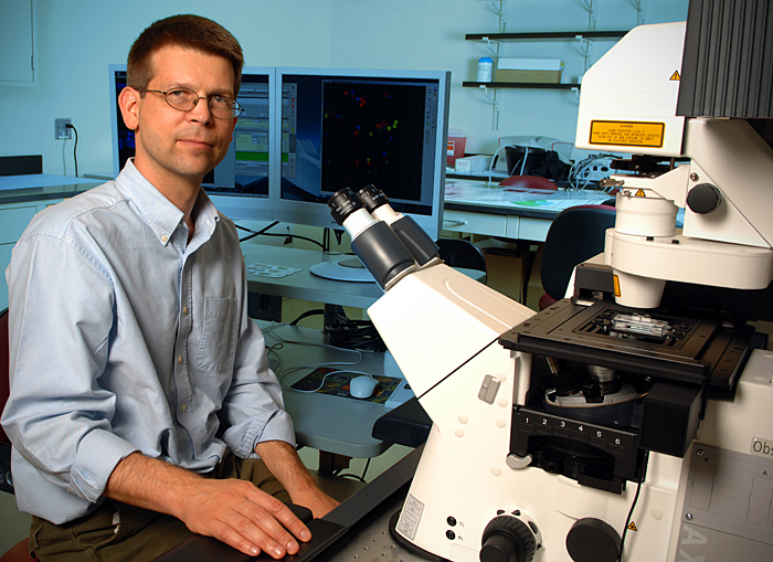
Czymmek has a bird's-eye view into the fascinating and rarely seen world of the microscopic, as director of the University of Delaware's Bio-imaging Center.
The center, a component of UD's Delaware Biotechnology Institute, is equipped with the latest technology for microscopic explorations into a diversity of intriguing subjects under investigation by University researchers, from plants that can decontaminate soils of toxic metal pollutants, to carbon nano-bombs for destroying cancer cells.
Czymmek, who also is an associate professor of biological sciences at UD, recently showcased the latest addition to the University's suite of high-tech imaging tools--a state-of-the-art laser-scanning confocal microscope. UD is among a handful of universities that own one of the million-dollar instruments.
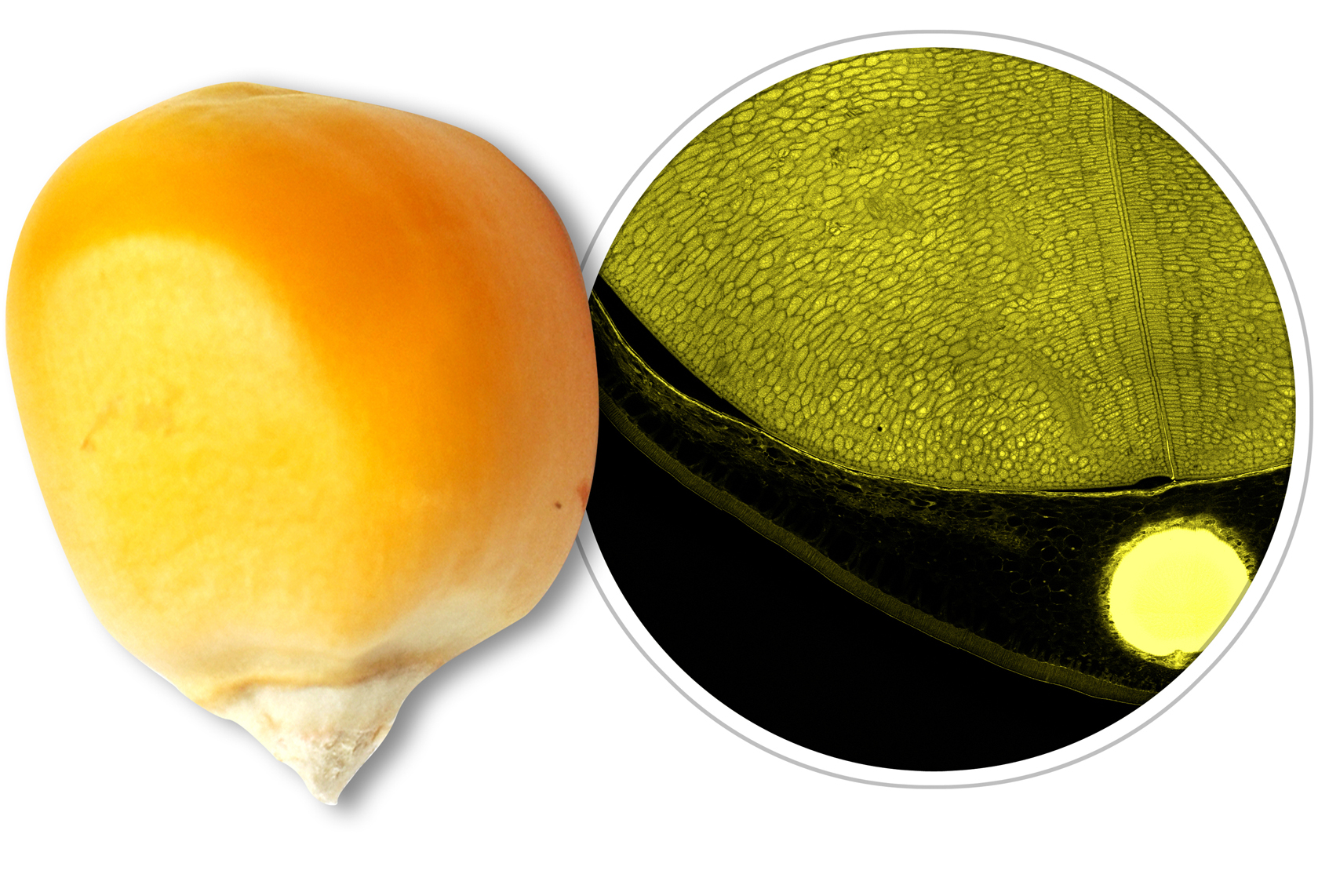
The microscope is particularly useful in examining thick samples such as muscle tissue at high resolution, Czymmek says, because a series of scans may be made at different depths within the sample and assembled automatically in minutes, yielding breathtakingly detailed, three-dimensional images, much like an MRI of the human body reveals.
“It has been my experience, that advances in analytical science often open the door to new scientific inventions and innovations,” said David Weir, director of the Delaware Biotechnology Institute. “The capability we now have with this new microscope, which allows us to observe natural processes as they occur and in great detail, will surely result in new, important discoveries.”
Currently, Czymmek and his staff--associate scientist Liz Adams and research associates Deborah Powell and Shannon Modla--are assisting UD researchers with a broad range of scientific projects on plants and fungi, vocal cords, bone health, biofilms, DNA repair, and gel-like synthetic polymers, among others.
The protein actin, shown here in living prostate cells, is responsible for cell motility. Notice the tiny feeler-like philipodia that help the cell sense its environment as it moves along. Video courtesy of Adam Aguiar and Robert Sikes, UD Department of Biological Sciences. |
An average of 175 users per year have been served at the center since it opened in 2001, according to Czymmek. UD faculty, staff and students, as well as research collaborators from industry and governmental agency partners, have all been trained in the safe and proper operation of the center's sophisticated “eyes.”
UD's Bio-Imaging Center also is an important resource for scientists beyond Delaware's borders, with colleagues from the National Institutes of Health, Johns Hopkins University, DuPont, Georgetown University, Merck and Virginia Commonwealth University attending microscopy training workshops hosted by Czymmek and his staff.
Czymmek, who refers to himself as a “jack of all trades,” has been using confocal microscopes on almost a daily basis since 1990 when they helped illuminate his doctoral studies of plant diseases and fungi.One of the things he most likes about his position at UD is its cross-disciplinary focus. He has assisted scientists in examining the hard exoskeleton of an insect, for example, to learn how to make new and improved materials.
A living bone cell shows the contraction and movement of the protein kinase from the cell cytoplasm to its surface when treated with a drug. Video courtesy of Victor Fomin and Randall Duncan, UD Department of Biological Sciences. |
“I like being able to help tie together the biology and engineering and help people figure out the best way to solve a problem,” he says.
With each new and improved tool for revealing hidden worlds, Czymmek and his staff gain a front-row seat into the formerly unknown and help put dozens of UD research studies literally into sharper focus.
“It's kind of like going out in space,” Czymmek says with a smile. “We get to see things that no one else has ever seen before.”
The Bio-imaging Center is open to UD researchers and collaborators on a fee-for-service basis. For more information, visit the center's Web page at [www.dbi.udel.edu/bioimaging/index.html].
Article by Tracey Bryant
Photo by Kathy F. Atkinson